
Advertisement
Research ArticleInflammationMetabolismNephrology
Open Access |  10.1172/JCI200755
Fumarate-induced succination of A-kinase anchor protein 12 exacerbates renal inflammation and fibrosis
Shuai Sun,1,2,3 Xu-yang Yan,2,3 Yu-hang Dong,4,5 Jian-min You,2,3 Zhen-yu Guo,2,3 Dong-xue Lv,4 Shuai-shuai Xie,4 Rui Hou,4 Xiang-yu Li,4 Ju-tao Yu,4 Xiao-yu Shen,4 Jie Wei,4 Zhen-yu Song,2,3 Zi-qi Chen,2,3 Yun-long Zhu,2,3 Xing-xin Xu,6 Juan Jin,4 Jia-gen Wen,4 Hao Wang,7 Xiao-ming Meng,4 and Wei Wang2,3
1Department of Urology, The Fifth Affiliated Hospital of Anhui Medical University, Fuyang, China.
2Department of Urology, The First Affiliated Hospital of Anhui Medical University, Hefei, China.
3Anhui Province Key Laboratory of Urological and Andrological Diseases Research and Medical Transformation, Institute of Urology;
4Inflammation and Immune Mediated Diseases Laboratory of Anhui Province, the Key Laboratory of Anti-inflammatory of Immune Medicines, Ministry of Education, Anhui Institute of Innovative Drugs, School of Pharmacy; and
5Department of Toxicology, School of Public Health, Anhui Medical University, Hefei, China.
6Department of Nephrology, The First Affiliated Hospital of Anhui Medical University, Hefei, China.
7Department of Urology, The Affiliated Jiangning Hospital with Nanjing Medical University, Nanjing, Jiangsu, China.
Address correspondence to: Wei Wang, Department of Urology, The First Affiliated Hospital of Anhui Medical University, No. 218 Jixi Road, Hefei 230032, China. Phone: 86.17755130890; Email: wangweimw@ahmu.edu.cn. Or to: Xiao-ming Meng, School of Pharmacy, Anhui Medical University, No. 81 Meishan Road, Hefei 230032, China. Phone: 86.551.65172130; Email: mengxiaoming@ahmu.edu.cn.
Authorship note: SS, XYY, YHD, and JMY contributed equally to this work.
Find articles by Sun, S. in: PubMed | Google Scholar
1Department of Urology, The Fifth Affiliated Hospital of Anhui Medical University, Fuyang, China.
2Department of Urology, The First Affiliated Hospital of Anhui Medical University, Hefei, China.
3Anhui Province Key Laboratory of Urological and Andrological Diseases Research and Medical Transformation, Institute of Urology;
4Inflammation and Immune Mediated Diseases Laboratory of Anhui Province, the Key Laboratory of Anti-inflammatory of Immune Medicines, Ministry of Education, Anhui Institute of Innovative Drugs, School of Pharmacy; and
5Department of Toxicology, School of Public Health, Anhui Medical University, Hefei, China.
6Department of Nephrology, The First Affiliated Hospital of Anhui Medical University, Hefei, China.
7Department of Urology, The Affiliated Jiangning Hospital with Nanjing Medical University, Nanjing, Jiangsu, China.
Address correspondence to: Wei Wang, Department of Urology, The First Affiliated Hospital of Anhui Medical University, No. 218 Jixi Road, Hefei 230032, China. Phone: 86.17755130890; Email: wangweimw@ahmu.edu.cn. Or to: Xiao-ming Meng, School of Pharmacy, Anhui Medical University, No. 81 Meishan Road, Hefei 230032, China. Phone: 86.551.65172130; Email: mengxiaoming@ahmu.edu.cn.
Authorship note: SS, XYY, YHD, and JMY contributed equally to this work.
Find articles by Yan, X. in: PubMed | Google Scholar
1Department of Urology, The Fifth Affiliated Hospital of Anhui Medical University, Fuyang, China.
2Department of Urology, The First Affiliated Hospital of Anhui Medical University, Hefei, China.
3Anhui Province Key Laboratory of Urological and Andrological Diseases Research and Medical Transformation, Institute of Urology;
4Inflammation and Immune Mediated Diseases Laboratory of Anhui Province, the Key Laboratory of Anti-inflammatory of Immune Medicines, Ministry of Education, Anhui Institute of Innovative Drugs, School of Pharmacy; and
5Department of Toxicology, School of Public Health, Anhui Medical University, Hefei, China.
6Department of Nephrology, The First Affiliated Hospital of Anhui Medical University, Hefei, China.
7Department of Urology, The Affiliated Jiangning Hospital with Nanjing Medical University, Nanjing, Jiangsu, China.
Address correspondence to: Wei Wang, Department of Urology, The First Affiliated Hospital of Anhui Medical University, No. 218 Jixi Road, Hefei 230032, China. Phone: 86.17755130890; Email: wangweimw@ahmu.edu.cn. Or to: Xiao-ming Meng, School of Pharmacy, Anhui Medical University, No. 81 Meishan Road, Hefei 230032, China. Phone: 86.551.65172130; Email: mengxiaoming@ahmu.edu.cn.
Authorship note: SS, XYY, YHD, and JMY contributed equally to this work.
Find articles by Dong, Y. in: PubMed | Google Scholar
1Department of Urology, The Fifth Affiliated Hospital of Anhui Medical University, Fuyang, China.
2Department of Urology, The First Affiliated Hospital of Anhui Medical University, Hefei, China.
3Anhui Province Key Laboratory of Urological and Andrological Diseases Research and Medical Transformation, Institute of Urology;
4Inflammation and Immune Mediated Diseases Laboratory of Anhui Province, the Key Laboratory of Anti-inflammatory of Immune Medicines, Ministry of Education, Anhui Institute of Innovative Drugs, School of Pharmacy; and
5Department of Toxicology, School of Public Health, Anhui Medical University, Hefei, China.
6Department of Nephrology, The First Affiliated Hospital of Anhui Medical University, Hefei, China.
7Department of Urology, The Affiliated Jiangning Hospital with Nanjing Medical University, Nanjing, Jiangsu, China.
Address correspondence to: Wei Wang, Department of Urology, The First Affiliated Hospital of Anhui Medical University, No. 218 Jixi Road, Hefei 230032, China. Phone: 86.17755130890; Email: wangweimw@ahmu.edu.cn. Or to: Xiao-ming Meng, School of Pharmacy, Anhui Medical University, No. 81 Meishan Road, Hefei 230032, China. Phone: 86.551.65172130; Email: mengxiaoming@ahmu.edu.cn.
Authorship note: SS, XYY, YHD, and JMY contributed equally to this work.
Find articles by You, J. in: PubMed | Google Scholar
1Department of Urology, The Fifth Affiliated Hospital of Anhui Medical University, Fuyang, China.
2Department of Urology, The First Affiliated Hospital of Anhui Medical University, Hefei, China.
3Anhui Province Key Laboratory of Urological and Andrological Diseases Research and Medical Transformation, Institute of Urology;
4Inflammation and Immune Mediated Diseases Laboratory of Anhui Province, the Key Laboratory of Anti-inflammatory of Immune Medicines, Ministry of Education, Anhui Institute of Innovative Drugs, School of Pharmacy; and
5Department of Toxicology, School of Public Health, Anhui Medical University, Hefei, China.
6Department of Nephrology, The First Affiliated Hospital of Anhui Medical University, Hefei, China.
7Department of Urology, The Affiliated Jiangning Hospital with Nanjing Medical University, Nanjing, Jiangsu, China.
Address correspondence to: Wei Wang, Department of Urology, The First Affiliated Hospital of Anhui Medical University, No. 218 Jixi Road, Hefei 230032, China. Phone: 86.17755130890; Email: wangweimw@ahmu.edu.cn. Or to: Xiao-ming Meng, School of Pharmacy, Anhui Medical University, No. 81 Meishan Road, Hefei 230032, China. Phone: 86.551.65172130; Email: mengxiaoming@ahmu.edu.cn.
Authorship note: SS, XYY, YHD, and JMY contributed equally to this work.
Find articles by Guo, Z. in: PubMed | Google Scholar
1Department of Urology, The Fifth Affiliated Hospital of Anhui Medical University, Fuyang, China.
2Department of Urology, The First Affiliated Hospital of Anhui Medical University, Hefei, China.
3Anhui Province Key Laboratory of Urological and Andrological Diseases Research and Medical Transformation, Institute of Urology;
4Inflammation and Immune Mediated Diseases Laboratory of Anhui Province, the Key Laboratory of Anti-inflammatory of Immune Medicines, Ministry of Education, Anhui Institute of Innovative Drugs, School of Pharmacy; and
5Department of Toxicology, School of Public Health, Anhui Medical University, Hefei, China.
6Department of Nephrology, The First Affiliated Hospital of Anhui Medical University, Hefei, China.
7Department of Urology, The Affiliated Jiangning Hospital with Nanjing Medical University, Nanjing, Jiangsu, China.
Address correspondence to: Wei Wang, Department of Urology, The First Affiliated Hospital of Anhui Medical University, No. 218 Jixi Road, Hefei 230032, China. Phone: 86.17755130890; Email: wangweimw@ahmu.edu.cn. Or to: Xiao-ming Meng, School of Pharmacy, Anhui Medical University, No. 81 Meishan Road, Hefei 230032, China. Phone: 86.551.65172130; Email: mengxiaoming@ahmu.edu.cn.
Authorship note: SS, XYY, YHD, and JMY contributed equally to this work.
Find articles by Lv, D. in: PubMed | Google Scholar
1Department of Urology, The Fifth Affiliated Hospital of Anhui Medical University, Fuyang, China.
2Department of Urology, The First Affiliated Hospital of Anhui Medical University, Hefei, China.
3Anhui Province Key Laboratory of Urological and Andrological Diseases Research and Medical Transformation, Institute of Urology;
4Inflammation and Immune Mediated Diseases Laboratory of Anhui Province, the Key Laboratory of Anti-inflammatory of Immune Medicines, Ministry of Education, Anhui Institute of Innovative Drugs, School of Pharmacy; and
5Department of Toxicology, School of Public Health, Anhui Medical University, Hefei, China.
6Department of Nephrology, The First Affiliated Hospital of Anhui Medical University, Hefei, China.
7Department of Urology, The Affiliated Jiangning Hospital with Nanjing Medical University, Nanjing, Jiangsu, China.
Address correspondence to: Wei Wang, Department of Urology, The First Affiliated Hospital of Anhui Medical University, No. 218 Jixi Road, Hefei 230032, China. Phone: 86.17755130890; Email: wangweimw@ahmu.edu.cn. Or to: Xiao-ming Meng, School of Pharmacy, Anhui Medical University, No. 81 Meishan Road, Hefei 230032, China. Phone: 86.551.65172130; Email: mengxiaoming@ahmu.edu.cn.
Authorship note: SS, XYY, YHD, and JMY contributed equally to this work.
Find articles by Xie, S. in: PubMed | Google Scholar
1Department of Urology, The Fifth Affiliated Hospital of Anhui Medical University, Fuyang, China.
2Department of Urology, The First Affiliated Hospital of Anhui Medical University, Hefei, China.
3Anhui Province Key Laboratory of Urological and Andrological Diseases Research and Medical Transformation, Institute of Urology;
4Inflammation and Immune Mediated Diseases Laboratory of Anhui Province, the Key Laboratory of Anti-inflammatory of Immune Medicines, Ministry of Education, Anhui Institute of Innovative Drugs, School of Pharmacy; and
5Department of Toxicology, School of Public Health, Anhui Medical University, Hefei, China.
6Department of Nephrology, The First Affiliated Hospital of Anhui Medical University, Hefei, China.
7Department of Urology, The Affiliated Jiangning Hospital with Nanjing Medical University, Nanjing, Jiangsu, China.
Address correspondence to: Wei Wang, Department of Urology, The First Affiliated Hospital of Anhui Medical University, No. 218 Jixi Road, Hefei 230032, China. Phone: 86.17755130890; Email: wangweimw@ahmu.edu.cn. Or to: Xiao-ming Meng, School of Pharmacy, Anhui Medical University, No. 81 Meishan Road, Hefei 230032, China. Phone: 86.551.65172130; Email: mengxiaoming@ahmu.edu.cn.
Authorship note: SS, XYY, YHD, and JMY contributed equally to this work.
Find articles by Hou, R. in: PubMed | Google Scholar
1Department of Urology, The Fifth Affiliated Hospital of Anhui Medical University, Fuyang, China.
2Department of Urology, The First Affiliated Hospital of Anhui Medical University, Hefei, China.
3Anhui Province Key Laboratory of Urological and Andrological Diseases Research and Medical Transformation, Institute of Urology;
4Inflammation and Immune Mediated Diseases Laboratory of Anhui Province, the Key Laboratory of Anti-inflammatory of Immune Medicines, Ministry of Education, Anhui Institute of Innovative Drugs, School of Pharmacy; and
5Department of Toxicology, School of Public Health, Anhui Medical University, Hefei, China.
6Department of Nephrology, The First Affiliated Hospital of Anhui Medical University, Hefei, China.
7Department of Urology, The Affiliated Jiangning Hospital with Nanjing Medical University, Nanjing, Jiangsu, China.
Address correspondence to: Wei Wang, Department of Urology, The First Affiliated Hospital of Anhui Medical University, No. 218 Jixi Road, Hefei 230032, China. Phone: 86.17755130890; Email: wangweimw@ahmu.edu.cn. Or to: Xiao-ming Meng, School of Pharmacy, Anhui Medical University, No. 81 Meishan Road, Hefei 230032, China. Phone: 86.551.65172130; Email: mengxiaoming@ahmu.edu.cn.
Authorship note: SS, XYY, YHD, and JMY contributed equally to this work.
Find articles by Li, X. in: PubMed | Google Scholar
1Department of Urology, The Fifth Affiliated Hospital of Anhui Medical University, Fuyang, China.
2Department of Urology, The First Affiliated Hospital of Anhui Medical University, Hefei, China.
3Anhui Province Key Laboratory of Urological and Andrological Diseases Research and Medical Transformation, Institute of Urology;
4Inflammation and Immune Mediated Diseases Laboratory of Anhui Province, the Key Laboratory of Anti-inflammatory of Immune Medicines, Ministry of Education, Anhui Institute of Innovative Drugs, School of Pharmacy; and
5Department of Toxicology, School of Public Health, Anhui Medical University, Hefei, China.
6Department of Nephrology, The First Affiliated Hospital of Anhui Medical University, Hefei, China.
7Department of Urology, The Affiliated Jiangning Hospital with Nanjing Medical University, Nanjing, Jiangsu, China.
Address correspondence to: Wei Wang, Department of Urology, The First Affiliated Hospital of Anhui Medical University, No. 218 Jixi Road, Hefei 230032, China. Phone: 86.17755130890; Email: wangweimw@ahmu.edu.cn. Or to: Xiao-ming Meng, School of Pharmacy, Anhui Medical University, No. 81 Meishan Road, Hefei 230032, China. Phone: 86.551.65172130; Email: mengxiaoming@ahmu.edu.cn.
Authorship note: SS, XYY, YHD, and JMY contributed equally to this work.
Find articles by Yu, J. in: PubMed | Google Scholar
1Department of Urology, The Fifth Affiliated Hospital of Anhui Medical University, Fuyang, China.
2Department of Urology, The First Affiliated Hospital of Anhui Medical University, Hefei, China.
3Anhui Province Key Laboratory of Urological and Andrological Diseases Research and Medical Transformation, Institute of Urology;
4Inflammation and Immune Mediated Diseases Laboratory of Anhui Province, the Key Laboratory of Anti-inflammatory of Immune Medicines, Ministry of Education, Anhui Institute of Innovative Drugs, School of Pharmacy; and
5Department of Toxicology, School of Public Health, Anhui Medical University, Hefei, China.
6Department of Nephrology, The First Affiliated Hospital of Anhui Medical University, Hefei, China.
7Department of Urology, The Affiliated Jiangning Hospital with Nanjing Medical University, Nanjing, Jiangsu, China.
Address correspondence to: Wei Wang, Department of Urology, The First Affiliated Hospital of Anhui Medical University, No. 218 Jixi Road, Hefei 230032, China. Phone: 86.17755130890; Email: wangweimw@ahmu.edu.cn. Or to: Xiao-ming Meng, School of Pharmacy, Anhui Medical University, No. 81 Meishan Road, Hefei 230032, China. Phone: 86.551.65172130; Email: mengxiaoming@ahmu.edu.cn.
Authorship note: SS, XYY, YHD, and JMY contributed equally to this work.
Find articles by Shen, X. in: PubMed | Google Scholar
1Department of Urology, The Fifth Affiliated Hospital of Anhui Medical University, Fuyang, China.
2Department of Urology, The First Affiliated Hospital of Anhui Medical University, Hefei, China.
3Anhui Province Key Laboratory of Urological and Andrological Diseases Research and Medical Transformation, Institute of Urology;
4Inflammation and Immune Mediated Diseases Laboratory of Anhui Province, the Key Laboratory of Anti-inflammatory of Immune Medicines, Ministry of Education, Anhui Institute of Innovative Drugs, School of Pharmacy; and
5Department of Toxicology, School of Public Health, Anhui Medical University, Hefei, China.
6Department of Nephrology, The First Affiliated Hospital of Anhui Medical University, Hefei, China.
7Department of Urology, The Affiliated Jiangning Hospital with Nanjing Medical University, Nanjing, Jiangsu, China.
Address correspondence to: Wei Wang, Department of Urology, The First Affiliated Hospital of Anhui Medical University, No. 218 Jixi Road, Hefei 230032, China. Phone: 86.17755130890; Email: wangweimw@ahmu.edu.cn. Or to: Xiao-ming Meng, School of Pharmacy, Anhui Medical University, No. 81 Meishan Road, Hefei 230032, China. Phone: 86.551.65172130; Email: mengxiaoming@ahmu.edu.cn.
Authorship note: SS, XYY, YHD, and JMY contributed equally to this work.
Find articles by Wei, J. in: PubMed | Google Scholar
1Department of Urology, The Fifth Affiliated Hospital of Anhui Medical University, Fuyang, China.
2Department of Urology, The First Affiliated Hospital of Anhui Medical University, Hefei, China.
3Anhui Province Key Laboratory of Urological and Andrological Diseases Research and Medical Transformation, Institute of Urology;
4Inflammation and Immune Mediated Diseases Laboratory of Anhui Province, the Key Laboratory of Anti-inflammatory of Immune Medicines, Ministry of Education, Anhui Institute of Innovative Drugs, School of Pharmacy; and
5Department of Toxicology, School of Public Health, Anhui Medical University, Hefei, China.
6Department of Nephrology, The First Affiliated Hospital of Anhui Medical University, Hefei, China.
7Department of Urology, The Affiliated Jiangning Hospital with Nanjing Medical University, Nanjing, Jiangsu, China.
Address correspondence to: Wei Wang, Department of Urology, The First Affiliated Hospital of Anhui Medical University, No. 218 Jixi Road, Hefei 230032, China. Phone: 86.17755130890; Email: wangweimw@ahmu.edu.cn. Or to: Xiao-ming Meng, School of Pharmacy, Anhui Medical University, No. 81 Meishan Road, Hefei 230032, China. Phone: 86.551.65172130; Email: mengxiaoming@ahmu.edu.cn.
Authorship note: SS, XYY, YHD, and JMY contributed equally to this work.
Find articles by Song, Z. in: PubMed | Google Scholar
1Department of Urology, The Fifth Affiliated Hospital of Anhui Medical University, Fuyang, China.
2Department of Urology, The First Affiliated Hospital of Anhui Medical University, Hefei, China.
3Anhui Province Key Laboratory of Urological and Andrological Diseases Research and Medical Transformation, Institute of Urology;
4Inflammation and Immune Mediated Diseases Laboratory of Anhui Province, the Key Laboratory of Anti-inflammatory of Immune Medicines, Ministry of Education, Anhui Institute of Innovative Drugs, School of Pharmacy; and
5Department of Toxicology, School of Public Health, Anhui Medical University, Hefei, China.
6Department of Nephrology, The First Affiliated Hospital of Anhui Medical University, Hefei, China.
7Department of Urology, The Affiliated Jiangning Hospital with Nanjing Medical University, Nanjing, Jiangsu, China.
Address correspondence to: Wei Wang, Department of Urology, The First Affiliated Hospital of Anhui Medical University, No. 218 Jixi Road, Hefei 230032, China. Phone: 86.17755130890; Email: wangweimw@ahmu.edu.cn. Or to: Xiao-ming Meng, School of Pharmacy, Anhui Medical University, No. 81 Meishan Road, Hefei 230032, China. Phone: 86.551.65172130; Email: mengxiaoming@ahmu.edu.cn.
Authorship note: SS, XYY, YHD, and JMY contributed equally to this work.
Find articles by Chen, Z. in: PubMed | Google Scholar
1Department of Urology, The Fifth Affiliated Hospital of Anhui Medical University, Fuyang, China.
2Department of Urology, The First Affiliated Hospital of Anhui Medical University, Hefei, China.
3Anhui Province Key Laboratory of Urological and Andrological Diseases Research and Medical Transformation, Institute of Urology;
4Inflammation and Immune Mediated Diseases Laboratory of Anhui Province, the Key Laboratory of Anti-inflammatory of Immune Medicines, Ministry of Education, Anhui Institute of Innovative Drugs, School of Pharmacy; and
5Department of Toxicology, School of Public Health, Anhui Medical University, Hefei, China.
6Department of Nephrology, The First Affiliated Hospital of Anhui Medical University, Hefei, China.
7Department of Urology, The Affiliated Jiangning Hospital with Nanjing Medical University, Nanjing, Jiangsu, China.
Address correspondence to: Wei Wang, Department of Urology, The First Affiliated Hospital of Anhui Medical University, No. 218 Jixi Road, Hefei 230032, China. Phone: 86.17755130890; Email: wangweimw@ahmu.edu.cn. Or to: Xiao-ming Meng, School of Pharmacy, Anhui Medical University, No. 81 Meishan Road, Hefei 230032, China. Phone: 86.551.65172130; Email: mengxiaoming@ahmu.edu.cn.
Authorship note: SS, XYY, YHD, and JMY contributed equally to this work.
Find articles by Zhu, Y. in: PubMed | Google Scholar
1Department of Urology, The Fifth Affiliated Hospital of Anhui Medical University, Fuyang, China.
2Department of Urology, The First Affiliated Hospital of Anhui Medical University, Hefei, China.
3Anhui Province Key Laboratory of Urological and Andrological Diseases Research and Medical Transformation, Institute of Urology;
4Inflammation and Immune Mediated Diseases Laboratory of Anhui Province, the Key Laboratory of Anti-inflammatory of Immune Medicines, Ministry of Education, Anhui Institute of Innovative Drugs, School of Pharmacy; and
5Department of Toxicology, School of Public Health, Anhui Medical University, Hefei, China.
6Department of Nephrology, The First Affiliated Hospital of Anhui Medical University, Hefei, China.
7Department of Urology, The Affiliated Jiangning Hospital with Nanjing Medical University, Nanjing, Jiangsu, China.
Address correspondence to: Wei Wang, Department of Urology, The First Affiliated Hospital of Anhui Medical University, No. 218 Jixi Road, Hefei 230032, China. Phone: 86.17755130890; Email: wangweimw@ahmu.edu.cn. Or to: Xiao-ming Meng, School of Pharmacy, Anhui Medical University, No. 81 Meishan Road, Hefei 230032, China. Phone: 86.551.65172130; Email: mengxiaoming@ahmu.edu.cn.
Authorship note: SS, XYY, YHD, and JMY contributed equally to this work.
Find articles by Xu, X. in: PubMed | Google Scholar
1Department of Urology, The Fifth Affiliated Hospital of Anhui Medical University, Fuyang, China.
2Department of Urology, The First Affiliated Hospital of Anhui Medical University, Hefei, China.
3Anhui Province Key Laboratory of Urological and Andrological Diseases Research and Medical Transformation, Institute of Urology;
4Inflammation and Immune Mediated Diseases Laboratory of Anhui Province, the Key Laboratory of Anti-inflammatory of Immune Medicines, Ministry of Education, Anhui Institute of Innovative Drugs, School of Pharmacy; and
5Department of Toxicology, School of Public Health, Anhui Medical University, Hefei, China.
6Department of Nephrology, The First Affiliated Hospital of Anhui Medical University, Hefei, China.
7Department of Urology, The Affiliated Jiangning Hospital with Nanjing Medical University, Nanjing, Jiangsu, China.
Address correspondence to: Wei Wang, Department of Urology, The First Affiliated Hospital of Anhui Medical University, No. 218 Jixi Road, Hefei 230032, China. Phone: 86.17755130890; Email: wangweimw@ahmu.edu.cn. Or to: Xiao-ming Meng, School of Pharmacy, Anhui Medical University, No. 81 Meishan Road, Hefei 230032, China. Phone: 86.551.65172130; Email: mengxiaoming@ahmu.edu.cn.
Authorship note: SS, XYY, YHD, and JMY contributed equally to this work.
Find articles by Jin, J. in: PubMed | Google Scholar
1Department of Urology, The Fifth Affiliated Hospital of Anhui Medical University, Fuyang, China.
2Department of Urology, The First Affiliated Hospital of Anhui Medical University, Hefei, China.
3Anhui Province Key Laboratory of Urological and Andrological Diseases Research and Medical Transformation, Institute of Urology;
4Inflammation and Immune Mediated Diseases Laboratory of Anhui Province, the Key Laboratory of Anti-inflammatory of Immune Medicines, Ministry of Education, Anhui Institute of Innovative Drugs, School of Pharmacy; and
5Department of Toxicology, School of Public Health, Anhui Medical University, Hefei, China.
6Department of Nephrology, The First Affiliated Hospital of Anhui Medical University, Hefei, China.
7Department of Urology, The Affiliated Jiangning Hospital with Nanjing Medical University, Nanjing, Jiangsu, China.
Address correspondence to: Wei Wang, Department of Urology, The First Affiliated Hospital of Anhui Medical University, No. 218 Jixi Road, Hefei 230032, China. Phone: 86.17755130890; Email: wangweimw@ahmu.edu.cn. Or to: Xiao-ming Meng, School of Pharmacy, Anhui Medical University, No. 81 Meishan Road, Hefei 230032, China. Phone: 86.551.65172130; Email: mengxiaoming@ahmu.edu.cn.
Authorship note: SS, XYY, YHD, and JMY contributed equally to this work.
Find articles by Wen, J. in: PubMed | Google Scholar
1Department of Urology, The Fifth Affiliated Hospital of Anhui Medical University, Fuyang, China.
2Department of Urology, The First Affiliated Hospital of Anhui Medical University, Hefei, China.
3Anhui Province Key Laboratory of Urological and Andrological Diseases Research and Medical Transformation, Institute of Urology;
4Inflammation and Immune Mediated Diseases Laboratory of Anhui Province, the Key Laboratory of Anti-inflammatory of Immune Medicines, Ministry of Education, Anhui Institute of Innovative Drugs, School of Pharmacy; and
5Department of Toxicology, School of Public Health, Anhui Medical University, Hefei, China.
6Department of Nephrology, The First Affiliated Hospital of Anhui Medical University, Hefei, China.
7Department of Urology, The Affiliated Jiangning Hospital with Nanjing Medical University, Nanjing, Jiangsu, China.
Address correspondence to: Wei Wang, Department of Urology, The First Affiliated Hospital of Anhui Medical University, No. 218 Jixi Road, Hefei 230032, China. Phone: 86.17755130890; Email: wangweimw@ahmu.edu.cn. Or to: Xiao-ming Meng, School of Pharmacy, Anhui Medical University, No. 81 Meishan Road, Hefei 230032, China. Phone: 86.551.65172130; Email: mengxiaoming@ahmu.edu.cn.
Authorship note: SS, XYY, YHD, and JMY contributed equally to this work.
Find articles by Wang, H. in: PubMed | Google Scholar
1Department of Urology, The Fifth Affiliated Hospital of Anhui Medical University, Fuyang, China.
2Department of Urology, The First Affiliated Hospital of Anhui Medical University, Hefei, China.
3Anhui Province Key Laboratory of Urological and Andrological Diseases Research and Medical Transformation, Institute of Urology;
4Inflammation and Immune Mediated Diseases Laboratory of Anhui Province, the Key Laboratory of Anti-inflammatory of Immune Medicines, Ministry of Education, Anhui Institute of Innovative Drugs, School of Pharmacy; and
5Department of Toxicology, School of Public Health, Anhui Medical University, Hefei, China.
6Department of Nephrology, The First Affiliated Hospital of Anhui Medical University, Hefei, China.
7Department of Urology, The Affiliated Jiangning Hospital with Nanjing Medical University, Nanjing, Jiangsu, China.
Address correspondence to: Wei Wang, Department of Urology, The First Affiliated Hospital of Anhui Medical University, No. 218 Jixi Road, Hefei 230032, China. Phone: 86.17755130890; Email: wangweimw@ahmu.edu.cn. Or to: Xiao-ming Meng, School of Pharmacy, Anhui Medical University, No. 81 Meishan Road, Hefei 230032, China. Phone: 86.551.65172130; Email: mengxiaoming@ahmu.edu.cn.
Authorship note: SS, XYY, YHD, and JMY contributed equally to this work.
Find articles by
Meng, X.
in:
PubMed
|
Google Scholar
|

1Department of Urology, The Fifth Affiliated Hospital of Anhui Medical University, Fuyang, China.
2Department of Urology, The First Affiliated Hospital of Anhui Medical University, Hefei, China.
3Anhui Province Key Laboratory of Urological and Andrological Diseases Research and Medical Transformation, Institute of Urology;
4Inflammation and Immune Mediated Diseases Laboratory of Anhui Province, the Key Laboratory of Anti-inflammatory of Immune Medicines, Ministry of Education, Anhui Institute of Innovative Drugs, School of Pharmacy; and
5Department of Toxicology, School of Public Health, Anhui Medical University, Hefei, China.
6Department of Nephrology, The First Affiliated Hospital of Anhui Medical University, Hefei, China.
7Department of Urology, The Affiliated Jiangning Hospital with Nanjing Medical University, Nanjing, Jiangsu, China.
Address correspondence to: Wei Wang, Department of Urology, The First Affiliated Hospital of Anhui Medical University, No. 218 Jixi Road, Hefei 230032, China. Phone: 86.17755130890; Email: wangweimw@ahmu.edu.cn. Or to: Xiao-ming Meng, School of Pharmacy, Anhui Medical University, No. 81 Meishan Road, Hefei 230032, China. Phone: 86.551.65172130; Email: mengxiaoming@ahmu.edu.cn.
Authorship note: SS, XYY, YHD, and JMY contributed equally to this work.
Find articles by Wang, W. in: PubMed | Google Scholar
Published June 30, 2026 - More info
J Clin Invest. 2026;136(14):e200755. https://doi.org/10.1172/JCI200755.
© 2026 Sun et al. This work is licensed under the Creative Commons Attribution 4.0 International License. To view a copy of this license, visit http://creativecommons.org/licenses/by/4.0/.
Received: September 30, 2025; Accepted: May 21, 2026


-
Results
Reduced FH expression is associated with renal fibrosis severity and kidney dysfunction. To investigate the involvement of FH in renal fibrosis, we initially conducted single-cell RNA-sequencing (RNA-seq) analysis using data from the NCBI Gene Expression Omnibus (GEO) database. Our analysis revealed that FH was downregulated in TECs in the unilateral ureteral obstruction (UUO) mouse model (Figure 1, A–C). To define the localization of FH expression in kidney tissue, we performed double immunofluorescence (IF) staining for FH and Lotus tetragonolobus lectin (LTL), a marker of proximal tubules. FH was predominantly expressed in proximal TECs and was reduced in kidney tissue from patients with hydronephrosis (Figure 1D). Immunohistochemistry (IHC) and Western blot analyses further showed that FH expression was markedly reduced in the kidney tissue from patients with hydronephrosis and CKD (Figure 1, E and G, and Supplemental Figure 1A; supplemental material available online with this article; https://doi.org/10.1172/JCI200755DS1). FH expression was positively correlated with the estimated glomerular filtration rate (eGFR) and negatively correlated with serum urea and creatinine levels, as well as the degree of renal fibrosis (Figure 1F and Supplemental Figure 1B).
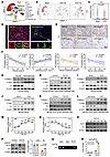 Figure 1
FH expression is reduced in CKD and is associated with KLF9-mediated repression. (A) Uniform manifold approximation and projection plot showing distinct renal cell clusters in the public single-cell RNA-seq dataset NCBI Gene Expression Omnibus (GEO) GSE140023. PT, proximal tubule; LOH, loop of Henle; CDT, convoluted distal tubule. (B and C) Feature plot and violin plot showing Fh1 expression in renal proximal TECs under sham, UUO 2-day, and UUO 7-day conditions. (D) Representative IF staining of FH and LTL in normal kidney and hydronephrotic kidney tissue. Scale bar, 100 μm. (E) Representative IHC staining of FH in human kidney tissue from healthy controls and patients with CKD stages 1–5 (n = 30). Scale bar, 100 μm. (F) Correlation analyses showing negative associations of FH-positive area with renal fibrosis area, serum urea, and serum creatinine, and a positive association with eGFR in human kidney samples (n = 23/30). (G) Western blot analysis of FH, α–smooth muscle actin (α-SMA), and collagen I in kidney tissues from healthy controls and patients with hydronephrosis (n = 6). (H) Western blot analysis of α-SMA and collagen I in primary human renal TECs after TGF-β1 stimulation (n = 3). (I and J) Western blot analysis of FH, α-SMA, and collagen I in UUO and UIRI mouse models (n = 6). (K and L) Western blot analysis of FH, α-SMA, and collagen I in mTECs subjected to TGF-β1 or H/R treatment (n = 3). (M and N) qRT-PCR analysis of Klf9 and Fh1 mRNA expression at the indicated time points after TGF-β1 treatment (n = 3). (O) Western blot analysis of FH and KLF9 protein expression at the indicated time points after TGF-β1 treatment (n = 3). (P) Western blot analysis of FH protein expression after Klf9 knockdown (KD) in TGF-β1–stimulated mTECs (n = 3). (Q and R) ChIP assay showing increased KLF9 occupancy at the upstream regulatory region of Fh1 after TGF-β1 stimulation (n = 3). Data are shown as mean ± SEM. Statistical analysis was performed using Spearman’s correlation analysis (F), 1-way ANOVA followed by Tukey’s multiple comparisons test (M and N), and 2-way ANOVA followed by Tukey’s multiple comparisons test (P and R). **P < 0.01, and ***P < 0.001.
Subsequently, we isolated primary human renal TECs from normal kidney tissue and found that TGF-β1 treatment reduced FH expression (Figure 1H and Supplemental Figure 1C). FH protein expression was also reduced in mouse models of renal fibrosis induced by UUO or unilateral ischemia/reperfusion injury (UIRI) (Figure 1, I and J, and Supplemental Figure 1, D–G). Similarly, in vitro fibrosis models induced by TGF-β1 or hypoxia/reoxygenation (H/R) showed reduced FH protein levels (Figure 1, K and L, and Supplemental Figure 1, H–J).
To identify candidate transcription factors potentially involved in Fh1 regulation, we performed an exploratory inspection of the genomic region upstream of the mouse Fh1 transcription start site using publicly available annotations in the UCSC Genome Browser. This inspection showed multiple annotated transcription factor–associated motifs across the upstream regulatory region (Supplemental Figure 2A). Among the annotated candidate transcription factors, B-cell lymphoma 6 (BCL6), paired box 3 (PAX3), and Krüppel-like factor 9 (KLF9) were selected for further analysis based on published evidence linking them to renal pathophysiology. Quantitative reverse transcription polymerase chain reaction (qRT-PCR) analysis conducted in both UUO and UIRI mouse models demonstrated that Bcl6 expression was downregulated, Pax3 expression remained unchanged, and Klf9 expression was upregulated under injury conditions (Supplemental Figure 2, B–D). To prioritize these candidates functionally, we performed siRNA-mediated knockdown (KD) of Bcl6 and Klf9 in TGF-β1–treated TECs. Silencing Bcl6 had no detectable effect on Fh1 expression, whereas Klf9 KD partially restored both FH protein and Fh1 mRNA levels in the injured cells (Figure 1P and Supplemental Figure 2, E and F), supporting prioritization of KLF9 for further study.
KLF9 is a transcriptional repressor that can inhibit gene transcription by binding to GC-rich promoter elements (25, 26). In a TGF-β1–stimulated in vitro model, time course analyses demonstrated that Klf9 mRNA and KLF9 protein levels increased before the decline in FH expression (Figure 1, M–O, and Supplemental Figure 2G), consistent with an upstream regulatory role. Consistently, single-cell RNA-seq analysis of UUO kidneys revealed increased Klf9 expression specifically in TECs during fibrotic progression (Supplemental Figure 2, I and J), which was confirmed by increased KLF9 expression in fibrotic kidneys from UUO mice (Supplemental Figure 2H). Finally, chromatin immunoprecipitation (ChIP) assays demonstrated enhanced KLF9 occupancy at the upstream regulatory region of Fh1 in fibrotic kidneys (Figure 1, Q and R), supporting a role for KLF9 as a transcriptional regulator of Fh1 during renal fibrosis. In parallel, Western blot analyses revealed that KLF9 KD reduced the expression of multiple fibrosis- and inflammation-related markers (Supplemental Figure 2K), indicating that KLF9 plays a functional role in regulating renal inflammatory and fibrotic responses.
FH deficiency exacerbates UUO- and UIRI-induced renal injury and fibrosis. To investigate the functional role of FH in renal fibrosis, we generated Fh1-knockout mice using a CRISPR/Cas9 genome-editing strategy targeting exons 3–9 of Fh1 (Figure 2A). Successful targeting was confirmed by PCR-based genotyping and agarose gel electrophoresis (Supplemental Figure 3, A and B). Consistent with Mouse Genome Informatics annotations, homozygous deletion of Fh1 resulted in early embryonic lethality, with embryos failing to progress beyond the egg cylinder stage (approximately embryonic day 6.0). Therefore, all subsequent experiments were performed using Fh1+/– mice. Assessment of baseline renal phenotypes revealed no significant differences in renal function or histological features between Fh1+/– mice and their wild-type (WT) littermates (Supplemental Figure 3, D–F). In contrast, IHC staining and Western blot analyses confirmed a marked reduction in FH protein expression in the kidneys of Fh1+/– mice compared with WT controls (Figure 2B and Supplemental Figure 3C).
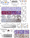 Figure 2
Fh1+/– mice show exacerbated renal injury and fibrosis. (A) Schematic illustration of the Fh1-knockout strategy. Fh1+/–, Fh1 heterozygous. (B) Western blot analysis of FH in kidneys from Fh1+/+ and Fh1+/– mice. (C) Experimental design of the UUO model. (D) Representative PAS, Masson’s trichrome, and α-SMA IHC staining of kidneys from Fh1+/+ and Fh1+/– mice subjected to sham operation or UUO (n = 6). Scale bars, 50 μm. (E) Western blot analysis of α-SMA and collagen I in kidneys from Fh1+/+ and Fh1+/– mice after sham operation or UUO (n = 6). (F) Experimental design for FH overexpression (OE) rescue in Fh1+/– mice. (G) Representative Masson’s trichrome and α-SMA IHC staining of kidneys from WT mice, Fh1+/– mice, and Fh1+/– mice with FH OE after UUO (n = 6). Scale bar, 50 μm. (H) Western blot analysis of α-SMA and collagen I in kidneys from WT mice, Fh1+/– mice, and Fh1+/– mice with FH OE after UUO (n = 6). Data are shown as mean ± SEM. Statistical analysis was performed using 2-tailed unpaired Student’s t test (B) and 2-way ANOVA followed by Tukey’s multiple comparisons test (D and E). **P < 0.01, and ***P < 0.001.
To assess the impact of the Fh1 deficiency on renal injury and fibrosis, we established a 7-day UUO model (Figure 2C). Periodic acid–Schiff (PAS) and Masson’s trichrome staining showed that renal injury and fibrosis were more pronounced in Fh1+/– mice compared with WT littermates (Figure 2D). IHC staining and Western blot analyses demonstrated increased expression of α-SMA and collagen I in Fh1+/– mice (Figure 2, D and E, and Supplemental Figure 3G), confirming that the absence of FH aggravated UUO-induced fibrosis. Additionally, we conducted a 21-day UIRI study in Fh1+/– mice and observed similar results. Fh1 deficiency exacerbated UIRI-induced kidney injury and fibrosis (Supplemental Figure 3H and Supplemental Figure 4A). To determine whether restoration of FH could rescue this phenotype, we utilized AAV2/9-mediated FH overexpression to restore FH expression in Fh1+/– mice and then induced UUO (Figure 2F). PAS, Masson’s trichrome, and IHC staining showed that restoration of FH reduced renal injury and fibrosis (Figure 2G and Supplemental Figure 4B). Western blot analyses further supported these findings (Figure 2H and Supplemental Figure 4C).
Proximal tubule–specific Fh1 conditional knockin attenuates UUO- and UIRI-induced renal injury and fibrosis. To further elucidate the role of FH in proximal TECs during renal fibrosis, we generated a proximal tubule–specific Fh1 conditional knockin (cKI) mouse model (Figure 3A). In this model, Fh1 FF mice carried a homozygous ROSA26-targeted Fh1 knock-in allele but did not express the transgene in the absence of Cre recombinase. In contrast, Fh1 cKI mice expressed Ggt1-Cre, enabling proximal TEC-specific activation of the knock-in Fh1 allele. All mice were genotyped using PCR followed by agarose gel electrophoresis (Figure 3B and Supplemental Figure 5A). IHC staining and Western blot analyses further confirmed successful generation of the Fh1 cKI mice (Supplemental Figure 5, B and C). Notably, Western blot revealed 2 FH protein bands: one corresponding to endogenous FH and the other to the 3×FLAG-tagged knock-in FH, consistent with the design of the cKI construct. Importantly, UUO markedly reduced FH protein levels in Fh1 FF kidneys. In contrast, FH expression in Fh1 cKI kidneys remained relatively preserved after injury compared with littermate controls, despite the overall suppressive effect of UUO on FH expression (Supplemental Figure 5D).
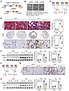 Figure 3
Conditional knockin of Fh1 in renal proximal tubules attenuates renal injury and fibrosis. (A) Schematic illustration of the conditional knockin (cKI) strategy for Fh1 expression in renal proximal tubules. (B) PCR-based genotyping of mice by agarose gel electrophoresis. (C) Experimental design for the UUO model. (D) Representative Masson’s trichrome and α-SMA IHC staining of kidneys from Fh1 FF and Fh1 cKI mice subjected to sham operation or UUO (n = 6). Scale bar, 50 μm. (E) Western blot analysis of α-SMA and collagen I in kidneys from Fh1 FF and Fh1 cKI mice after sham operation or UUO (n = 6). (F) Experimental design of the UIRI model. (G) Representative collagen I IHC staining of kidneys from Fh1 FF and Fh1 cKI mice subjected to sham surgery or UIRI (n = 6). Scale bar, 50 μm. (H) Western blot analysis of α-SMA and collagen I in kidneys from Fh1 FF and Fh1 cKI mice after sham operation or UIRI (n = 6). Data are shown as mean ± SEM. Statistical analysis was performed using 2-way ANOVA followed by Tukey’s multiple comparisons test (D, E, and H). ***P < 0.001.
In Fh1 cKI mice, UUO-induced tubular injury and extracellular collagen deposition were attenuated compared with Fh1 FF mice, as shown by hematoxylin and eosin (H&E) and Masson’s trichrome staining (Figure 3, C and D, and Supplemental Figure 5E). Western blot and IHC analyses further showed reduced expression of α-SMA and collagen I in Fh1 cKI mice following UUO (Figure 3, D and E, and Supplemental Figure 5E). We next extended these findings to the UIRI model (Figure 3F). Consistently, proximal tubule–specific Fh1 cKI attenuated UIRI-induced kidney injury and fibrosis (Figure 3, G and H, and Supplemental Figure 6A).
FH regulates fibrotic responses in TGF-β1– and H/R-treated TECs. TEC injury plays a crucial role in the pathogenesis of CKD (27). To validate the in vivo findings, we used siRNA transfection to silence Fh1 in murine tubular epithelial cells (mTECs) (Supplemental Figure 7A). qRT-PCR analysis and Western blot analyses confirmed that Fh1 siRNA reduced both Fh1 mRNA and FH protein levels (Supplemental Figure 7, B and C). Following TGF-β1 treatment, Acta2 and Col1a1 mRNA levels were increased in Fh1-KD cells compared with those transfected with control siRNA (Supplemental Figure 7D). Western blot and IF staining further showed that Fh1 KD increased α-SMA and collagen I protein expression (Supplemental Figure 7, E and F). Similar results were observed in the H/R-induced fibrogenesis model in mTECs (Supplemental Figure 7, G and H), supporting a role for Fh1 KD in promoting renal fibrogenic responses under TGF-β1 and H/R stimulation.
Subsequently, we overexpressed Fh1 in mTECs by plasmid transfection (Supplemental Figure 8A). qRT-PCR and Western blot analyses confirmed that Fh1 OE increased Fh1 mRNA and FH protein levels (Supplemental Figure 8, B and C). In parallel, qRT-PCR, Western blot, and IF staining showed that Fh1 OE reduced α-SMA and collagen I expression in TGF-β1– and H/R-stimulated mTECs (Supplemental Figure 8, D–H). We next treated the cells with an FH inhibitor (Supplemental Figure 9A). Compared with cells treated with TGF-β1 alone, FH inhibitor treatment increased α-SMA and collagen I protein expression (Supplemental Figure 9, B and C).
FH deficiency promotes renal inflammation. Recent studies have highlighted an important role for FH in innate immunity and inflammation (21, 22, 28). To investigate the mechanisms through which FH influences renal fibrosis, we conducted RNA-seq analysis (Figure 4A). Kyoto Encyclopedia of Genes and Genomes (KEGG) enrichment analysis and Gene Set Enrichment Analysis (GSEA) showed that FH deficiency was associated with enrichment of inflammatory pathways, including NF-κB, tumor necrosis factor (TNF), and interleukin-17 (IL-17) signaling (Figure 4, B–E). We next performed RNA-seq in Fh1-KD cells treated with TGF-β1 (Figure 4F). KEGG enrichment analysis showed that pathways upregulated following Fh1 KD in the fibrotic cell model were predominantly inflammatory, including PI3K/AKT and TNF signaling (Figure 4G and Supplemental Figure 10A). Together, these findings supported a role for FH loss in promoting inflammatory pathway activation during renal fibrosis.
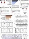 Figure 4
FH deficiency exacerbates renal inflammatory responses. (A) Schematic overview of the RNA-seq workflow. (B) KEGG pathway enrichment analysis showing the top 10 upregulated pathways in Fh1-KD cells. (C–E) GSEA showing enrichment of NF-κB, IL-17, and TNF, signaling pathways in Fh1-KD cells. (F) Schematic overview of the RNA-seq workflow following TGF-β1 stimulation. (G) KEGG pathway enrichment analysis showing the top 10 upregulated pathways in Fh1-KD cells after TGF-β1 treatment. (H and I) Western blot analysis of p-p65 in cells transfected with empty vector (EV), Fh1-OE, and Fh1-KD cells with or without TGF-β1 stimulation (n = 3). (J) Western blot analysis of p-p65 in kidneys from Fh1 FF and Fh1 cKI mice subjected to sham operation or UUO (n = 6). (K–M) qRT-PCR analysis of inflammatory gene expression in EV, Fh1-OE, and Fh1-KD cells with or without TGF-β1 treatment (n = 3) and in kidneys from Fh1 FF and Fh1 cKI mice after sham operation or UUO (n = 6). (N) Representative F4/80 IHC staining of kidneys from Fh1 FF and Fh1 cKI mice subjected to sham operation or UUO (n = 6). Scale bar, 50 μm. Data are shown as mean ± SEM. Statistical analysis was performed using 2-way ANOVA followed by Tukey’s multiple comparisons test (K–N). *P < 0.05, **P < 0.01, and ***P < 0.001.
To further examine the effect of FH on inflammatory signaling, we performed Western blot, which showed that Fh1 OE reduced NF-κB p65 phosphorylation (Figure 4H and Supplemental Figure 10B). Consistent with these findings, qRT-PCR analysis revealed that chemokine ligand 2 (Ccl2), Il6, and Tnfa mRNA levels were reduced in Fh1-OE cells (Figure 4K). Conversely, Fh1 KD increased NF-κB p65 phosphorylation and the expression of pro-inflammatory genes, including Ccl2, Il6, and Tnfa (Figure 4, I and L, and Supplemental Figure 10C). In the UUO model, Fh1 cKI mice showed reduced macrophage infiltration in the renal interstitium, decreased p65 phosphorylation, and lower expression of inflammatory genes (Figures 4, J, M and N, and Supplemental Figure 10D). In contrast, Fh1+/– mice showed increased macrophage infiltration and inflammatory cytokine expression (Supplemental Figure 10, E and F).
Fumarate accumulation promotes renal inflammation and fibrosis. FH catalyzes the reversible conversion of fumarate to malate (16). To determine whether FH deficiency was associated with fumarate accumulation during renal fibrosis, we measured fumarate levels in human samples and experimental models. In patients with CKD, serum and urinary fumarate levels were inversely correlated with eGFR (Figure 5A). Fumarate levels were also increased in the kidneys and serum of UUO mice (Figure 5B), as well as in an in vitro fibrosis model (Figure 5C). To test whether fumarate accumulation promoted renal inflammation and fibrosis, we treated cells with monomethyl fumarate (MMF), a cell-permeable fumarate derivative that increases intracellular fumarate levels without markedly altering its reactivity (22). In the fibrotic setting, MMF treatment increased α-SMA and collagen I protein expression and enhanced NF-κB p65 activation (Figure 5D and Supplemental Figure 11A). qRT-PCR analysis further showed that the mRNA levels of inflammatory genes increased with MMF treatment (Figure 5E). In TGF-β1–treated cells, Fh1 OE reduced inflammatory gene expression, whereas MMF administration reversed this effect (Figure 5G). Western blot analyses further showed similar results (Figure 5F and Supplemental Figure 11B). In vivo, mice administered MMF and then subjected to UUO developed more severe renal fibrosis and inflammatory responses than saline-treated controls, as shown by PAS, Sirius red, and IHC staining, as well as Western blot and qRT-PCR analyses (Figure 5, H–J, and Supplemental Figure 11, C–E). Together, these findings indicated that fumarate accumulation resulting from FH deficiency promoted renal inflammation and fibrosis.
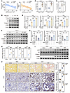 Figure 5
Fumarate accumulation promotes renal inflammation and fibrosis. (A) Spearman’s correlation analysis showing negative correlations between serum fumarate and eGFR (n = 30, r = –0.6440, P < 0.001) and between urinary fumarate and eGFR (n = 30, r = –0.5889, P < 0.001). (B) Fumarate levels in kidneys and serum from mice subjected to sham operation or UUO (n = 6). (C) Cystolic fumarate levels in mTECs treated with control, TGF-β1, or FH-IN-1 (n = 3). (D) Western blot analysis of α-SMA, collagen I, and p-p65 in cells treated with increasing concentrations of MMF with or without TGF-β1 stimulation (n = 3). (E) qRT-PCR analysis of Ccl2, Il6, and Tnfa expression under the same conditions as in D (n = 3). (F) Western blot analysis of α-SMA, collagen I, and p-p65 in EV, Fh1-OE, and Fh1-OE + MMF groups after TGF-β1 stimulation (n = 3). (G) qRT-PCR analysis of inflammatory gene expression in the same groups as in F (n = 3). (H) qRT-PCR analysis of inflammatory gene expression in kidneys from control and MMF-treated mice subjected to sham operation or UUO (n = 6). (I) Western blot analysis of α-SMA, collagen I, and p-p65 in kidneys from control and MMF-treated mice after sham operation or UUO (n = 6). (J) Representative Sirius red staining and IHC staining for α-SMA and F4/80 in kidneys from control and MMF-treated mice after sham operation or UUO (n = 6). Scale bar, 50 μm. Data are shown as mean ± SEM. Statistical analysis was performed using Spearman’s correlation analysis (A), 2-tailed unpaired Student’s t test (B), 1-way ANOVA followed by Tukey’s multiple comparisons test (C, E, and G), and 2-way ANOVA followed by Tukey’s multiple comparisons test (H and J). *P < 0.05, **P < 0.01, and ***P < 0.001.
Fumarate directly succinates AKAP12 at cysteine 670. As an electrophile metabolite, fumarate can undergo Michael addition with nucleophilic cysteine residues on proteins, resulting in formation of S-(2-succinyl) cysteine (2SC), a modification known as succination (29, 30). IHC staining showed increased 2SC levels in kidneys from Fh1-deficient mice subjected to UUO (Supplemental Figure 12A). We next used a desthiobiotin-iodoacetamide (DBIA) probe in a competitive cysteine-labeling strategy, coupled with liquid chromatography-tandem mass spectrometry (LC-MS/MS), to identify candidate cysteine sites potentially modified by fumarate (Figure 6A). This analysis identified 26 high-confidence modification sites across 24 proteins (Figure 6, B and C). Based on prior RNA-seq results and their potential relevance to inflammatory signaling, IMPDH2, HSPBP1, and AKAP12 were prioritized for further analysis (Supplemental Figure 12, B–E). We then individually knocked down Impdh2, Hspbp1, and Akap12 by siRNA transfection (Supplemental Figure 12F). Among these candidates, Akap12 knockdown produced the most pronounced effect on fibrogenic responses in vitro (Supplemental Figure 12, G–I).
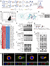 Figure 6
Fumarate promotes renal inflammation and fibrosis through succination of AKAP12 at C670. (A) Schematic illustration of the in vitro experimental design. (B) Volcano plot showing altered proteins and cysteine sites (Ctrl / Compd > 4, P < 0.05). Ctrl, control proteome; Compd, MMF-treated experimental proteome. (C) Hierarchical clustering heatmap showing differentially modified proteins and cysteine sites between control and fumarate-treated groups. (D) Molecular docking model predicting the interaction of fumarate with AKAP12 at C670 via Michael addition. (E) Western blot analysis showing reduced AKAP12 recovery in the FA-alkyne pulldown after MMF treatment (n = 3). (F) Western blot analysis of AKAP12 succination in cells treated with control, TGF-β1, MMF, or FH-IN-1 (n = 3). (G) Schematic diagram of plasmid construction for AKAP12 site-directed mutagenesis. (H) Western blot analysis of AKAP12 succination in AKAP12 WT and AKAP12 C670S cells with or without TGF-β1 treatment (n = 3). (I) Co-IP and Western blot analysis of AKAP12-PKCζ interaction and PKCζ phosphorylation in AKAP12 WT and AKAP12 C670S cells with or without TGF-β1 treatment (n = 3). (J) Western blot analysis of α-SMA, collagen I, p-IκB, p-P65, p-GSK-3β, and p-β-catenin in AKAP12 WT and AKAP12 C670S cells with or without TGF-β1 treatment (n = 3). (K) Representative IF images showing AKAP12–PKCζ colocalization in AKAP12 WT and AKAP12 C670S cells under the indicated conditions (n = 3). Scale bar, 10 μm.
AKAP12, also known as Src-suppressed C-kinase substrate, is a member of the A-kinase anchoring protein family (31, 32). It serves as a scaffold that anchors protein kinases, phosphatases, and other signaling molecules to specific subcellular compartments, thereby regulating intracellular signaling (33–35). However, its function in renal fibrosis remains unclear. Western blot and IF staining showed that Akap12 KD enhanced TGF-β1–induced expression of fibrotic markers (Supplemental Figure 12, J and K). Conversely, Akap12 OE attenuated these effects (Supplemental Figure 13, A and B). Molecular docking analysis using Schrödinger software predicted that fumarate could interact with AKAP12 at C670 via a Michael addition reaction, with a docking score of –6.169, supporting the possibility of succination at this site (Figure 6D). Consistently, exogenous fumarate treatment reduced AKAP12 enrichment in the FA-alkyne pulldown (Figure 6E). Moreover, treatment with TGF-β1, MMF, or FH-IN-1 increased AKAP12 succination, as assessed by 2SC immunoblotting (Figure 6F). Mutation of AKAP12 C670 from cysteine to serine markedly reduced its succination (Figure 6, G and H).
AKAP12 succination promotes renal inflammation and fibrosis through PKCζ/NF-κB and PKCζ/β-catenin signaling. AKAP12 has been reported to interact with PKCζ and restrain its activation (36, 37). PKCζ can promote NF-κB signaling through phosphorylation of IκB and p65 (38, 39). It has also been implicated in regulation of the GSK-3β/β-catenin axis during renal fibrosis (40). Western blot analyses showed that Fh1 KD in mTECs increased PKCζ phosphorylation and β-catenin expression, whereas Fh1 OE attenuated these changes (Supplemental Figure 13, C and D). Similar changes were observed in Fh1+/– and Fh1 cKI mice (Supplemental Figure 13, E and F). Consistent with our earlier findings, FH deficiency was also associated with activation of NF-κB signaling (Figure 4, H–M, and Supplemental Figure 10, B–E). Co-immunoprecipitation (co-IP) assays showed that TGF-β1 stimulation reduced the interaction between AKAP12 and PKCζ while increasing PKCζ phosphorylation, and C670S mutation of AKAP12 partially reversed these effects (Figure 6I and Supplemental Figure 14A). Double IF staining further supported these findings (Figure 6K). Moreover, C670S mutation reduced phosphorylation of IκB, p65 and GSK-3β; promoted phosphorylation and degradation of β-catenin; and reduced expression of α-SMA and collagen I, as well as the expression of inflammatory genes (Figure 6J and Supplemental Figure 14, B–D). Together, these findings support a mechanism in which succination of AKAP12 at C670 weakens its interaction with PKCζ, thereby attenuating the inhibitory effect of AKAP12 on PKCζ activation and promoting PKCζ/NF-κB and PKCζ/β-catenin signaling, ultimately enhancing renal inflammation and fibrosis.
Akap12 C670S mutation attenuates UUO- and UIRI-induced renal inflammation and fibrosis. To further elucidate the functional role of the AKAP12 C670 site in renal inflammation and fibrosis, we generated a C57BL/6J mouse model harboring the Akap12 C670S mutation, designated as Akap12CS/CS, using CRISPR/Cas9-mediated genome engineering (Figure 7, A and C). Akap12CS/CS mice showed no overt abnormalities compared with WT littermates. Assessment of baseline renal phenotypes revealed no significant differences in renal function or histological features between Akap12CS/CS mice and their WT littermates (Supplemental Figure 15, A–C).
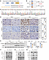 Figure 7
Akap12CS/CS mice are protected against UUO-induced renal inflammation and fibrosis. (A) Schematic illustration of the CRISPR/Cas9-mediated C670S mutation introduced into the mouse Akap12 locus. (B) Experimental design of the UUO model. (C) Representative Sanger sequencing traces confirming the Akap12CS/CS genotype. (D) Representative IHC staining for α-SMA, collagen I, and F4/80 in kidneys from WT and Akap12CS/CS mice subjected to sham operation or UUO (n = 6). Scale bar, 50 μm. (E) Western blot analysis of α-SMA and collagen I in kidneys from WT and Akap12CS/CS mice after sham operation or UUO (n = 6). (F) Western blot and co-IP analyses of PKCζ phosphorylation and AKAP12-PKCζ interaction in kidneys from WT and Akap12CS/CS mice after sham operation or UUO (n = 6). (G and H) Western blot analysis of p-IκB, p-p65, p-GSK-3β, and p-β-catenin in kidneys from WT and Akap12CS/CS mice after sham operation or UUO (n = 6). Data are shown as mean ± SEM. Statistical analysis was performed using 2-way ANOVA followed by Tukey’s multiple comparisons test (D and E). **P < 0.01, and ***P < 0.001.
Subsequently, we established a 7-day UUO model (Figure 7B). Compared with WT mice, Akap12CS/CS mice showed reduced renal injury, inflammation, and fibrosis, as shown by histological and molecular analyses (Figure 7, D and E, and Supplemental Figure 15, E and F). Immunoblot analysis revealed that AKAP12 protein levels were reduced following UUO-induced injury. Under identical injury conditions, AKAP12 protein abundance was comparable between Akap12CS/CS mice and WT littermates, suggesting that the C670S mutation did not affect overall AKAP12 expression (Supplemental Figure 15D). These findings suggest that the protective phenotype observed in Akap12CS/CS mice was not due to altered AKAP12 protein levels, but rather to the functional consequences of the C670S mutation. Similar results were observed in a 21-day UIRI model (Supplemental Figure 15, G and H, and Supplemental Figure 16, A and B). Co-IP analysis showed that UUO reduced the interaction between AKAP12 and PKCζ and increased PKCζ phosphorylation compared with sham controls. This injury-induced disruption of the AKAP12-PKCζ interaction was attenuated in Akap12CS/CS mice (Figure 7F and Supplemental Figure 16C). In parallel, Akap12CS/CS mice showed reduced phosphorylation of IκB, p65, and GSK-3β, together with enhanced phosphorylation and degradation of β-catenin, compared with WT mice subjected to UUO (Figure 7, G and H, and Supplemental Figure 16, D and E). Collectively, these findings indicate that the AKAP12 C670S mutation diminishes PKCζ/NF-κB and PKCζ/β-catenin signaling, thereby reducing UUO- and UIRI-induced renal inflammation and fibrosis.
FH deficiency exacerbates liver and pulmonary fibrosis. Next, we investigated the role of FH in fibrosis in other organs. We established a liver fibrosis model by intraperitoneal injection of CCl4 and a pulmonary fibrosis model by bleomycin administration in Fh1+/– mice (Figure 8, A and B). In both the liver fibrosis and pulmonary fibrosis models, H&E and Masson’s trichrome staining showed that Fh1+/– mice developed more severe fibrosis than WT mice (Figure 8, C and D). IHC staining further showed increased α-SMA, collagen I, F4/80, and 2SC signals in the liver and lung of Fh1+/– mice, consistent with increased fibrosis, inflammation, and succination (Supplemental Figure 17, A and B).
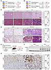 Figure 8
Fh1+/– mice show exacerbated liver and lung fibrosis, whereas AAV2/9-mediated FH OE alleviates renal fibrosis. (A and B) Experimental design for CCl4-induced liver fibrosis and bleomycin-induced pulmonary fibrosis models in Fh1+/+ and Fh1+/– mice. (C) Representative H&E and Masson’s trichrome staining of liver sections from Fh1+/+ and Fh1+/– mice treated with vehicle or CCl4 (n = 6). Scale bar, 100 μm. (D) Representative H&E and Masson’s trichrome staining of lung sections from Fh1+/+ and Fh1+/– mice treated with vehicle or bleomycin (n = 6). Scale bar, 100 μm. (E) Experimental design of renal in situ delivery of AAV2/9 encoding Fh1. (F) Western blot analysis of α-SMA and collagen I in kidneys from EV and Fh1-OE mice after sham operation or UIRI (n = 6). (G) Representative IHC staining for α-SMA and collagen I in kidneys from EV and Fh1-OE mice subjected to sham operation or UUO (n = 6). Scale bar, 50 μm. Data are shown as mean ± SEM. Statistical analysis was performed using 2-way ANOVA followed by Tukey’s multiple comparisons test (C, D, and G). **P < 0.01, and ***P < 0.001.
Renal in situ delivery of AAV2/9 encoding Fh1 alleviates renal fibrosis. To investigate the therapeutic potential of FH, we induced FH OE by renal in situ delivery of recombinant AAV2/9 vectors encoding Fh1 (Figure 8E and Supplemental Figure 18, A–C). PAS and Masson’s trichrome staining showed that, compared with WT mice, Fh1-OE mice exhibited reduced renal injury and fibrosis following UUO or UIRI (Supplemental Figure 18, E and G). These findings were further supported by Western blot and IHC analyses of fibrotic markers (Figure 8, F and G, and Supplemental Figure 18, D–F).


Copyright © 2026 American Society for Clinical Investigation
ISSN: 0021-9738 (print), 1558-8238 (online)