
Advertisement
Research ArticleImmunologyOncology
Open Access |  10.1172/JCI199525
TARBP2 facilitates renal cell carcinoma immune escape by suppressing 53BP1 phase separation to upregulate B7-H3
Minyu Chen,1,2 Mingjie Lin,1 Jiangquan Zhu,3 Kezhi Liu,1 Guannan Shu,3 Zhuting Tong,4 Mi Zhou,5 Liangmin Fu,6,7,8 Taowei Yang,1 Yihui Pan,9 Xinwei Zhou,1 Jinchen Luo,1 Wuyuan Liao,1,10 Qihao Li,1 Lican Liao,11 Hansen Lin,1 Kangbo Huang,12,13 Lizhen Zhang,1 Wei Chen,1 Junhang Luo,1,14 and Jiaxing Zhang11
1Department of Urology, The First Affiliated Hospital of Sun Yat-sen University, Guangzhou, China.
2Department of Urology, Sichuan Provincial People’s Hospital, University of Electronic Science and Technology of China, Chengdu, China.
3Department of Urology, Guangzhou Women and Children’s Medical Center, Guangzhou Medical University, Guangdong Provincial Clinical Research Center for Child Health, Guangzhou, China.
4Department of Radiation Oncology, The First Affiliated Hospital of Anhui Medical University, Hefei, China.
5Department of Oncology and Southwest Cancer Center, Southwest Hospital of The Third Military Medical University (Army Medical University), Chongqing, China.
6Department of Urology, The Second Xiangya Hospital of Central South University, Changsha, China.
7Key Laboratory of Diabetes Immunology (Central South University), Ministry of Education, National Clinical Research Center for Metabolic Disease, Changsha, China.
8Uro-Oncology Institute of Central South University, Changsha, China.
9Department of Urology, The Third Affiliated Hospital of Soochow University, Changzhou, China.
10Department of Urology, Cancer Hospital Chinese Academy of Medical Sciences, Shenzhen, China.
11Department of Oncology, The First Affiliated Hospital of Sun Yat-sen University, Guangzhou, China.
12Department of Urology and
13State Key Laboratory of Oncology in South China, Guangdong Provincial Clinical Research Center for Cancer, Sun Yat-sen University Cancer Center, Guangzhou, China.
14Institute of Precision Medicine, The First Affiliated Hospital of Sun Yat-sen University, Guangzhou, China.
Address for correspondence. Jiaxing Zhang, Junhang Luo, Wei Chen, or Lizhen Zhang, The First Affiliated Hospital of Sun Yat-sen University, No. 58, Zhongshan Second Road, Guangzhou, Guangdong, China. Email: zhangjx25@mail.sysu.edu.cn (J Zhang); luojunh@mail.sysu.edu.cn (Junhang Luo); chenw3@mail.sysu.edu.cn (WC); zhanglzh3@alumni.sysu.edu.cn (LZ).
Authorship note: MC, ML, J Zhu, KL, GS, and ZT contributed equally to this work as co–first authors.
Find articles by Chen, M. in: PubMed | Google Scholar
1Department of Urology, The First Affiliated Hospital of Sun Yat-sen University, Guangzhou, China.
2Department of Urology, Sichuan Provincial People’s Hospital, University of Electronic Science and Technology of China, Chengdu, China.
3Department of Urology, Guangzhou Women and Children’s Medical Center, Guangzhou Medical University, Guangdong Provincial Clinical Research Center for Child Health, Guangzhou, China.
4Department of Radiation Oncology, The First Affiliated Hospital of Anhui Medical University, Hefei, China.
5Department of Oncology and Southwest Cancer Center, Southwest Hospital of The Third Military Medical University (Army Medical University), Chongqing, China.
6Department of Urology, The Second Xiangya Hospital of Central South University, Changsha, China.
7Key Laboratory of Diabetes Immunology (Central South University), Ministry of Education, National Clinical Research Center for Metabolic Disease, Changsha, China.
8Uro-Oncology Institute of Central South University, Changsha, China.
9Department of Urology, The Third Affiliated Hospital of Soochow University, Changzhou, China.
10Department of Urology, Cancer Hospital Chinese Academy of Medical Sciences, Shenzhen, China.
11Department of Oncology, The First Affiliated Hospital of Sun Yat-sen University, Guangzhou, China.
12Department of Urology and
13State Key Laboratory of Oncology in South China, Guangdong Provincial Clinical Research Center for Cancer, Sun Yat-sen University Cancer Center, Guangzhou, China.
14Institute of Precision Medicine, The First Affiliated Hospital of Sun Yat-sen University, Guangzhou, China.
Address for correspondence. Jiaxing Zhang, Junhang Luo, Wei Chen, or Lizhen Zhang, The First Affiliated Hospital of Sun Yat-sen University, No. 58, Zhongshan Second Road, Guangzhou, Guangdong, China. Email: zhangjx25@mail.sysu.edu.cn (J Zhang); luojunh@mail.sysu.edu.cn (Junhang Luo); chenw3@mail.sysu.edu.cn (WC); zhanglzh3@alumni.sysu.edu.cn (LZ).
Authorship note: MC, ML, J Zhu, KL, GS, and ZT contributed equally to this work as co–first authors.
Find articles by Lin, M. in: PubMed | Google Scholar
1Department of Urology, The First Affiliated Hospital of Sun Yat-sen University, Guangzhou, China.
2Department of Urology, Sichuan Provincial People’s Hospital, University of Electronic Science and Technology of China, Chengdu, China.
3Department of Urology, Guangzhou Women and Children’s Medical Center, Guangzhou Medical University, Guangdong Provincial Clinical Research Center for Child Health, Guangzhou, China.
4Department of Radiation Oncology, The First Affiliated Hospital of Anhui Medical University, Hefei, China.
5Department of Oncology and Southwest Cancer Center, Southwest Hospital of The Third Military Medical University (Army Medical University), Chongqing, China.
6Department of Urology, The Second Xiangya Hospital of Central South University, Changsha, China.
7Key Laboratory of Diabetes Immunology (Central South University), Ministry of Education, National Clinical Research Center for Metabolic Disease, Changsha, China.
8Uro-Oncology Institute of Central South University, Changsha, China.
9Department of Urology, The Third Affiliated Hospital of Soochow University, Changzhou, China.
10Department of Urology, Cancer Hospital Chinese Academy of Medical Sciences, Shenzhen, China.
11Department of Oncology, The First Affiliated Hospital of Sun Yat-sen University, Guangzhou, China.
12Department of Urology and
13State Key Laboratory of Oncology in South China, Guangdong Provincial Clinical Research Center for Cancer, Sun Yat-sen University Cancer Center, Guangzhou, China.
14Institute of Precision Medicine, The First Affiliated Hospital of Sun Yat-sen University, Guangzhou, China.
Address for correspondence. Jiaxing Zhang, Junhang Luo, Wei Chen, or Lizhen Zhang, The First Affiliated Hospital of Sun Yat-sen University, No. 58, Zhongshan Second Road, Guangzhou, Guangdong, China. Email: zhangjx25@mail.sysu.edu.cn (J Zhang); luojunh@mail.sysu.edu.cn (Junhang Luo); chenw3@mail.sysu.edu.cn (WC); zhanglzh3@alumni.sysu.edu.cn (LZ).
Authorship note: MC, ML, J Zhu, KL, GS, and ZT contributed equally to this work as co–first authors.
Find articles by Zhu, J. in: PubMed | Google Scholar
1Department of Urology, The First Affiliated Hospital of Sun Yat-sen University, Guangzhou, China.
2Department of Urology, Sichuan Provincial People’s Hospital, University of Electronic Science and Technology of China, Chengdu, China.
3Department of Urology, Guangzhou Women and Children’s Medical Center, Guangzhou Medical University, Guangdong Provincial Clinical Research Center for Child Health, Guangzhou, China.
4Department of Radiation Oncology, The First Affiliated Hospital of Anhui Medical University, Hefei, China.
5Department of Oncology and Southwest Cancer Center, Southwest Hospital of The Third Military Medical University (Army Medical University), Chongqing, China.
6Department of Urology, The Second Xiangya Hospital of Central South University, Changsha, China.
7Key Laboratory of Diabetes Immunology (Central South University), Ministry of Education, National Clinical Research Center for Metabolic Disease, Changsha, China.
8Uro-Oncology Institute of Central South University, Changsha, China.
9Department of Urology, The Third Affiliated Hospital of Soochow University, Changzhou, China.
10Department of Urology, Cancer Hospital Chinese Academy of Medical Sciences, Shenzhen, China.
11Department of Oncology, The First Affiliated Hospital of Sun Yat-sen University, Guangzhou, China.
12Department of Urology and
13State Key Laboratory of Oncology in South China, Guangdong Provincial Clinical Research Center for Cancer, Sun Yat-sen University Cancer Center, Guangzhou, China.
14Institute of Precision Medicine, The First Affiliated Hospital of Sun Yat-sen University, Guangzhou, China.
Address for correspondence. Jiaxing Zhang, Junhang Luo, Wei Chen, or Lizhen Zhang, The First Affiliated Hospital of Sun Yat-sen University, No. 58, Zhongshan Second Road, Guangzhou, Guangdong, China. Email: zhangjx25@mail.sysu.edu.cn (J Zhang); luojunh@mail.sysu.edu.cn (Junhang Luo); chenw3@mail.sysu.edu.cn (WC); zhanglzh3@alumni.sysu.edu.cn (LZ).
Authorship note: MC, ML, J Zhu, KL, GS, and ZT contributed equally to this work as co–first authors.
Find articles by Liu, K. in: PubMed | Google Scholar
1Department of Urology, The First Affiliated Hospital of Sun Yat-sen University, Guangzhou, China.
2Department of Urology, Sichuan Provincial People’s Hospital, University of Electronic Science and Technology of China, Chengdu, China.
3Department of Urology, Guangzhou Women and Children’s Medical Center, Guangzhou Medical University, Guangdong Provincial Clinical Research Center for Child Health, Guangzhou, China.
4Department of Radiation Oncology, The First Affiliated Hospital of Anhui Medical University, Hefei, China.
5Department of Oncology and Southwest Cancer Center, Southwest Hospital of The Third Military Medical University (Army Medical University), Chongqing, China.
6Department of Urology, The Second Xiangya Hospital of Central South University, Changsha, China.
7Key Laboratory of Diabetes Immunology (Central South University), Ministry of Education, National Clinical Research Center for Metabolic Disease, Changsha, China.
8Uro-Oncology Institute of Central South University, Changsha, China.
9Department of Urology, The Third Affiliated Hospital of Soochow University, Changzhou, China.
10Department of Urology, Cancer Hospital Chinese Academy of Medical Sciences, Shenzhen, China.
11Department of Oncology, The First Affiliated Hospital of Sun Yat-sen University, Guangzhou, China.
12Department of Urology and
13State Key Laboratory of Oncology in South China, Guangdong Provincial Clinical Research Center for Cancer, Sun Yat-sen University Cancer Center, Guangzhou, China.
14Institute of Precision Medicine, The First Affiliated Hospital of Sun Yat-sen University, Guangzhou, China.
Address for correspondence. Jiaxing Zhang, Junhang Luo, Wei Chen, or Lizhen Zhang, The First Affiliated Hospital of Sun Yat-sen University, No. 58, Zhongshan Second Road, Guangzhou, Guangdong, China. Email: zhangjx25@mail.sysu.edu.cn (J Zhang); luojunh@mail.sysu.edu.cn (Junhang Luo); chenw3@mail.sysu.edu.cn (WC); zhanglzh3@alumni.sysu.edu.cn (LZ).
Authorship note: MC, ML, J Zhu, KL, GS, and ZT contributed equally to this work as co–first authors.
Find articles by Shu, G. in: PubMed | Google Scholar
1Department of Urology, The First Affiliated Hospital of Sun Yat-sen University, Guangzhou, China.
2Department of Urology, Sichuan Provincial People’s Hospital, University of Electronic Science and Technology of China, Chengdu, China.
3Department of Urology, Guangzhou Women and Children’s Medical Center, Guangzhou Medical University, Guangdong Provincial Clinical Research Center for Child Health, Guangzhou, China.
4Department of Radiation Oncology, The First Affiliated Hospital of Anhui Medical University, Hefei, China.
5Department of Oncology and Southwest Cancer Center, Southwest Hospital of The Third Military Medical University (Army Medical University), Chongqing, China.
6Department of Urology, The Second Xiangya Hospital of Central South University, Changsha, China.
7Key Laboratory of Diabetes Immunology (Central South University), Ministry of Education, National Clinical Research Center for Metabolic Disease, Changsha, China.
8Uro-Oncology Institute of Central South University, Changsha, China.
9Department of Urology, The Third Affiliated Hospital of Soochow University, Changzhou, China.
10Department of Urology, Cancer Hospital Chinese Academy of Medical Sciences, Shenzhen, China.
11Department of Oncology, The First Affiliated Hospital of Sun Yat-sen University, Guangzhou, China.
12Department of Urology and
13State Key Laboratory of Oncology in South China, Guangdong Provincial Clinical Research Center for Cancer, Sun Yat-sen University Cancer Center, Guangzhou, China.
14Institute of Precision Medicine, The First Affiliated Hospital of Sun Yat-sen University, Guangzhou, China.
Address for correspondence. Jiaxing Zhang, Junhang Luo, Wei Chen, or Lizhen Zhang, The First Affiliated Hospital of Sun Yat-sen University, No. 58, Zhongshan Second Road, Guangzhou, Guangdong, China. Email: zhangjx25@mail.sysu.edu.cn (J Zhang); luojunh@mail.sysu.edu.cn (Junhang Luo); chenw3@mail.sysu.edu.cn (WC); zhanglzh3@alumni.sysu.edu.cn (LZ).
Authorship note: MC, ML, J Zhu, KL, GS, and ZT contributed equally to this work as co–first authors.
Find articles by Tong, Z. in: PubMed | Google Scholar
1Department of Urology, The First Affiliated Hospital of Sun Yat-sen University, Guangzhou, China.
2Department of Urology, Sichuan Provincial People’s Hospital, University of Electronic Science and Technology of China, Chengdu, China.
3Department of Urology, Guangzhou Women and Children’s Medical Center, Guangzhou Medical University, Guangdong Provincial Clinical Research Center for Child Health, Guangzhou, China.
4Department of Radiation Oncology, The First Affiliated Hospital of Anhui Medical University, Hefei, China.
5Department of Oncology and Southwest Cancer Center, Southwest Hospital of The Third Military Medical University (Army Medical University), Chongqing, China.
6Department of Urology, The Second Xiangya Hospital of Central South University, Changsha, China.
7Key Laboratory of Diabetes Immunology (Central South University), Ministry of Education, National Clinical Research Center for Metabolic Disease, Changsha, China.
8Uro-Oncology Institute of Central South University, Changsha, China.
9Department of Urology, The Third Affiliated Hospital of Soochow University, Changzhou, China.
10Department of Urology, Cancer Hospital Chinese Academy of Medical Sciences, Shenzhen, China.
11Department of Oncology, The First Affiliated Hospital of Sun Yat-sen University, Guangzhou, China.
12Department of Urology and
13State Key Laboratory of Oncology in South China, Guangdong Provincial Clinical Research Center for Cancer, Sun Yat-sen University Cancer Center, Guangzhou, China.
14Institute of Precision Medicine, The First Affiliated Hospital of Sun Yat-sen University, Guangzhou, China.
Address for correspondence. Jiaxing Zhang, Junhang Luo, Wei Chen, or Lizhen Zhang, The First Affiliated Hospital of Sun Yat-sen University, No. 58, Zhongshan Second Road, Guangzhou, Guangdong, China. Email: zhangjx25@mail.sysu.edu.cn (J Zhang); luojunh@mail.sysu.edu.cn (Junhang Luo); chenw3@mail.sysu.edu.cn (WC); zhanglzh3@alumni.sysu.edu.cn (LZ).
Authorship note: MC, ML, J Zhu, KL, GS, and ZT contributed equally to this work as co–first authors.
Find articles by Zhou, M. in: PubMed | Google Scholar
1Department of Urology, The First Affiliated Hospital of Sun Yat-sen University, Guangzhou, China.
2Department of Urology, Sichuan Provincial People’s Hospital, University of Electronic Science and Technology of China, Chengdu, China.
3Department of Urology, Guangzhou Women and Children’s Medical Center, Guangzhou Medical University, Guangdong Provincial Clinical Research Center for Child Health, Guangzhou, China.
4Department of Radiation Oncology, The First Affiliated Hospital of Anhui Medical University, Hefei, China.
5Department of Oncology and Southwest Cancer Center, Southwest Hospital of The Third Military Medical University (Army Medical University), Chongqing, China.
6Department of Urology, The Second Xiangya Hospital of Central South University, Changsha, China.
7Key Laboratory of Diabetes Immunology (Central South University), Ministry of Education, National Clinical Research Center for Metabolic Disease, Changsha, China.
8Uro-Oncology Institute of Central South University, Changsha, China.
9Department of Urology, The Third Affiliated Hospital of Soochow University, Changzhou, China.
10Department of Urology, Cancer Hospital Chinese Academy of Medical Sciences, Shenzhen, China.
11Department of Oncology, The First Affiliated Hospital of Sun Yat-sen University, Guangzhou, China.
12Department of Urology and
13State Key Laboratory of Oncology in South China, Guangdong Provincial Clinical Research Center for Cancer, Sun Yat-sen University Cancer Center, Guangzhou, China.
14Institute of Precision Medicine, The First Affiliated Hospital of Sun Yat-sen University, Guangzhou, China.
Address for correspondence. Jiaxing Zhang, Junhang Luo, Wei Chen, or Lizhen Zhang, The First Affiliated Hospital of Sun Yat-sen University, No. 58, Zhongshan Second Road, Guangzhou, Guangdong, China. Email: zhangjx25@mail.sysu.edu.cn (J Zhang); luojunh@mail.sysu.edu.cn (Junhang Luo); chenw3@mail.sysu.edu.cn (WC); zhanglzh3@alumni.sysu.edu.cn (LZ).
Authorship note: MC, ML, J Zhu, KL, GS, and ZT contributed equally to this work as co–first authors.
Find articles by Fu, L. in: PubMed | Google Scholar
1Department of Urology, The First Affiliated Hospital of Sun Yat-sen University, Guangzhou, China.
2Department of Urology, Sichuan Provincial People’s Hospital, University of Electronic Science and Technology of China, Chengdu, China.
3Department of Urology, Guangzhou Women and Children’s Medical Center, Guangzhou Medical University, Guangdong Provincial Clinical Research Center for Child Health, Guangzhou, China.
4Department of Radiation Oncology, The First Affiliated Hospital of Anhui Medical University, Hefei, China.
5Department of Oncology and Southwest Cancer Center, Southwest Hospital of The Third Military Medical University (Army Medical University), Chongqing, China.
6Department of Urology, The Second Xiangya Hospital of Central South University, Changsha, China.
7Key Laboratory of Diabetes Immunology (Central South University), Ministry of Education, National Clinical Research Center for Metabolic Disease, Changsha, China.
8Uro-Oncology Institute of Central South University, Changsha, China.
9Department of Urology, The Third Affiliated Hospital of Soochow University, Changzhou, China.
10Department of Urology, Cancer Hospital Chinese Academy of Medical Sciences, Shenzhen, China.
11Department of Oncology, The First Affiliated Hospital of Sun Yat-sen University, Guangzhou, China.
12Department of Urology and
13State Key Laboratory of Oncology in South China, Guangdong Provincial Clinical Research Center for Cancer, Sun Yat-sen University Cancer Center, Guangzhou, China.
14Institute of Precision Medicine, The First Affiliated Hospital of Sun Yat-sen University, Guangzhou, China.
Address for correspondence. Jiaxing Zhang, Junhang Luo, Wei Chen, or Lizhen Zhang, The First Affiliated Hospital of Sun Yat-sen University, No. 58, Zhongshan Second Road, Guangzhou, Guangdong, China. Email: zhangjx25@mail.sysu.edu.cn (J Zhang); luojunh@mail.sysu.edu.cn (Junhang Luo); chenw3@mail.sysu.edu.cn (WC); zhanglzh3@alumni.sysu.edu.cn (LZ).
Authorship note: MC, ML, J Zhu, KL, GS, and ZT contributed equally to this work as co–first authors.
Find articles by Yang, T. in: PubMed | Google Scholar
1Department of Urology, The First Affiliated Hospital of Sun Yat-sen University, Guangzhou, China.
2Department of Urology, Sichuan Provincial People’s Hospital, University of Electronic Science and Technology of China, Chengdu, China.
3Department of Urology, Guangzhou Women and Children’s Medical Center, Guangzhou Medical University, Guangdong Provincial Clinical Research Center for Child Health, Guangzhou, China.
4Department of Radiation Oncology, The First Affiliated Hospital of Anhui Medical University, Hefei, China.
5Department of Oncology and Southwest Cancer Center, Southwest Hospital of The Third Military Medical University (Army Medical University), Chongqing, China.
6Department of Urology, The Second Xiangya Hospital of Central South University, Changsha, China.
7Key Laboratory of Diabetes Immunology (Central South University), Ministry of Education, National Clinical Research Center for Metabolic Disease, Changsha, China.
8Uro-Oncology Institute of Central South University, Changsha, China.
9Department of Urology, The Third Affiliated Hospital of Soochow University, Changzhou, China.
10Department of Urology, Cancer Hospital Chinese Academy of Medical Sciences, Shenzhen, China.
11Department of Oncology, The First Affiliated Hospital of Sun Yat-sen University, Guangzhou, China.
12Department of Urology and
13State Key Laboratory of Oncology in South China, Guangdong Provincial Clinical Research Center for Cancer, Sun Yat-sen University Cancer Center, Guangzhou, China.
14Institute of Precision Medicine, The First Affiliated Hospital of Sun Yat-sen University, Guangzhou, China.
Address for correspondence. Jiaxing Zhang, Junhang Luo, Wei Chen, or Lizhen Zhang, The First Affiliated Hospital of Sun Yat-sen University, No. 58, Zhongshan Second Road, Guangzhou, Guangdong, China. Email: zhangjx25@mail.sysu.edu.cn (J Zhang); luojunh@mail.sysu.edu.cn (Junhang Luo); chenw3@mail.sysu.edu.cn (WC); zhanglzh3@alumni.sysu.edu.cn (LZ).
Authorship note: MC, ML, J Zhu, KL, GS, and ZT contributed equally to this work as co–first authors.
Find articles by Pan, Y. in: PubMed | Google Scholar
1Department of Urology, The First Affiliated Hospital of Sun Yat-sen University, Guangzhou, China.
2Department of Urology, Sichuan Provincial People’s Hospital, University of Electronic Science and Technology of China, Chengdu, China.
3Department of Urology, Guangzhou Women and Children’s Medical Center, Guangzhou Medical University, Guangdong Provincial Clinical Research Center for Child Health, Guangzhou, China.
4Department of Radiation Oncology, The First Affiliated Hospital of Anhui Medical University, Hefei, China.
5Department of Oncology and Southwest Cancer Center, Southwest Hospital of The Third Military Medical University (Army Medical University), Chongqing, China.
6Department of Urology, The Second Xiangya Hospital of Central South University, Changsha, China.
7Key Laboratory of Diabetes Immunology (Central South University), Ministry of Education, National Clinical Research Center for Metabolic Disease, Changsha, China.
8Uro-Oncology Institute of Central South University, Changsha, China.
9Department of Urology, The Third Affiliated Hospital of Soochow University, Changzhou, China.
10Department of Urology, Cancer Hospital Chinese Academy of Medical Sciences, Shenzhen, China.
11Department of Oncology, The First Affiliated Hospital of Sun Yat-sen University, Guangzhou, China.
12Department of Urology and
13State Key Laboratory of Oncology in South China, Guangdong Provincial Clinical Research Center for Cancer, Sun Yat-sen University Cancer Center, Guangzhou, China.
14Institute of Precision Medicine, The First Affiliated Hospital of Sun Yat-sen University, Guangzhou, China.
Address for correspondence. Jiaxing Zhang, Junhang Luo, Wei Chen, or Lizhen Zhang, The First Affiliated Hospital of Sun Yat-sen University, No. 58, Zhongshan Second Road, Guangzhou, Guangdong, China. Email: zhangjx25@mail.sysu.edu.cn (J Zhang); luojunh@mail.sysu.edu.cn (Junhang Luo); chenw3@mail.sysu.edu.cn (WC); zhanglzh3@alumni.sysu.edu.cn (LZ).
Authorship note: MC, ML, J Zhu, KL, GS, and ZT contributed equally to this work as co–first authors.
Find articles by Zhou, X. in: PubMed | Google Scholar
1Department of Urology, The First Affiliated Hospital of Sun Yat-sen University, Guangzhou, China.
2Department of Urology, Sichuan Provincial People’s Hospital, University of Electronic Science and Technology of China, Chengdu, China.
3Department of Urology, Guangzhou Women and Children’s Medical Center, Guangzhou Medical University, Guangdong Provincial Clinical Research Center for Child Health, Guangzhou, China.
4Department of Radiation Oncology, The First Affiliated Hospital of Anhui Medical University, Hefei, China.
5Department of Oncology and Southwest Cancer Center, Southwest Hospital of The Third Military Medical University (Army Medical University), Chongqing, China.
6Department of Urology, The Second Xiangya Hospital of Central South University, Changsha, China.
7Key Laboratory of Diabetes Immunology (Central South University), Ministry of Education, National Clinical Research Center for Metabolic Disease, Changsha, China.
8Uro-Oncology Institute of Central South University, Changsha, China.
9Department of Urology, The Third Affiliated Hospital of Soochow University, Changzhou, China.
10Department of Urology, Cancer Hospital Chinese Academy of Medical Sciences, Shenzhen, China.
11Department of Oncology, The First Affiliated Hospital of Sun Yat-sen University, Guangzhou, China.
12Department of Urology and
13State Key Laboratory of Oncology in South China, Guangdong Provincial Clinical Research Center for Cancer, Sun Yat-sen University Cancer Center, Guangzhou, China.
14Institute of Precision Medicine, The First Affiliated Hospital of Sun Yat-sen University, Guangzhou, China.
Address for correspondence. Jiaxing Zhang, Junhang Luo, Wei Chen, or Lizhen Zhang, The First Affiliated Hospital of Sun Yat-sen University, No. 58, Zhongshan Second Road, Guangzhou, Guangdong, China. Email: zhangjx25@mail.sysu.edu.cn (J Zhang); luojunh@mail.sysu.edu.cn (Junhang Luo); chenw3@mail.sysu.edu.cn (WC); zhanglzh3@alumni.sysu.edu.cn (LZ).
Authorship note: MC, ML, J Zhu, KL, GS, and ZT contributed equally to this work as co–first authors.
Find articles by Luo, J. in: PubMed | Google Scholar
1Department of Urology, The First Affiliated Hospital of Sun Yat-sen University, Guangzhou, China.
2Department of Urology, Sichuan Provincial People’s Hospital, University of Electronic Science and Technology of China, Chengdu, China.
3Department of Urology, Guangzhou Women and Children’s Medical Center, Guangzhou Medical University, Guangdong Provincial Clinical Research Center for Child Health, Guangzhou, China.
4Department of Radiation Oncology, The First Affiliated Hospital of Anhui Medical University, Hefei, China.
5Department of Oncology and Southwest Cancer Center, Southwest Hospital of The Third Military Medical University (Army Medical University), Chongqing, China.
6Department of Urology, The Second Xiangya Hospital of Central South University, Changsha, China.
7Key Laboratory of Diabetes Immunology (Central South University), Ministry of Education, National Clinical Research Center for Metabolic Disease, Changsha, China.
8Uro-Oncology Institute of Central South University, Changsha, China.
9Department of Urology, The Third Affiliated Hospital of Soochow University, Changzhou, China.
10Department of Urology, Cancer Hospital Chinese Academy of Medical Sciences, Shenzhen, China.
11Department of Oncology, The First Affiliated Hospital of Sun Yat-sen University, Guangzhou, China.
12Department of Urology and
13State Key Laboratory of Oncology in South China, Guangdong Provincial Clinical Research Center for Cancer, Sun Yat-sen University Cancer Center, Guangzhou, China.
14Institute of Precision Medicine, The First Affiliated Hospital of Sun Yat-sen University, Guangzhou, China.
Address for correspondence. Jiaxing Zhang, Junhang Luo, Wei Chen, or Lizhen Zhang, The First Affiliated Hospital of Sun Yat-sen University, No. 58, Zhongshan Second Road, Guangzhou, Guangdong, China. Email: zhangjx25@mail.sysu.edu.cn (J Zhang); luojunh@mail.sysu.edu.cn (Junhang Luo); chenw3@mail.sysu.edu.cn (WC); zhanglzh3@alumni.sysu.edu.cn (LZ).
Authorship note: MC, ML, J Zhu, KL, GS, and ZT contributed equally to this work as co–first authors.
Find articles by Liao, W. in: PubMed | Google Scholar
1Department of Urology, The First Affiliated Hospital of Sun Yat-sen University, Guangzhou, China.
2Department of Urology, Sichuan Provincial People’s Hospital, University of Electronic Science and Technology of China, Chengdu, China.
3Department of Urology, Guangzhou Women and Children’s Medical Center, Guangzhou Medical University, Guangdong Provincial Clinical Research Center for Child Health, Guangzhou, China.
4Department of Radiation Oncology, The First Affiliated Hospital of Anhui Medical University, Hefei, China.
5Department of Oncology and Southwest Cancer Center, Southwest Hospital of The Third Military Medical University (Army Medical University), Chongqing, China.
6Department of Urology, The Second Xiangya Hospital of Central South University, Changsha, China.
7Key Laboratory of Diabetes Immunology (Central South University), Ministry of Education, National Clinical Research Center for Metabolic Disease, Changsha, China.
8Uro-Oncology Institute of Central South University, Changsha, China.
9Department of Urology, The Third Affiliated Hospital of Soochow University, Changzhou, China.
10Department of Urology, Cancer Hospital Chinese Academy of Medical Sciences, Shenzhen, China.
11Department of Oncology, The First Affiliated Hospital of Sun Yat-sen University, Guangzhou, China.
12Department of Urology and
13State Key Laboratory of Oncology in South China, Guangdong Provincial Clinical Research Center for Cancer, Sun Yat-sen University Cancer Center, Guangzhou, China.
14Institute of Precision Medicine, The First Affiliated Hospital of Sun Yat-sen University, Guangzhou, China.
Address for correspondence. Jiaxing Zhang, Junhang Luo, Wei Chen, or Lizhen Zhang, The First Affiliated Hospital of Sun Yat-sen University, No. 58, Zhongshan Second Road, Guangzhou, Guangdong, China. Email: zhangjx25@mail.sysu.edu.cn (J Zhang); luojunh@mail.sysu.edu.cn (Junhang Luo); chenw3@mail.sysu.edu.cn (WC); zhanglzh3@alumni.sysu.edu.cn (LZ).
Authorship note: MC, ML, J Zhu, KL, GS, and ZT contributed equally to this work as co–first authors.
Find articles by Li, Q. in: PubMed | Google Scholar
1Department of Urology, The First Affiliated Hospital of Sun Yat-sen University, Guangzhou, China.
2Department of Urology, Sichuan Provincial People’s Hospital, University of Electronic Science and Technology of China, Chengdu, China.
3Department of Urology, Guangzhou Women and Children’s Medical Center, Guangzhou Medical University, Guangdong Provincial Clinical Research Center for Child Health, Guangzhou, China.
4Department of Radiation Oncology, The First Affiliated Hospital of Anhui Medical University, Hefei, China.
5Department of Oncology and Southwest Cancer Center, Southwest Hospital of The Third Military Medical University (Army Medical University), Chongqing, China.
6Department of Urology, The Second Xiangya Hospital of Central South University, Changsha, China.
7Key Laboratory of Diabetes Immunology (Central South University), Ministry of Education, National Clinical Research Center for Metabolic Disease, Changsha, China.
8Uro-Oncology Institute of Central South University, Changsha, China.
9Department of Urology, The Third Affiliated Hospital of Soochow University, Changzhou, China.
10Department of Urology, Cancer Hospital Chinese Academy of Medical Sciences, Shenzhen, China.
11Department of Oncology, The First Affiliated Hospital of Sun Yat-sen University, Guangzhou, China.
12Department of Urology and
13State Key Laboratory of Oncology in South China, Guangdong Provincial Clinical Research Center for Cancer, Sun Yat-sen University Cancer Center, Guangzhou, China.
14Institute of Precision Medicine, The First Affiliated Hospital of Sun Yat-sen University, Guangzhou, China.
Address for correspondence. Jiaxing Zhang, Junhang Luo, Wei Chen, or Lizhen Zhang, The First Affiliated Hospital of Sun Yat-sen University, No. 58, Zhongshan Second Road, Guangzhou, Guangdong, China. Email: zhangjx25@mail.sysu.edu.cn (J Zhang); luojunh@mail.sysu.edu.cn (Junhang Luo); chenw3@mail.sysu.edu.cn (WC); zhanglzh3@alumni.sysu.edu.cn (LZ).
Authorship note: MC, ML, J Zhu, KL, GS, and ZT contributed equally to this work as co–first authors.
Find articles by Liao, L. in: PubMed | Google Scholar
1Department of Urology, The First Affiliated Hospital of Sun Yat-sen University, Guangzhou, China.
2Department of Urology, Sichuan Provincial People’s Hospital, University of Electronic Science and Technology of China, Chengdu, China.
3Department of Urology, Guangzhou Women and Children’s Medical Center, Guangzhou Medical University, Guangdong Provincial Clinical Research Center for Child Health, Guangzhou, China.
4Department of Radiation Oncology, The First Affiliated Hospital of Anhui Medical University, Hefei, China.
5Department of Oncology and Southwest Cancer Center, Southwest Hospital of The Third Military Medical University (Army Medical University), Chongqing, China.
6Department of Urology, The Second Xiangya Hospital of Central South University, Changsha, China.
7Key Laboratory of Diabetes Immunology (Central South University), Ministry of Education, National Clinical Research Center for Metabolic Disease, Changsha, China.
8Uro-Oncology Institute of Central South University, Changsha, China.
9Department of Urology, The Third Affiliated Hospital of Soochow University, Changzhou, China.
10Department of Urology, Cancer Hospital Chinese Academy of Medical Sciences, Shenzhen, China.
11Department of Oncology, The First Affiliated Hospital of Sun Yat-sen University, Guangzhou, China.
12Department of Urology and
13State Key Laboratory of Oncology in South China, Guangdong Provincial Clinical Research Center for Cancer, Sun Yat-sen University Cancer Center, Guangzhou, China.
14Institute of Precision Medicine, The First Affiliated Hospital of Sun Yat-sen University, Guangzhou, China.
Address for correspondence. Jiaxing Zhang, Junhang Luo, Wei Chen, or Lizhen Zhang, The First Affiliated Hospital of Sun Yat-sen University, No. 58, Zhongshan Second Road, Guangzhou, Guangdong, China. Email: zhangjx25@mail.sysu.edu.cn (J Zhang); luojunh@mail.sysu.edu.cn (Junhang Luo); chenw3@mail.sysu.edu.cn (WC); zhanglzh3@alumni.sysu.edu.cn (LZ).
Authorship note: MC, ML, J Zhu, KL, GS, and ZT contributed equally to this work as co–first authors.
Find articles by Lin, H. in: PubMed | Google Scholar
1Department of Urology, The First Affiliated Hospital of Sun Yat-sen University, Guangzhou, China.
2Department of Urology, Sichuan Provincial People’s Hospital, University of Electronic Science and Technology of China, Chengdu, China.
3Department of Urology, Guangzhou Women and Children’s Medical Center, Guangzhou Medical University, Guangdong Provincial Clinical Research Center for Child Health, Guangzhou, China.
4Department of Radiation Oncology, The First Affiliated Hospital of Anhui Medical University, Hefei, China.
5Department of Oncology and Southwest Cancer Center, Southwest Hospital of The Third Military Medical University (Army Medical University), Chongqing, China.
6Department of Urology, The Second Xiangya Hospital of Central South University, Changsha, China.
7Key Laboratory of Diabetes Immunology (Central South University), Ministry of Education, National Clinical Research Center for Metabolic Disease, Changsha, China.
8Uro-Oncology Institute of Central South University, Changsha, China.
9Department of Urology, The Third Affiliated Hospital of Soochow University, Changzhou, China.
10Department of Urology, Cancer Hospital Chinese Academy of Medical Sciences, Shenzhen, China.
11Department of Oncology, The First Affiliated Hospital of Sun Yat-sen University, Guangzhou, China.
12Department of Urology and
13State Key Laboratory of Oncology in South China, Guangdong Provincial Clinical Research Center for Cancer, Sun Yat-sen University Cancer Center, Guangzhou, China.
14Institute of Precision Medicine, The First Affiliated Hospital of Sun Yat-sen University, Guangzhou, China.
Address for correspondence. Jiaxing Zhang, Junhang Luo, Wei Chen, or Lizhen Zhang, The First Affiliated Hospital of Sun Yat-sen University, No. 58, Zhongshan Second Road, Guangzhou, Guangdong, China. Email: zhangjx25@mail.sysu.edu.cn (J Zhang); luojunh@mail.sysu.edu.cn (Junhang Luo); chenw3@mail.sysu.edu.cn (WC); zhanglzh3@alumni.sysu.edu.cn (LZ).
Authorship note: MC, ML, J Zhu, KL, GS, and ZT contributed equally to this work as co–first authors.
Find articles by Huang, K. in: PubMed | Google Scholar
1Department of Urology, The First Affiliated Hospital of Sun Yat-sen University, Guangzhou, China.
2Department of Urology, Sichuan Provincial People’s Hospital, University of Electronic Science and Technology of China, Chengdu, China.
3Department of Urology, Guangzhou Women and Children’s Medical Center, Guangzhou Medical University, Guangdong Provincial Clinical Research Center for Child Health, Guangzhou, China.
4Department of Radiation Oncology, The First Affiliated Hospital of Anhui Medical University, Hefei, China.
5Department of Oncology and Southwest Cancer Center, Southwest Hospital of The Third Military Medical University (Army Medical University), Chongqing, China.
6Department of Urology, The Second Xiangya Hospital of Central South University, Changsha, China.
7Key Laboratory of Diabetes Immunology (Central South University), Ministry of Education, National Clinical Research Center for Metabolic Disease, Changsha, China.
8Uro-Oncology Institute of Central South University, Changsha, China.
9Department of Urology, The Third Affiliated Hospital of Soochow University, Changzhou, China.
10Department of Urology, Cancer Hospital Chinese Academy of Medical Sciences, Shenzhen, China.
11Department of Oncology, The First Affiliated Hospital of Sun Yat-sen University, Guangzhou, China.
12Department of Urology and
13State Key Laboratory of Oncology in South China, Guangdong Provincial Clinical Research Center for Cancer, Sun Yat-sen University Cancer Center, Guangzhou, China.
14Institute of Precision Medicine, The First Affiliated Hospital of Sun Yat-sen University, Guangzhou, China.
Address for correspondence. Jiaxing Zhang, Junhang Luo, Wei Chen, or Lizhen Zhang, The First Affiliated Hospital of Sun Yat-sen University, No. 58, Zhongshan Second Road, Guangzhou, Guangdong, China. Email: zhangjx25@mail.sysu.edu.cn (J Zhang); luojunh@mail.sysu.edu.cn (Junhang Luo); chenw3@mail.sysu.edu.cn (WC); zhanglzh3@alumni.sysu.edu.cn (LZ).
Authorship note: MC, ML, J Zhu, KL, GS, and ZT contributed equally to this work as co–first authors.
Find articles by Zhang, L. in: PubMed | Google Scholar
1Department of Urology, The First Affiliated Hospital of Sun Yat-sen University, Guangzhou, China.
2Department of Urology, Sichuan Provincial People’s Hospital, University of Electronic Science and Technology of China, Chengdu, China.
3Department of Urology, Guangzhou Women and Children’s Medical Center, Guangzhou Medical University, Guangdong Provincial Clinical Research Center for Child Health, Guangzhou, China.
4Department of Radiation Oncology, The First Affiliated Hospital of Anhui Medical University, Hefei, China.
5Department of Oncology and Southwest Cancer Center, Southwest Hospital of The Third Military Medical University (Army Medical University), Chongqing, China.
6Department of Urology, The Second Xiangya Hospital of Central South University, Changsha, China.
7Key Laboratory of Diabetes Immunology (Central South University), Ministry of Education, National Clinical Research Center for Metabolic Disease, Changsha, China.
8Uro-Oncology Institute of Central South University, Changsha, China.
9Department of Urology, The Third Affiliated Hospital of Soochow University, Changzhou, China.
10Department of Urology, Cancer Hospital Chinese Academy of Medical Sciences, Shenzhen, China.
11Department of Oncology, The First Affiliated Hospital of Sun Yat-sen University, Guangzhou, China.
12Department of Urology and
13State Key Laboratory of Oncology in South China, Guangdong Provincial Clinical Research Center for Cancer, Sun Yat-sen University Cancer Center, Guangzhou, China.
14Institute of Precision Medicine, The First Affiliated Hospital of Sun Yat-sen University, Guangzhou, China.
Address for correspondence. Jiaxing Zhang, Junhang Luo, Wei Chen, or Lizhen Zhang, The First Affiliated Hospital of Sun Yat-sen University, No. 58, Zhongshan Second Road, Guangzhou, Guangdong, China. Email: zhangjx25@mail.sysu.edu.cn (J Zhang); luojunh@mail.sysu.edu.cn (Junhang Luo); chenw3@mail.sysu.edu.cn (WC); zhanglzh3@alumni.sysu.edu.cn (LZ).
Authorship note: MC, ML, J Zhu, KL, GS, and ZT contributed equally to this work as co–first authors.
Find articles by Chen, W. in: PubMed | Google Scholar
1Department of Urology, The First Affiliated Hospital of Sun Yat-sen University, Guangzhou, China.
2Department of Urology, Sichuan Provincial People’s Hospital, University of Electronic Science and Technology of China, Chengdu, China.
3Department of Urology, Guangzhou Women and Children’s Medical Center, Guangzhou Medical University, Guangdong Provincial Clinical Research Center for Child Health, Guangzhou, China.
4Department of Radiation Oncology, The First Affiliated Hospital of Anhui Medical University, Hefei, China.
5Department of Oncology and Southwest Cancer Center, Southwest Hospital of The Third Military Medical University (Army Medical University), Chongqing, China.
6Department of Urology, The Second Xiangya Hospital of Central South University, Changsha, China.
7Key Laboratory of Diabetes Immunology (Central South University), Ministry of Education, National Clinical Research Center for Metabolic Disease, Changsha, China.
8Uro-Oncology Institute of Central South University, Changsha, China.
9Department of Urology, The Third Affiliated Hospital of Soochow University, Changzhou, China.
10Department of Urology, Cancer Hospital Chinese Academy of Medical Sciences, Shenzhen, China.
11Department of Oncology, The First Affiliated Hospital of Sun Yat-sen University, Guangzhou, China.
12Department of Urology and
13State Key Laboratory of Oncology in South China, Guangdong Provincial Clinical Research Center for Cancer, Sun Yat-sen University Cancer Center, Guangzhou, China.
14Institute of Precision Medicine, The First Affiliated Hospital of Sun Yat-sen University, Guangzhou, China.
Address for correspondence. Jiaxing Zhang, Junhang Luo, Wei Chen, or Lizhen Zhang, The First Affiliated Hospital of Sun Yat-sen University, No. 58, Zhongshan Second Road, Guangzhou, Guangdong, China. Email: zhangjx25@mail.sysu.edu.cn (J Zhang); luojunh@mail.sysu.edu.cn (Junhang Luo); chenw3@mail.sysu.edu.cn (WC); zhanglzh3@alumni.sysu.edu.cn (LZ).
Authorship note: MC, ML, J Zhu, KL, GS, and ZT contributed equally to this work as co–first authors.
Find articles by Luo, J. in: PubMed | Google Scholar
1Department of Urology, The First Affiliated Hospital of Sun Yat-sen University, Guangzhou, China.
2Department of Urology, Sichuan Provincial People’s Hospital, University of Electronic Science and Technology of China, Chengdu, China.
3Department of Urology, Guangzhou Women and Children’s Medical Center, Guangzhou Medical University, Guangdong Provincial Clinical Research Center for Child Health, Guangzhou, China.
4Department of Radiation Oncology, The First Affiliated Hospital of Anhui Medical University, Hefei, China.
5Department of Oncology and Southwest Cancer Center, Southwest Hospital of The Third Military Medical University (Army Medical University), Chongqing, China.
6Department of Urology, The Second Xiangya Hospital of Central South University, Changsha, China.
7Key Laboratory of Diabetes Immunology (Central South University), Ministry of Education, National Clinical Research Center for Metabolic Disease, Changsha, China.
8Uro-Oncology Institute of Central South University, Changsha, China.
9Department of Urology, The Third Affiliated Hospital of Soochow University, Changzhou, China.
10Department of Urology, Cancer Hospital Chinese Academy of Medical Sciences, Shenzhen, China.
11Department of Oncology, The First Affiliated Hospital of Sun Yat-sen University, Guangzhou, China.
12Department of Urology and
13State Key Laboratory of Oncology in South China, Guangdong Provincial Clinical Research Center for Cancer, Sun Yat-sen University Cancer Center, Guangzhou, China.
14Institute of Precision Medicine, The First Affiliated Hospital of Sun Yat-sen University, Guangzhou, China.
Address for correspondence. Jiaxing Zhang, Junhang Luo, Wei Chen, or Lizhen Zhang, The First Affiliated Hospital of Sun Yat-sen University, No. 58, Zhongshan Second Road, Guangzhou, Guangdong, China. Email: zhangjx25@mail.sysu.edu.cn (J Zhang); luojunh@mail.sysu.edu.cn (Junhang Luo); chenw3@mail.sysu.edu.cn (WC); zhanglzh3@alumni.sysu.edu.cn (LZ).
Authorship note: MC, ML, J Zhu, KL, GS, and ZT contributed equally to this work as co–first authors.
Find articles by Zhang, J. in: PubMed | Google Scholar
Published November 2, 2026 - More info
J Clin Invest. 2026;136(21):e199525. https://doi.org/10.1172/JCI199525.
© 2026 Chen et al. This work is licensed under the Creative Commons Attribution 4.0 International License. To view a copy of this license, visit http://creativecommons.org/licenses/by/4.0/.
Received: August 26, 2025; Accepted: September 3, 2026

-
Results
Identification of TARBP2 as a biomarker of poor prognosis and ICB resistance in RCC. Integrated analyses of RNA-seq data from The Cancer Genome Atlas–Kidney Renal Clear Cell Carcinoma (TCGA-KIRC) cohort, the CheckMate ICB treatment cohort, and pretreatment FFPE samples from patients with RCC receiving ICB therapy were conducted to identify genes associated with poor prognosis and ICB resistance. TARBP2 and GINS3 emerged as candidate genes exhibiting 3 consistent unfavorable characteristics, including association with poor prognosis in both TCGA-KIRC and CheckMate cohorts based on a Cox proportional hazards model, elevated expression in patients with progressive disease following ICB therapy, and increased expression in pretreatment FFPE samples from ICB nonresponders (Figure 1, A and B, and Supplemental Table 1; supplemental material available online with this article; https://doi.org/10.1172/JCI199525DS1). Among these, TARBP2 expression was associated with stage III–IV disease in the TCGA-KIRC cohort, whereas GINS3 did not exhibit a similar association (Figure 1C). In the CheckMate cohort, both genes showed a trend toward higher expression in cases of progressive disease (Figure 1D and Supplemental Figure 1, A–C). However, Kaplan-Meier survival analyses revealed that elevated expression of TARBP2 was associated with reduced overall survival (OS) in the TCGA-KIRC cohort and reduced OS and progression-free survival in the CheckMate cohort. In contrast, GINS3 showed a weaker association restricted mainly to progression-free survival in the CheckMate cohort (Supplemental Figure 1, D–I). Furthermore, TARBP2 but not GINS3 retained prognostic value across independent RCC (E-MTAB-1980) and melanoma anti–PD-1 cohorts (Figure 1, E and F, and Supplemental Figure 1, J and K) (18). Therefore, TARBP2 was selected for subsequent analyses, and its prognostic relevance was validated in an independent RCC cohort from Sun Yat-sen University (SYSU) using IHC staining (Figure 1G and Supplemental Table 2).
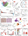 Figure 1
TARBP2 correlates with poor prognosis and ICB resistance in RCC and is predominantly expressed in malignant RCC cells. (A) Venn diagram depicting overlapped genes in data mining. DEGs, differentially expressed genes. (B) Scatterplot illustrating differentially expressed genes in RCC ICB pretreatment FFPE samples between ICB responders and nonresponders (n = 3 samples from different patients per group). (C) TARBP2 (top) and GINS3 (bottom) expression in low-stage (I–II, n = 323) and high-stage (III–IV, n = 206) RCC from the TCGA-KIRC cohort. Boxes represent the IQR, with the center line indicating the median. Whiskers extend to values within 1.5-fold of the IQR, and points beyond the whiskers are shown as outliers. (D) Waterfall plot of TARBP2 expression (z score) across progressive disease (PD), stable disease (SD), partial response (PR), and complete response (CR) samples in the CheckMate cohort (n = 172 samples). (E and F) OS of RCC patients with low or high TARBP2 expression in the E-MTAB-1980 cohort (E) and Riaz2017_PD1_Melanoma cohort (F). (G) Top: Representative IHC images of RCC tissues with low or high TARBP2 H-score in the SYSU cohort. Scale bar: 100 μm. Bottom: OS of RCC patients with low (n = 60) or high (n = 60) TARBP2 H-score in the SYSU cohort. (H) Uniform manifold approximation and projection (UMAP) of 192,626 cells classified into 12 cell populations. (I) UMAP of reclustered epithelial and RCC cells. (J) Differentiation trajectories from epithelial cells to E-RCC cells and subsequently to RCC cells. (K) Dynamic TARBP2 expression over pseudotime. (L) TARBP2 expression levels and proportions of TARBP2+ cells across cell types in all samples regardless of ICB treatment status. (M) Immune response activation scores of E-RCC and RCC cells. Statistics were analyzed by Mann-Whitney test in C and M and by log-rank test in E and F. ****P < 0.0001; **P < 0.01; *P < 0.05.
To further characterize TARBP2 at single-cell resolution, integrated scRNA-seq datasets (19, 20) comprising 36 tissue samples from 13 patients with advanced RCC were analyzed (Supplemental Figure 1L). Following quality control and data integration, 192,626 high-quality cells were classified into 12 major cell populations based on canonical marker gene expression profiles (Figure 1H and Supplemental Figure 1M). To resolve epithelial and malignant compartments, epithelial and RCC cells were further subclustered into 15 transcriptionally distinct subpopulations (Supplemental Figure 2A). Multimodal analyses incorporating tissue origin, RCC marker expression, copy number variation (CNV) profiles, and pseudotime trajectory inference were used to define cellular states. Clusters 15, 14, 13, and 10 were identified as epithelial cells based on enrichment in adjacent normal tissues, low expression of CA9 and NDUFA4L2, absence of CNV amplification, and early pseudotime localization. Clusters 4, 3, and 1 exhibited elevated CNV burden and late pseudotime positioning, consistent with malignant RCC identity. Other clusters retained epithelial transcriptional features and were therefore designated as epithelial-RCC (E-RCC) cells, consistent with the preservation of epithelial traits during early tumor development (Figure 1I and Supplemental Figure 2, B–I) (21). Trajectory reconstruction demonstrated a continuous transition from epithelial cells to E-RCC cells and subsequently to malignant RCC cells (Figure 1J), accompanied by a progressive increase in TARBP2 expression (Figure 1K). Specifically, TARBP2 expression was highest in malignant RCC cells irrespective of ICB treatment status (Figure 1L and Supplemental Figure 2, J and K). Furthermore, RCC cells with elevated TARBP2 expression displayed reduced immune response activation scores compared with E-RCC cells (Figure 1M), suggesting an association between TARBP2 expression, tumor progression, and the establishment of an immunosuppressive tumor microenvironment.
TARBP2 inhibits antitumor immunity in RCC. To determine whether TARBP2 regulates antitumor immune responses in RCC, Tarbp2 was silenced in the murine RCC cell line Renca using lentiviral shRNA constructs (Supplemental Figure 3A). Results from cell counting kit-8 (CCK-8) and colony formation assays demonstrated that Tarbp2 knockdown did not affect the in vitro proliferation capacity of Renca cells (Supplemental Figure 3, B and C). In vivo experiments demonstrated that Tarbp2 knockdown did not affect the growth of Renca-derived subcutaneous tumors in immunodeficient NCG mice (Figure 2, A–D) but significantly suppressed tumor growth in immunocompetent BALB/c mice (Figure 2, E–H). These findings indicate that the tumor-promoting effects of TARBP2 are dependent on interactions with the host immune system rather than the intrinsic proliferative capacity of RCC cells.
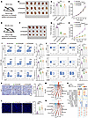 Figure 2
TARBP2 impairs immune-mediated tumor suppression in RCC. (A) Schematic of subcutaneous implantation of control or Tarbp2-knockdown Renca cells into NCG mice. (B–D) Tumor images (B), weights (C), and growth curves (D) of the indicated groups in NCG mice (n = 6 mice per group). (E) Schematic of subcutaneous implantation of control or Tarbp2-knockdown Renca cells into BALB/c mice. (F–H) Tumor images (F), weights (G), and growth curves (H) of the indicated groups in BALB/c mice (n = 5 mice per group). (I) Proportions of CD8+ T cells, CD4+ T cells, and CD3–CD49b+ NK cells in CD45+ cells from the indicated Renca-derived tumors in BALB/c mice (n = 5 mice per group). (J) Proportions of granzyme B+ (GZMB) cells in CD8+ T cells, CD4+ T cells, and NK cells from the indicated Renca-derived tumors in BALB/c mice (n = 5 mice per group). (K and L) Cell proliferation and apoptosis in tumor sections from BALB/c mice, as assessed by Ki67 (K) and TUNEL (L) staining, respectively (n = 5 mice per group). Scale bars: 50 μm. (M and N) The potential intercellular communication based on the calculation of interactive ligands-receptors between RCC/E-RCC cells and other cells in ICB-untreated (M) and ICB-treated (N) patient samples. Lines link cell types expressing the cognate receptors or ligands. Line thickness represents the inferred communication strength. (O) Dot plot showing immune checkpoint–related ligand-receptor pairs between RCC/E-RCC cells and T/NK cells. All data are presented as mean ± SD. Statistics were analyzed by 1-way ANOVA followed by Dunnett’s multiple-comparison test for comparisons with the control group in C, G, and I–L and by 2-way ANOVA followed by Bonferroni’s multiple-comparison test in D and H. ****P < 0.0001; ***P < 0.001; **P < 0.01; *P < 0.05.
To further characterize the immunological alterations associated with Tarbp2 knockdown, immune cell composition in Renca-derived tumors from BALB/c mice was analyzed by flow cytometry (Supplemental Figure 4A). Tarbp2 knockdown significantly increased the intratumoral accumulation of CD8+ T cells, CD4+ T cells, and NK cells (Figure 2I), whereas the abundance of B cells and tumor-associated macrophages remained largely unchanged (Supplemental Figure 4B). Notably, although the proportion of CD4+FOXP3+ Tregs among CD4+ T cells was decreased, no significant alteration was observed in the percentage of Tregs within CD45+ cells (Supplemental Figure 4C). Moreover, the effector function of CD8+ T cells, CD4+ T cells, and NK cells, as indicated by granzyme B and IFN-γ, was enhanced in the Tarbp2-knockdown group (Figure 2J and Supplemental Figure 4D). Ki67 and TUNEL staining revealed that Tarbp2 knockdown exhibited no significant effect on proliferation but markedly increased apoptosis rates in tumor sections from BALB/c mice (Figure 2, K and L). Given that both independent shRNAs targeting Tarbp2 produced consistent phenotypic effects, shTarbp2#3, which showed the highest silencing efficiency (Supplemental Figure 3A), was used for subsequent experiments and is hereafter designated as shTarbp2.
To investigate tumor-immune interactions, we performed cell–cell communication analysis using the integrated scRNA-seq data. The results illustrated that RCC cells with elevated TARBP2 expression exhibited enhanced communication with T and NK cells compared with E-RCC cells, regardless of ICB treatment status (Figure 2, M and N). Further ligand-receptor analysis revealed enhanced coinhibitory signaling mediated by immune checkpoint ligand-receptor pairs in RCC-T/NK cell interactions relative to E-RCC-T/NK cell interactions. Representative examples included the LGALS9-HAVCR2 pair in RCC-T cell communication and the HLA-E-CD94:NKG2A pair in RCC-NK cell crosstalk (Figure 2O).
Collectively, these results indicate that elevated TARBP2 in RCC cells inhibits antitumor immunity.
TARBP2 attenuates NK cell cytotoxic activity in RCC. Having discovered that TARBP2 in RCC cells suppresses T and NK cell function within the tumor microenvironment, we next investigated which immune cell populations are primarily targeted by TARBP2. CD8+ T, CD4+ T, or NK cells were selectively depleted using specific antibodies in BALB/c mice bearing subcutaneous tumors (Figure 3A). The tumor growth suppression observed in mice implanted with shTarbp2 Renca cells was most strongly reversed by NK cell depletion, followed by partial reversal upon CD8+ T cell depletion, whereas CD4+ T cell depletion did not significantly affect tumor control in this model (Figure 3, B–D). These results demonstrate that the enhanced antitumor immunity induced by Tarbp2 knockdown is dependent on CD8+ T cells and particularly NK cells, leading us to focus on NK cells.
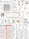 Figure 3
TARBP2 suppresses NK cell–mediated antitumor immunity in RCC. (A) Experimental design of Renca-derived tumors with or without Tarbp2 knockdown in BALB/c mice treated with isotype control, CD4+ T cell–depleting antibody (αCD4), CD8+ T cell–depleting antibody (αCD8), or NK cell–depleting antibody (αNK). (B–D) Tumor images (B), weights (C), and growth curves (D) (n = 5 mice per group). (E) Workflow for isolating CD3–NKp46+ cells from Renca-derived tumors for scRNA-seq. (F) Proportions of clusters in Tarbp2-knockdown versus control tumors. (G) Dot plots of key marker genes defining clusters from FACS-sorted CD3–NKp46+ cells, including NK/NKT cell markers, surface receptors, transcription factors (TFs), integrins, cytotoxic molecules, cytokines, inhibitory molecules, and proliferation markers. (H) Expression of Cd69, Ifng, Ccl4, and Tigit. (I) Gene Ontology (GO) and KEGG enrichment analyses in clusters 2 and 3. (J) NK activation and TGF-β signaling signature scores across clusters. (K) Comparison and correlation of the NK activation and TGF-β signaling signature scores in clusters 2 and 3. (L) Heatmap showing transcription factor regulon activity associated with NK cell activation and proliferation over pseudotime. (M) Apoptotic rates (%, annexin V+) of primary RCC cells after incubation with NK-92MI or peripheral NK cells at different E/T ratios for 8 h (n = 3 independent experiments). (N) Proportions of CD107a+, TNF-α+, and IFN-γ+ NK cells after incubation with primary RCC cells (E/T = 1:1, 8 h; n = 3 independent experiments). All data are presented as mean ± SD. Statistics were analyzed by 1-way ANOVA followed by Tukey’s multiple-comparison test in C, by 2-way ANOVA followed by Bonferroni’s multiple-comparison test in D, by χ2 test in F, by Mann-Whitney test for violin plots and Spearman’s correlation for scatterplot in K, and by 1-way ANOVA followed by Dunnett’s multiple-comparison test in M and N. ****P < 0.0001; ***P < 0.001; **P < 0.01; *P < 0.05.
To characterize NK cell alterations associated with Tarbp2 knockdown, we employed scRNA-seq on CD3–NKp46+ cells isolated by FACS from control and shTarbp2 Renca-derived tumors in BALB/c mice. After quality control, 37,915 high-quality cells were retained and classified into 8 clusters (Figure 3E). Cluster composition analysis revealed a reduction of cluster 2 and an increase of cluster 3 in the shTarbp2 group (Figure 3F). We further conducted a systematic examination of specific markers to better depict the transcriptomic profiles in each cell cluster (Figure 3, G and H, and Supplemental Figure 5, A–H). Clusters 1–6 were defined as bona fide NK cells based on expression of Nkg7, Ncr1, and Eomes and negligible expression of Cd3e, Cd8a, and Il7r. Cluster 8 was annotated as an innate lymphoid cell (ILC) cluster due to expression of Ncr1, Il7r, and Rora and negligible expression of Cd3e, Cd8a, and Eomes (22, 23). Expression of Tbx21 but not Eomes in the ILC cluster indicated the predominance of ILC1s (22). ILC cluster identification was further confirmed by utilizing published NK and ILC1-specific signatures (Supplemental Figure 5I) (24, 25). Cluster 7 was identified as cycling cells based on Mki67 and Birc5 expression. Subsequent reintegration and label transfer analysis following regression of cell cycle genes revealed a ubiquitous distribution of cycling cells across all NK/ILC clusters, indicating that cycling cells do not represent a distinct phenotype (Supplemental Figure 5J). Of note, cycling cells exhibited a significantly higher proportion of NK cluster 5 and a markedly lower proportion of NK cluster 6 compared with noncycling cells (Supplemental Figure 5K), suggesting that NK cluster 5 possesses greater proliferation potential, whereas NK cluster 6 exhibits opposing functional characteristics. Within NK subsets, cluster 6 exhibited a dysfunctional tissue-resident phenotype characterized by upregulation of Klrg1, Itga1, and Havcr2, alongside downregulation of Itga2 and Itgam. In contrast, clusters 2 and 3 displayed elevated expression of Itga2 and Itgam, corresponding to conventional cytotoxic NK cells (Figure 3G) (26). We further compared the transcriptomic profiles of clusters 2 and 3. Cluster 3 exhibited upregulated expression of Ifng and the activation marker Cd69 (27), along with enhanced Ccl4 expression, which facilitates DC recruitment to augment antitumor immune responses (28). In addition, the immune checkpoint gene Tigit was downregulated in cluster 3 (Figure 3H). Gene Ontology and Kyoto Encyclopedia of Genes and Genomes (KEGG) analyses illustrated enrichment of pathways related to immune activation in cluster 3 (Figure 3I). Moreover, the enrichment scores of the NK activation signature previously reported (29) were higher in cluster 3 compared with cluster 2, while TGF-β signaling, the activation of which suppresses NK cell proliferation and cytotoxicity (30), showed an inverse trend (Figure 3, J and K). Trajectory analysis of NK clusters 1–6 revealed that clusters 2 and 3 had transition potential toward the dysfunctional state characterized by cluster 6 (Supplemental Figure 5L). Gene regulatory network analysis was applied to further investigate the potential drivers of this transition. Our analysis revealed concomitant downregulation of multiple transcription factor regulons associated with NK cell activation and proliferation over pseudotime, including AP-1 complex members (e.g., Jun, Fos) and their upstream regulator Ikzf1 (31), along with NF-κB family members (e.g., Rel, Nfkb1) (29). The heatmap demonstrated that cluster 3, positioned at earlier stages of pseudotime relative to cluster 2, exhibited higher levels of these regulons (Figure 3L). Since Tarbp2 knockdown in RCC cells promoted the accumulation of cluster 3, these results suggest that TARBP2 in RCC cells may direct NK cell differentiation toward a dysfunctional trajectory.
We next investigated whether TARBP2 in RCC cells drives NK cell dysfunction through secreted factors or direct cell–cell contact. To this end, we used 0.4 μm Transwell inserts, which permit the diffusion of soluble mediators but prevent cell migration, to coculture Renca cells with murine primary NK cells (Supplemental Figure 6A). Under indirect coculture conditions, in which Renca and NK cells were separated by the Transwell inserts, shTarbp2 did not alter Renca cell apoptosis or NK cell activation (e.g., Cd69), inhibitory (e.g., Tigit), and maturation (e.g., Itgam) marker expression. In contrast, under direct coculture conditions allowing physical contact between Renca and NK cells, shTarbp2 significantly increased Renca cell apoptosis, enhanced NK cell activation and maturation marker expression, and reduced inhibitory marker expression, consistent with the scRNA-seq findings (Supplemental Figure 6, B and C). These results suggest that TARBP2 in RCC cells may regulate NK cell function in a contact-dependent manner.
To investigate whether TARBP2 suppresses NK cell cytotoxicity in vitro, primary RCC cells derived from patients were cocultured with NK-92MI cells and FACS-sorted peripheral blood NK cells (Supplemental Figure 6A and Supplemental Figure 7A). TARBP2 knockdown in primary RCC cells (targets [T]) enhanced the cytotoxicity of NK cells (effectors [E]) at different E/T ratios (Figure 3M). The proportions of CD107a+, TNF-α+, and IFN-γ+ NK cells were increased after coculture with TARBP2-knockdown RCC cells (Figure 3N and Supplemental Figure 7, B and C).
In summary, TARBP2 in RCC cells inhibits NK cell cytotoxicity and may exert this effect, at least in part, through direct cell–cell contact with NK cells.
TARBP2 drives NK cell dysfunction in RCC through the p53/B7-H3 axis. We next investigated the molecular mechanisms underlying TARBP2-mediated modulation of NK cell function. Tumor cells from shTarbp2 and control Renca-derived tumors in BALB/c mice were isolated using a magnetic-activated cell sorting (MACS) tumor cell isolation kit, and the transcriptomic profiles of these cells were obtained by RNA-seq (Supplemental Figure 8A). Bioinformatics analysis demonstrated substantial transcriptional alterations following Tarbp2 knockdown, with 877 upregulated and 961 downregulated genes meeting the thresholds of |log2(fold change)| > 1 and P < 0.05 (Supplemental Figure 8B). Given that TARBP2-mediated regulation of NK cell function is dependent on direct cell–cell contact and that contact-dependent regulation of NK cell function is predominantly mediated by membrane-bound ligand-receptor interactions (32), cell surface molecules were prioritized as candidate mediators of TARBP2-driven modulation of NK cell function. Notably, B7h3, an immune checkpoint previously reported to attenuate NK cell–mediated tumor control (15, 33, 34) and whose human homolog B7-H3 was also highly expressed in pretreatment FFPE samples from ICB nonresponders, emerged as the most significantly downregulated surface molecule following Tarbp2 knockdown (Figure 4, A and B, and Supplemental Figure 8, C and D). Flow cytometry and immunofluorescence analyses further confirmed decreased plasma membrane–localized B7-H3 levels in MACS-isolated tumor cells from shTarbp2 tumors (Figure 4C and Supplemental Figure 8E). A positive correlation between TARBP2 and B7-H3 expression was observed across multiple cancer types in the TCGA database (Figure 4D). This TARBP2 and B7-H3 coexpression pattern was also identified in the CheckMate ICB treatment cohort, the Clinical Proteomic Tumor Analysis Consortium RCC dataset, and Cancer Cell Line Encyclopedia RCC cell dataset (Figure 4E). The correlation was further validated by IHC in RCC samples (Figure 4F). We next performed KEGG pathway enrichment analysis of differentially expressed genes between tumor cells isolated from shTarbp2 and control Renca-derived tumors, and enrichment of the p53 signaling pathway was observed (Supplemental Figure 8F). GSEA further confirmed activation of p53 signaling following Tarbp2 knockdown (Figure 4G). Given prior evidence that p53 negatively regulates B7-H3 expression (35), we hypothesized that TARBP2 drives B7-H3 expression by suppressing the p53 pathway.
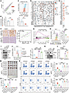 Figure 4
TARBP2/p53/B7-H3 axis regulates NK cell antitumor function in RCC. (A) Differentially expressed genes between tumor cells isolated from shTarbp2 and control Renca-derived tumors (n = 5 mice/group). (B) B7-H3 expression in RCC ICB pretreatment FFPE samples from responders and nonresponders (n = 3 patients/group). (C) Cell surface B7-H3 on isolated tumor cells (n = 5 mice/group). (D and E) Spearman’s correlations between TARBP2 and 38 common immune checkpoint genes across 33 TCGA cancer types (D) or RCC-related datasets (E). POS, positive; NEG, negative. (F) Representative IHC images of RCC tissues stained for TARBP2 and B7-H3. Scale bar: 100 μm (left). Spearman’s correlation between TARBP2 and B7-H3 H-scores (right; n = 40 samples). (G) GSEA of the p53 signaling pathway in isolated tumor cells (n = 5 mice/group). (H) Cell surface B7-H3 levels in TARBP2-knockdown and control 769-P cells (n = 3 independent experiments). (I) Total B7-H3, p53, and p21 protein levels in TARBP2-knockdown and control 769-P cells. GAPDH served as loading control. (J and K) Cell surface (J) and total (K) B7-H3 levels in TP53-knockdown 769-P cells with TARBP2 knockdown or control. (L) B7-H3 promoter–driven reporter activity in 769-P cells under control, TARBP2-knockdown, or TARBP2/TP53–co-knockdown conditions (n = 4 independent experiments). Relative luciferase activities were normalized to vector control samples. (M and N) Tumor weights (M) and growth curves (N) (n = 5 mice/group). (O) Proportions of CD3–CD49b+ NK cells within CD45+ cells and frequencies of CD107a+ cells among NK cells in indicated tumors (n = 5 mice/group). All data are presented as mean ± SD. Statistics were analyzed by unpaired 2-tailed Student’s t test in B and C, by 1-way ANOVA followed by Dunnett’s multiple-comparison test in H, by 1-way ANOVA followed by Tukey’s multiple-comparison test in J and L, and by 2-way ANOVA followed by Tukey’s multiple-comparison test in N and O. ****P < 0.0001; ***P < 0.001; **P < 0.01; *P < 0.05.
To validate this hypothesis, we conducted in vitro experiments using RCC cell lines with WT TP53 (769-P, ACHN, Caki-1). Based on endogenous TARBP2 levels (Supplemental Figure 8, G and H), we knocked down TARBP2 in 769-P and ACHN cells and overexpressed TARBP2 in Caki-1 cells. TARBP2 knockdown reduced both total and cell surface B7-H3 protein levels, while increasing p53 and its downstream target p21 (Figure 4, H and I, and Supplemental Figure 8, I and J). In contrast, TARBP2 overexpression produced the opposite effects (Supplemental Figure 8, K and L). For all subsequent rescue experiments, shTARBP2#2 was used and is hereafter referred to as shTARBP2. Rescue experiments showed that TP53 knockdown reversed the reduction in B7-H3 protein levels induced by shTARBP2 (Figure 4, J and K, and Supplemental Figure 9, A and B). p53 is a transcription factor encoded by TP53, one of the most frequently mutated genes in human cancers. Most TP53 mutations are missense mutations located within the DNA-binding domain and impair the DNA-binding activity of p53 (36). Based on this, WT TP53 or 3 common DNA-binding–deficient mutants (R175H, R248W, and R273H) were overexpressed in TARBP2-overexpressing Caki-1 cells. Only WT TP53 reversed TARBP2-induced upregulation of B7-H3, whereas mutant constructs failed to do so (Supplemental Figure 9, C and D). Dual-luciferase reporter assays assessing B7-H3 promoter–driven transcriptional activity showed trends consistent with the changes in B7-H3 protein levels. TP53 knockdown reversed the decrease in B7-H3 promoter–driven transcriptional activity induced by shTARBP2, whereas only WT TP53 overexpression reversed the increase in transcriptional activity induced by TARBP2 overexpression (Figure 4L and Supplemental Figure 9, E and F). These findings suggest that TARBP2 promotes B7-H3 expression by suppressing the transcriptional regulatory activity of p53.
To further determine whether p53 directly binds to the B7-H3 promoter region, the human B7-H3 promoter sequence was analyzed using the JASPAR database. Potential p53-binding sites were identified within the B7-H3 promoter region (Supplemental Figure 9G). ChIP-qPCR demonstrated significant enrichment of p53 at the B7-H3 promoter. TP53 knockdown abolished the increase in p53 enrichment induced by shTARBP2, whereas only WT TP53 overexpression rescued the decrease caused by TARBP2 overexpression (Supplemental Figure 9, H–J). These findings indicate that TARBP2 promotes B7-H3 expression by inhibiting p53 binding to the B7-H3 promoter region, thereby attenuating the transcriptional repression of B7-H3 by p53.
To investigate the impact of the TARBP2/p53/B7-H3 axis on tumor growth and NK cell function in vivo, we performed genetic interventions targeting Tarbp2, Trp53, and B7h3 in Renca cells, followed by subcutaneous implantation in BALB/c mice. Combined knockdown of Tarbp2 and Trp53 abrogated Tarbp2 knockdown–induced tumor growth suppression, NK cell accumulation, and increases in CD107a+, TNF-α+, and IFN-γ+ NK cell proportions. Combined knockdown of Tarbp2, Trp53, and B7h3 further reversed these effects observed in the double knockdown setting (Supplemental Figure 10, A–D). To determine the dependency of B7-H3, B7h3-knockout Renca cells were generated via CRISPR/Cas9 technology. These genetically modified cells subsequently underwent Tarbp2 knockdown or overexpression for assessment of phenotypic alterations in BALB/c mice. No significant differences in tumor growth, NK cell accumulation, CD107a degranulation, or cytokine production were observed among B7h3-knockout groups (Figure 4, M–O, and Supplemental Figure 10, E and F).
In summary, TARBP2 in RCC cells impairs the antitumor potential of NK cells by relieving p53-mediated transcriptional repression of B7-H3.
TARBP2 impairs 53BP1-p53 complex formation and chromatin accumulation. Previous studies demonstrated that TARBP2 forms a complex with Dicer to participate in miRNA processing and RISC assembly, thereby facilitating posttranscriptional gene silencing (7, 37). TARBP2 has also been reported to regulate mRNA stability (9, 10). However, TARBP2 knockdown or overexpression did not alter TP53 mRNA levels (Supplemental Figure 11, A–C). Moreover, Dicer silencing in TARBP2-overexpressing Caki-1 cells did not reverse TARBP2-induced upregulation of B7-H3 or the downregulation of p53 and its downstream target p21 (Supplemental Figure 11, D and E). Notably, RCC samples from the SYSU cohort showed higher overall TARBP2 expression in high-grade (G3-4) tumors than in low-grade (G1-2) tumors, particularly in the nucleus (Figure 5A and Supplemental Figure 12A). Patients with higher nuclear TARBP2 expression exhibited poorer OS (Supplemental Figure 12B). Given that TARBP2 functions as a multifunctional protein capable of interacting with diverse partner proteins and modulating their functional activities (11, 12), nuclear and cytoplasmic fractions of 769-P cells were isolated and subjected to co-IP coupled with mass spectrometry analysis to characterize the TARBP2 protein interactome. Following subtraction of control IgG-bound proteins, nuclear-specific interactors were ranked by descending confidence score. Among the top candidates, 53BP1, a p53-binding regulator of p53 signaling (38), was identified within the top 5 hits (Figure 5B and Supplemental Figure 12C). Co-IP assays using nuclear fractions confirmed the interaction between endogenous TARBP2 and 53BP1 in 769-P and ACHN cells (Figure 5, C and D, and Supplemental Figure 12, D and E). Overexpressed FLAG-tagged TARBP2 or HA-tagged 53BP1 coimmunoprecipitated with endogenous 53BP1 or TARBP2 in Caki-1 cells, respectively (Supplemental Figure 12, F and G). Moreover, FLAG-tagged TARBP2 coimmunoprecipitated with HA-tagged 53BP1 and vice versa when coexpressed in Caki-1 cells (Figure 5E). 3D structured illumination microscopy demonstrated nuclear colocalization of TARBP2 and 53BP1 (Figure 5F and Supplemental Figure 12H).
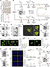 Figure 5
TARBP2 disrupts 53BP1-p53 complex formation and chromatin accumulation. (A) Top: Representative IHC staining of TARBP2. Scale bar: 50 μm. Bottom: H-score of nuclear TARBP2 in low- and high-grade RCC tissues from the SYSU cohort (n = 120 samples). Boxes represent the IQR, center lines indicate the median, whiskers extend to 1.5-fold of the IQR, and dots indicate outliers. (B) Workflow of nucleocytoplasmic separation, co-IP, and mass spectrometry identifying nuclear TARBP2–53BP1 interaction. (C–E) Co-IP assays showing interaction between endogenous TARBP2 and 53BP1 in 769-P cells (C and D) and between overexpressed TARBP2-FLAG and 53BP1-HA in Caki-1 cells (E). EV, empty vector. (F) 3D structured illumination microscopy rendering of 769-P cells stained to indicate TARBP2 (green) and 53BP1 (red) colocalization (yellow). Scale bars: 5 μm and 1 μm (insets). The x–y plane represents lateral cross-sectional views parallel to the substrate surface, and the z axis indicates axial dimension perpendicular to the substrate surface. (G and H) Total 53BP1 levels in 769-P (G) or Caki-1 (H) cells. GAPDH served as loading control. (I and J) Representative confocal images (left) and quantification of 53BP1 foci (right) in 769-P (I) or Caki-1 (J) cells (n > 120 nuclei/group). Dashed lines outline nuclei. Scale bars: 5 μm. (K and L) 53BP1 and p53 levels in chromatin fractions of 769-P (K) or Caki-1 (L) cells. Histone H3 and H4 served as chromatin loading controls. (M) Representative images (left) and quantification (right) of endogenous TARBP2-53BP1 and 53BP1-p53 interactions in 769-P cells (n > 100 nuclei/group). DAPI (blue) marks nuclei, and red dots indicate positive signals. Scale bar: 5 μm. (N) Sequential co-IP assays demonstrating distinct endogenous TARBP2-53BP1 and 53BP1-p53 complexes. Solid lines indicate medians in I (right), J (right), and M (right). Statistics were analyzed by unpaired 2-tailed Student’s t test in A, by Kruskal-Wallis test followed by Dunn’s multiple-comparison test in I and M, and by Mann-Whitney test in J. ****P < 0.0001; ***P < 0.001; *P < 0.05.
We further investigated whether TARBP2 modulates the function of 53BP1. Knockdown or overexpression of TARBP2 did not affect endogenous DNA damage levels (Supplemental Figure 12, I–K) or total 53BP1 protein abundance (Figure 5, G and H, and Supplemental Figure 12L). However, TARBP2 knockdown significantly enhanced the formation of chromatin-associated 53BP1 nuclear bodies known as 53BP1 foci, while TARBP2 overexpression exhibited the opposite effect (Figure 5, I and J, and Supplemental Figure 12M). Consistent with prior evidence that 53BP1 chromatin retention independent of extensive DNA damage enhances p53 chromatin binding and augments p53 responses (39), increased p53 chromatin accumulation correlated with 53BP1 enrichment under TARBP2 knockdown, whereas an inverse relationship was observed under TARBP2 overexpression (Figure 5, K and L, and Supplemental Figure 12N). Furthermore, the interaction between 53BP1 and p53 is critical for p53-mediated gene regulatory events (38). A proximity ligation assay confirmed that TARBP2 knockdown disrupted formation of the TARBP2-53BP1 complex while increasing the number of 53BP1-p53 puncta, whereas TARBP2 overexpression exhibited the opposite effect (Figure 5M and Supplemental Figure 12, O and P). Sequential co-IP using nuclear extracts further clarified the interaction hierarchy. Initial TARBP2 immunoprecipitation identified 53BP1 as an interacting partner, while p53 was undetectable in the TARBP2-bound complex. Subsequent p53 immunoprecipitation of the TARBP2-unbound fraction demonstrated an association between 53BP1 and p53, with TARBP2 remaining absent from the precipitated complex (Figure 5N). These results suggest that TARBP2 interferes with 53BP1-p53 complex formation and suppresses chromatin accumulation of both 53BP1 and p53.
TARBP2 restricts 53BP1 phase separation to attenuate p53 activation and upregulate B7-H3. To identify the region of 53BP1 required for interaction with TARBP2, we transfected FLAG-tagged TARBP2-overexpressing Caki-1 cells with a series of plasmids expressing HA-tagged truncation mutants of 53BP1 (Figure 6A). Co-IP assays revealed that the oligomerization domain (OD), whose protein sequence is conserved across multiple species (Figure 6B), was required for stable interaction between TARBP2 and 53BP1 (Figure 6C). Additionally, TARBP2 did not affect chromatin accumulation of the 53BP1 fragment lacking the OD (Figure 6, D and E, and Supplemental Figure 13A). Previous studies have demonstrated that deletion of the OD disrupts liquid–liquid phase separation (LLPS) of 53BP1 (39–41) and that impaired LLPS compromises 53BP1-mediated p53 activation (39, 40). Therefore, we hypothesized that TARBP2 interacts with the OD of 53BP1 to suppress 53BP1 LLPS, thereby regulating the p53/B7-H3 axis.
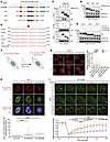 Figure 6
TARBP2 inhibits 53BP1 oligomerization-driven LLPS. (A) Schematic of 53BP1 truncation mutants. (B) Cross-species conservation of the 53BP1 OD protein sequence. (C) Co-IP assays showing the interactions between overexpressed TARBP2-FLAG and indicated 53BP1 truncation mutants in the nuclei of Caki-1 cells. (D and E) Immunoblot analysis of 53BP1 truncation mutants in chromatin fractions from Caki-1 cells with TARBP2 overexpression or control (D) and 769-P cells with TARBP2 knockdown or control (E). Histone H3 and H4 served as chromatin loading controls. (F) Schematic of optoDroplet formation of Cry2-mCherry-53BP1 fusion proteins with or without the OD. (G) Representative images (left) and quantification (right) of optoDroplet formation of Cry2-mCherry-53BP1 fusion proteins with or without the OD in 769-P cells with TARBP2 knockdown or control treatment (n > 120 nuclei per group). Dashed lines outline nuclei. Scale bar: 5 μm. (H) Cry2-mCherry-53BP1 (containing the OD) optoDroplet induction followed by p53 (green) and DAPI (blue) staining in 769-P cells with TARBP2 knockdown or control treatment (n > 100 nuclei per group). Scale bar: 5 μm. (I) Representative images (top) and quantification (bottom) of fluorescence recovery after photobleaching analysis of GFP-53BP1 puncta in 769-P cells with TARBP2 knockdown or control treatment (n = 12 events from independent cells per group). Scale bar: 5 μm. Red circles indicate bleached puncta. Time zero (0 second) on the x axis marks the execution of photobleaching. Solid lines indicate medians in G (right) and H (bottom). Data are presented as mean ± SD in I (bottom). Statistics were analyzed by Kruskal-Wallis test followed by Dunn’s multiple-comparison test in G and H and by 2-way ANOVA followed by Bonferroni’s multiple-comparison test in I. ****P < 0.0001.
To verify this hypothesis, mCherry-labeled Arabidopsis thaliana cryptochrome 2–based (Cry2-based) fusion constructs containing 53BP1 with or without the OD were used to evaluate blue light–induced 53BP1 oligomerization and formation of phase-separated condensates, termed optoDroplets (Figure 6F and Supplemental Figure 13B) (40, 42). Formation of 53BP1 optoDroplets strictly required the OD, and TARBP2 perturbation influenced the assembly of OD-containing Cry2-mCherry-53BP1 into these phase-separated structures (Figure 6G and Supplemental Figure 13, C and D). Furthermore, p53 accumulation within OD-containing Cry2-mCherry-53BP1 optoDroplets increased in TARBP2-knockdown cells and decreased in TARBP2-overexpressing cells (Figure 6H and Supplemental Figure 13E). Fluorescence recovery after photobleaching analysis was performed to evaluate the LLPS capacity of GFP-tagged 53BP1 under TARBP2 perturbation in live cells. Remarkably, TARBP2 knockdown enhanced fluorescence recovery of GFP-53BP1 puncta after photobleaching, whereas its overexpression produced the opposite effect (Figure 6I and Supplemental Figure 13F).
We next assessed the role of LLPS in regulating the p53/B7-H3 axis. TARBP2-knockdown and control cells were treated with 1,6-hexanediol, an aliphatic alcohol commonly used to disrupt LLPS. 1,6-Hexanediol treatment restored the reduction in total and cell surface B7-H3 levels induced by TARBP2 knockdown, accompanied by reversal of upregulation of the p53 downstream target p21 (Supplemental Figure 14, A–D). To determine whether TARBP2-mediated regulation of the p53/B7-H3 axis depends on 53BP1 OD-driven LLPS, Cry2-mCherry-53BP1 constructs with or without the OD were expressed in TARBP2-overexpressing Caki-1 cells, followed by blue light stimulation. The OD-containing construct reversed the TARBP2-induced increase in total and cell surface B7-H3 levels while also restoring p21 expression. This rescue effect was further enhanced upon blue light stimulation. In contrast, the OD-deficient construct showed no such effect (Supplemental Figure 14, E and F).
Collectively, these results demonstrate that TARBP2 restricts 53BP1 OD-driven LLPS, thereby impairing 53BP1-mediated p53 activation and ultimately promoting B7-H3 upregulation.
Targeting TARBP2 sensitizes RCC to adoptive NK cell therapy and enhances ICB efficacy. To evaluate the clinical relevance of TARBP2 in predicting responses to ICB therapy, we performed multiplex IHC on pretreatment tumor tissues from patients with RCC classified as responders or nonresponders to ICB therapy. TARBP2 expression was predominantly localized in pan-cytokeratin+ tumor cells, and significantly higher TARBP2 expression was observed in tumor cells from nonresponders compared with responders (Supplemental Figure 15, A and B). Furthermore, tumors from nonresponders showed elevated nuclear TARBP2 expression and increased B7-H3 expression, accompanied by reduced accumulation of CD3+ T cells and particularly CD3–CD56+ NK cells (Figure 7, A and B).
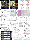 Figure 7
Targeting TARBP2 sensitizes RCC to adoptive NK cell therapy and ICB therapy. (A) Representative multiplex IHC staining of pan-cytokeratin (PanCK), TARBP2, B7-H3, CD3, CD56, and DAPI in RCC samples from ICB responders and nonresponders. Scale bars: 50 μm and 50 μm (insets). (B) Quantification of nuclear TARBP2+, B7-H3+, CD3+, and CD3–CD56+ cells in RCC samples from ICB responders and nonresponders (n = 5 patients/group). (C) Illustration of the RCC PDX model in immunodeficient NCG mice treated with adoptive NK cell transfer and siRNA at the indicated time points. (D–F) Tumor images (D), weights (E), and growth curves (F) in the indicated PDX groups (n = 5 mice/group). (G) Representative PDX tumor sections from NCG mice treated with siRNA and adoptive NK-92MI cell transfer stained with H&E, TARBP2, B7-H3, and Cleaved Caspase-3. Scale bar: 50 μm. (H) Proportion of CD107a+ cells among CD3–CD56+ NK cells in PDX tumors (n = 5 mice/group). (I) Schematic of Renca-derived orthotopic tumors in BALB/c mice treated with adoptive NK cell transfer, anti–PD-1 antibody, and anti–B7-H3 antibody at the indicated time points. (J and K) Quantification of bioluminescence intensity (J) and representative bioluminescence images (K) of orthotopic tumors from the indicated groups (n = 5 mice/group). (L) Proportions of CD3–CD49b+ NK cells among CD45+ cells, CD107a+ cells among NK cells, and the ratio of PKH26+ transferred NK cells to PKH26− endogenous NK cells in orthotopic tumors from the indicated groups (n = 5 mice/group). All data are presented as mean ± SD. Statistics were analyzed by unpaired 2-tailed Student’s t test in B and H, by 1-way ANOVA followed by Tukey’s multiple-comparison test in E and L, and by 2-way ANOVA followed by Bonferroni’s multiple-comparison test in F and J. ****P < 0.0001; ***P < 0.001; **P < 0.01; *P < 0.05.
To investigate whether TARBP2 inhibition enhances NK cell–mediated antitumor activity in a clinically relevant human RCC model, immunodeficient NCG mice were engrafted with RCC patient-derived xenografts (PDXs) and subsequently received adoptive transfer of NK-92MI cells. Localized administration of TARBP2-targeting siRNA (siTARBP2) was conducted to abrogate TARBP2 expression in PDX tumors (Figure 7C). Consistent with observations in the NCG Renca-derived tumor model (Figure 2, A–D), siTARBP2 alone exerted negligible effects on tumor growth in the absence of NK-92MI cell transfer. Although NK-92MI cell monotherapy produced moderate antitumor effects, combination treatment with siTARBP2 achieved the greatest suppression of PDX tumor growth (Figure 7, D–F). The expression levels of TARBP2 and B7-H3, along with apoptosis levels indicated by Cleaved Caspase-3 staining in PDX tumor tissues, were assessed by IHC (Figure 7G and Supplemental Figure 16, A and B). Additionally, flow cytometry analysis demonstrated that cotreatment with siTARBP2 significantly increased the proportions of CD107a+, TNF-α+, and IFN-γ+ NK cells within PDX tumor microenvironments compared with NK-92MI monotherapy (Figure 7H and Supplemental Figure 16C).
To further determine whether TARBP2 inhibition synergizes with adoptive NK cell therapy to enhance ICB efficacy, we established orthotopic RCC tumors in immunocompetent BALB/c mice using luciferase-expressing Renca cells transfected with either Tarbp2 knockdown or control vectors. Tumor-bearing mice were then assigned to treatment groups and received adoptive NK cell transfer alone or in combination with anti–PD-1, anti–B7-H3, or anti–PD-1 plus anti–B7-H3 antibodies. NK cells were labeled with PKH26 prior to transfer to distinguish transferred NK cells from endogenous murine NK cells within tumors (Figure 7I). Consistent with the NCG PDX model, Tarbp2 knockdown combined with NK cell transfer significantly suppressed tumor growth. The addition of anti–PD-1 or anti–B7-H3 further improved tumor control, with dual blockade achieving the greatest therapeutic efficacy. Notably, Tarbp2 knockdown significantly enhanced treatment efficacy across all treatment groups (Figure 7, J and K, and Supplemental Figure 17). No evident toxicity was observed, as indicated by body weight monitoring and histological examination of organ sections (Supplemental Figure 18, A and B). Flow cytometry analysis of orthotopic tumors showed that, among all groups receiving adoptive NK cell transfer, no significant differences were observed in the ratio of PKH26+ transferred NK cells to PKH26– endogenous NK cells within the intratumoral NK cell population. However, Tarbp2 knockdown significantly increased the proportion of NK cells among intratumoral immune cells and enhanced NK cell effector functions across all treatment groups. These effects were most pronounced in mice receiving Tarbp2 knockdown together with adoptive NK cell transfer and dual checkpoint blockade (Figure 7L and Supplemental Figure 19, A and B).
Collectively, these findings demonstrate that TARBP2 inhibition sensitizes RCC to adoptive NK cell therapy, with further therapeutic enhancement achieved by concurrent ICB treatment.


Copyright © 2026 American Society for Clinical Investigation
ISSN: 0021-9738 (print), 1558-8238 (online)