
Advertisement
Research ArticleGeneticsOncology
Open Access |  10.1172/JCI199454
Polycomb scaffolding subunit EED supports the SCLC neuroendocrine identity and EGFR-mutant LUAD-to-SCLC transformation
Yixiang Li,1 Yasmin N. Laimon,2 Hyeonseo Cho,1 Marina Vivero,2 Gabriel Roberti De Oliveira,2 Maxwell D. Seager,2 Andrew Delcea,2 Varunika Savla,2 Yuting Chen,3 Yavuz T. Durmaz,1 Xintao Qiu,1,4 Shweta Kukreja,1,4 Rong Li,1,4 Talal El Zarif,1 Wesley Lu,1,5 McKayla Van Orden,1,5 Jacob E. Berchuck,6 Roderick T. Bronson,7 Shuqiang Li,1,5 Hongbin Ji,3,8,9,10 David A. Barbie,1 Katerina Politi,11 Matthew L. Freedman,1,4 Henry W. Long,1,4 Sabina Signoretti,2,12 and Matthew G. Oser1,13
1Department of Medical Oncology, Dana-Farber Cancer Institute and Brigham and Women’s Hospital, and
2Department of Pathology, Brigham and Women’s Hospital, Harvard Medical School, Boston, Massachusetts, USA.
3State Key Laboratory of Cell Biology, Shanghai Institute of Biochemistry and Cell Biology, Center for Excellence in Molecular Cell Science, Chinese Academy of Sciences, Shanghai, China.
4Center for Functional Cancer Epigenetics and
5Translational Immunogenomics Lab, Dana-Farber Cancer Institute, Boston, Massachusetts, USA.
6Winship Cancer Institute, Emory University School of Medicine, Atlanta, Georgia, USA.
7Division of Immunology, Department of Microbiology and Immunobiology, Harvard Medical School, Boston, Massachusetts, USA.
8University of Chinese Academy of Sciences, Beijing, China.
9School of Life Science, Hangzhou Institute for Advanced Study, University of Chinese Academy of Sciences, Hangzhou, China.
10School of Life Science and Technology, Shanghai Tech University, Shanghai, China.
11Departments of Pathology and Internal Medicine (Section of Medical Oncology), Yale School of Medicine and Yale Cancer Center, New Haven, Connecticut, USA.
12Department of Oncologic Pathology, Dana-Farber Cancer Institute, Boston, Massachusetts, USA.
13Department of Medicine, Brigham and Women’s Hospital, Harvard Medical School, Boston, Massachusetts, USA.
Address correspondence to: Matthew G. Oser, Dana-Farber Cancer Institute, 450 Brookline Avenue, LC 4117, Boston, Massachusetts 02215, USA. Phone: 617.632.6036; Email: matthew_oser@dfci.harvard.edu.
Find articles by Li, Y. in: PubMed | Google Scholar
1Department of Medical Oncology, Dana-Farber Cancer Institute and Brigham and Women’s Hospital, and
2Department of Pathology, Brigham and Women’s Hospital, Harvard Medical School, Boston, Massachusetts, USA.
3State Key Laboratory of Cell Biology, Shanghai Institute of Biochemistry and Cell Biology, Center for Excellence in Molecular Cell Science, Chinese Academy of Sciences, Shanghai, China.
4Center for Functional Cancer Epigenetics and
5Translational Immunogenomics Lab, Dana-Farber Cancer Institute, Boston, Massachusetts, USA.
6Winship Cancer Institute, Emory University School of Medicine, Atlanta, Georgia, USA.
7Division of Immunology, Department of Microbiology and Immunobiology, Harvard Medical School, Boston, Massachusetts, USA.
8University of Chinese Academy of Sciences, Beijing, China.
9School of Life Science, Hangzhou Institute for Advanced Study, University of Chinese Academy of Sciences, Hangzhou, China.
10School of Life Science and Technology, Shanghai Tech University, Shanghai, China.
11Departments of Pathology and Internal Medicine (Section of Medical Oncology), Yale School of Medicine and Yale Cancer Center, New Haven, Connecticut, USA.
12Department of Oncologic Pathology, Dana-Farber Cancer Institute, Boston, Massachusetts, USA.
13Department of Medicine, Brigham and Women’s Hospital, Harvard Medical School, Boston, Massachusetts, USA.
Address correspondence to: Matthew G. Oser, Dana-Farber Cancer Institute, 450 Brookline Avenue, LC 4117, Boston, Massachusetts 02215, USA. Phone: 617.632.6036; Email: matthew_oser@dfci.harvard.edu.
Find articles by Laimon, Y. in: PubMed | Google Scholar
1Department of Medical Oncology, Dana-Farber Cancer Institute and Brigham and Women’s Hospital, and
2Department of Pathology, Brigham and Women’s Hospital, Harvard Medical School, Boston, Massachusetts, USA.
3State Key Laboratory of Cell Biology, Shanghai Institute of Biochemistry and Cell Biology, Center for Excellence in Molecular Cell Science, Chinese Academy of Sciences, Shanghai, China.
4Center for Functional Cancer Epigenetics and
5Translational Immunogenomics Lab, Dana-Farber Cancer Institute, Boston, Massachusetts, USA.
6Winship Cancer Institute, Emory University School of Medicine, Atlanta, Georgia, USA.
7Division of Immunology, Department of Microbiology and Immunobiology, Harvard Medical School, Boston, Massachusetts, USA.
8University of Chinese Academy of Sciences, Beijing, China.
9School of Life Science, Hangzhou Institute for Advanced Study, University of Chinese Academy of Sciences, Hangzhou, China.
10School of Life Science and Technology, Shanghai Tech University, Shanghai, China.
11Departments of Pathology and Internal Medicine (Section of Medical Oncology), Yale School of Medicine and Yale Cancer Center, New Haven, Connecticut, USA.
12Department of Oncologic Pathology, Dana-Farber Cancer Institute, Boston, Massachusetts, USA.
13Department of Medicine, Brigham and Women’s Hospital, Harvard Medical School, Boston, Massachusetts, USA.
Address correspondence to: Matthew G. Oser, Dana-Farber Cancer Institute, 450 Brookline Avenue, LC 4117, Boston, Massachusetts 02215, USA. Phone: 617.632.6036; Email: matthew_oser@dfci.harvard.edu.
Find articles by Cho, H. in: PubMed | Google Scholar
1Department of Medical Oncology, Dana-Farber Cancer Institute and Brigham and Women’s Hospital, and
2Department of Pathology, Brigham and Women’s Hospital, Harvard Medical School, Boston, Massachusetts, USA.
3State Key Laboratory of Cell Biology, Shanghai Institute of Biochemistry and Cell Biology, Center for Excellence in Molecular Cell Science, Chinese Academy of Sciences, Shanghai, China.
4Center for Functional Cancer Epigenetics and
5Translational Immunogenomics Lab, Dana-Farber Cancer Institute, Boston, Massachusetts, USA.
6Winship Cancer Institute, Emory University School of Medicine, Atlanta, Georgia, USA.
7Division of Immunology, Department of Microbiology and Immunobiology, Harvard Medical School, Boston, Massachusetts, USA.
8University of Chinese Academy of Sciences, Beijing, China.
9School of Life Science, Hangzhou Institute for Advanced Study, University of Chinese Academy of Sciences, Hangzhou, China.
10School of Life Science and Technology, Shanghai Tech University, Shanghai, China.
11Departments of Pathology and Internal Medicine (Section of Medical Oncology), Yale School of Medicine and Yale Cancer Center, New Haven, Connecticut, USA.
12Department of Oncologic Pathology, Dana-Farber Cancer Institute, Boston, Massachusetts, USA.
13Department of Medicine, Brigham and Women’s Hospital, Harvard Medical School, Boston, Massachusetts, USA.
Address correspondence to: Matthew G. Oser, Dana-Farber Cancer Institute, 450 Brookline Avenue, LC 4117, Boston, Massachusetts 02215, USA. Phone: 617.632.6036; Email: matthew_oser@dfci.harvard.edu.
Find articles by Vivero, M. in: PubMed | Google Scholar
1Department of Medical Oncology, Dana-Farber Cancer Institute and Brigham and Women’s Hospital, and
2Department of Pathology, Brigham and Women’s Hospital, Harvard Medical School, Boston, Massachusetts, USA.
3State Key Laboratory of Cell Biology, Shanghai Institute of Biochemistry and Cell Biology, Center for Excellence in Molecular Cell Science, Chinese Academy of Sciences, Shanghai, China.
4Center for Functional Cancer Epigenetics and
5Translational Immunogenomics Lab, Dana-Farber Cancer Institute, Boston, Massachusetts, USA.
6Winship Cancer Institute, Emory University School of Medicine, Atlanta, Georgia, USA.
7Division of Immunology, Department of Microbiology and Immunobiology, Harvard Medical School, Boston, Massachusetts, USA.
8University of Chinese Academy of Sciences, Beijing, China.
9School of Life Science, Hangzhou Institute for Advanced Study, University of Chinese Academy of Sciences, Hangzhou, China.
10School of Life Science and Technology, Shanghai Tech University, Shanghai, China.
11Departments of Pathology and Internal Medicine (Section of Medical Oncology), Yale School of Medicine and Yale Cancer Center, New Haven, Connecticut, USA.
12Department of Oncologic Pathology, Dana-Farber Cancer Institute, Boston, Massachusetts, USA.
13Department of Medicine, Brigham and Women’s Hospital, Harvard Medical School, Boston, Massachusetts, USA.
Address correspondence to: Matthew G. Oser, Dana-Farber Cancer Institute, 450 Brookline Avenue, LC 4117, Boston, Massachusetts 02215, USA. Phone: 617.632.6036; Email: matthew_oser@dfci.harvard.edu.
Find articles by De Oliveira, G. in: PubMed | Google Scholar
1Department of Medical Oncology, Dana-Farber Cancer Institute and Brigham and Women’s Hospital, and
2Department of Pathology, Brigham and Women’s Hospital, Harvard Medical School, Boston, Massachusetts, USA.
3State Key Laboratory of Cell Biology, Shanghai Institute of Biochemistry and Cell Biology, Center for Excellence in Molecular Cell Science, Chinese Academy of Sciences, Shanghai, China.
4Center for Functional Cancer Epigenetics and
5Translational Immunogenomics Lab, Dana-Farber Cancer Institute, Boston, Massachusetts, USA.
6Winship Cancer Institute, Emory University School of Medicine, Atlanta, Georgia, USA.
7Division of Immunology, Department of Microbiology and Immunobiology, Harvard Medical School, Boston, Massachusetts, USA.
8University of Chinese Academy of Sciences, Beijing, China.
9School of Life Science, Hangzhou Institute for Advanced Study, University of Chinese Academy of Sciences, Hangzhou, China.
10School of Life Science and Technology, Shanghai Tech University, Shanghai, China.
11Departments of Pathology and Internal Medicine (Section of Medical Oncology), Yale School of Medicine and Yale Cancer Center, New Haven, Connecticut, USA.
12Department of Oncologic Pathology, Dana-Farber Cancer Institute, Boston, Massachusetts, USA.
13Department of Medicine, Brigham and Women’s Hospital, Harvard Medical School, Boston, Massachusetts, USA.
Address correspondence to: Matthew G. Oser, Dana-Farber Cancer Institute, 450 Brookline Avenue, LC 4117, Boston, Massachusetts 02215, USA. Phone: 617.632.6036; Email: matthew_oser@dfci.harvard.edu.
Find articles by Seager, M. in: PubMed | Google Scholar
1Department of Medical Oncology, Dana-Farber Cancer Institute and Brigham and Women’s Hospital, and
2Department of Pathology, Brigham and Women’s Hospital, Harvard Medical School, Boston, Massachusetts, USA.
3State Key Laboratory of Cell Biology, Shanghai Institute of Biochemistry and Cell Biology, Center for Excellence in Molecular Cell Science, Chinese Academy of Sciences, Shanghai, China.
4Center for Functional Cancer Epigenetics and
5Translational Immunogenomics Lab, Dana-Farber Cancer Institute, Boston, Massachusetts, USA.
6Winship Cancer Institute, Emory University School of Medicine, Atlanta, Georgia, USA.
7Division of Immunology, Department of Microbiology and Immunobiology, Harvard Medical School, Boston, Massachusetts, USA.
8University of Chinese Academy of Sciences, Beijing, China.
9School of Life Science, Hangzhou Institute for Advanced Study, University of Chinese Academy of Sciences, Hangzhou, China.
10School of Life Science and Technology, Shanghai Tech University, Shanghai, China.
11Departments of Pathology and Internal Medicine (Section of Medical Oncology), Yale School of Medicine and Yale Cancer Center, New Haven, Connecticut, USA.
12Department of Oncologic Pathology, Dana-Farber Cancer Institute, Boston, Massachusetts, USA.
13Department of Medicine, Brigham and Women’s Hospital, Harvard Medical School, Boston, Massachusetts, USA.
Address correspondence to: Matthew G. Oser, Dana-Farber Cancer Institute, 450 Brookline Avenue, LC 4117, Boston, Massachusetts 02215, USA. Phone: 617.632.6036; Email: matthew_oser@dfci.harvard.edu.
Find articles by Delcea, A. in: PubMed | Google Scholar
1Department of Medical Oncology, Dana-Farber Cancer Institute and Brigham and Women’s Hospital, and
2Department of Pathology, Brigham and Women’s Hospital, Harvard Medical School, Boston, Massachusetts, USA.
3State Key Laboratory of Cell Biology, Shanghai Institute of Biochemistry and Cell Biology, Center for Excellence in Molecular Cell Science, Chinese Academy of Sciences, Shanghai, China.
4Center for Functional Cancer Epigenetics and
5Translational Immunogenomics Lab, Dana-Farber Cancer Institute, Boston, Massachusetts, USA.
6Winship Cancer Institute, Emory University School of Medicine, Atlanta, Georgia, USA.
7Division of Immunology, Department of Microbiology and Immunobiology, Harvard Medical School, Boston, Massachusetts, USA.
8University of Chinese Academy of Sciences, Beijing, China.
9School of Life Science, Hangzhou Institute for Advanced Study, University of Chinese Academy of Sciences, Hangzhou, China.
10School of Life Science and Technology, Shanghai Tech University, Shanghai, China.
11Departments of Pathology and Internal Medicine (Section of Medical Oncology), Yale School of Medicine and Yale Cancer Center, New Haven, Connecticut, USA.
12Department of Oncologic Pathology, Dana-Farber Cancer Institute, Boston, Massachusetts, USA.
13Department of Medicine, Brigham and Women’s Hospital, Harvard Medical School, Boston, Massachusetts, USA.
Address correspondence to: Matthew G. Oser, Dana-Farber Cancer Institute, 450 Brookline Avenue, LC 4117, Boston, Massachusetts 02215, USA. Phone: 617.632.6036; Email: matthew_oser@dfci.harvard.edu.
Find articles by Savla, V. in: PubMed | Google Scholar
1Department of Medical Oncology, Dana-Farber Cancer Institute and Brigham and Women’s Hospital, and
2Department of Pathology, Brigham and Women’s Hospital, Harvard Medical School, Boston, Massachusetts, USA.
3State Key Laboratory of Cell Biology, Shanghai Institute of Biochemistry and Cell Biology, Center for Excellence in Molecular Cell Science, Chinese Academy of Sciences, Shanghai, China.
4Center for Functional Cancer Epigenetics and
5Translational Immunogenomics Lab, Dana-Farber Cancer Institute, Boston, Massachusetts, USA.
6Winship Cancer Institute, Emory University School of Medicine, Atlanta, Georgia, USA.
7Division of Immunology, Department of Microbiology and Immunobiology, Harvard Medical School, Boston, Massachusetts, USA.
8University of Chinese Academy of Sciences, Beijing, China.
9School of Life Science, Hangzhou Institute for Advanced Study, University of Chinese Academy of Sciences, Hangzhou, China.
10School of Life Science and Technology, Shanghai Tech University, Shanghai, China.
11Departments of Pathology and Internal Medicine (Section of Medical Oncology), Yale School of Medicine and Yale Cancer Center, New Haven, Connecticut, USA.
12Department of Oncologic Pathology, Dana-Farber Cancer Institute, Boston, Massachusetts, USA.
13Department of Medicine, Brigham and Women’s Hospital, Harvard Medical School, Boston, Massachusetts, USA.
Address correspondence to: Matthew G. Oser, Dana-Farber Cancer Institute, 450 Brookline Avenue, LC 4117, Boston, Massachusetts 02215, USA. Phone: 617.632.6036; Email: matthew_oser@dfci.harvard.edu.
Find articles by Chen, Y. in: PubMed | Google Scholar
1Department of Medical Oncology, Dana-Farber Cancer Institute and Brigham and Women’s Hospital, and
2Department of Pathology, Brigham and Women’s Hospital, Harvard Medical School, Boston, Massachusetts, USA.
3State Key Laboratory of Cell Biology, Shanghai Institute of Biochemistry and Cell Biology, Center for Excellence in Molecular Cell Science, Chinese Academy of Sciences, Shanghai, China.
4Center for Functional Cancer Epigenetics and
5Translational Immunogenomics Lab, Dana-Farber Cancer Institute, Boston, Massachusetts, USA.
6Winship Cancer Institute, Emory University School of Medicine, Atlanta, Georgia, USA.
7Division of Immunology, Department of Microbiology and Immunobiology, Harvard Medical School, Boston, Massachusetts, USA.
8University of Chinese Academy of Sciences, Beijing, China.
9School of Life Science, Hangzhou Institute for Advanced Study, University of Chinese Academy of Sciences, Hangzhou, China.
10School of Life Science and Technology, Shanghai Tech University, Shanghai, China.
11Departments of Pathology and Internal Medicine (Section of Medical Oncology), Yale School of Medicine and Yale Cancer Center, New Haven, Connecticut, USA.
12Department of Oncologic Pathology, Dana-Farber Cancer Institute, Boston, Massachusetts, USA.
13Department of Medicine, Brigham and Women’s Hospital, Harvard Medical School, Boston, Massachusetts, USA.
Address correspondence to: Matthew G. Oser, Dana-Farber Cancer Institute, 450 Brookline Avenue, LC 4117, Boston, Massachusetts 02215, USA. Phone: 617.632.6036; Email: matthew_oser@dfci.harvard.edu.
Find articles by Durmaz, Y. in: PubMed | Google Scholar
1Department of Medical Oncology, Dana-Farber Cancer Institute and Brigham and Women’s Hospital, and
2Department of Pathology, Brigham and Women’s Hospital, Harvard Medical School, Boston, Massachusetts, USA.
3State Key Laboratory of Cell Biology, Shanghai Institute of Biochemistry and Cell Biology, Center for Excellence in Molecular Cell Science, Chinese Academy of Sciences, Shanghai, China.
4Center for Functional Cancer Epigenetics and
5Translational Immunogenomics Lab, Dana-Farber Cancer Institute, Boston, Massachusetts, USA.
6Winship Cancer Institute, Emory University School of Medicine, Atlanta, Georgia, USA.
7Division of Immunology, Department of Microbiology and Immunobiology, Harvard Medical School, Boston, Massachusetts, USA.
8University of Chinese Academy of Sciences, Beijing, China.
9School of Life Science, Hangzhou Institute for Advanced Study, University of Chinese Academy of Sciences, Hangzhou, China.
10School of Life Science and Technology, Shanghai Tech University, Shanghai, China.
11Departments of Pathology and Internal Medicine (Section of Medical Oncology), Yale School of Medicine and Yale Cancer Center, New Haven, Connecticut, USA.
12Department of Oncologic Pathology, Dana-Farber Cancer Institute, Boston, Massachusetts, USA.
13Department of Medicine, Brigham and Women’s Hospital, Harvard Medical School, Boston, Massachusetts, USA.
Address correspondence to: Matthew G. Oser, Dana-Farber Cancer Institute, 450 Brookline Avenue, LC 4117, Boston, Massachusetts 02215, USA. Phone: 617.632.6036; Email: matthew_oser@dfci.harvard.edu.
Find articles by
Qiu, X.
in:
PubMed
|
Google Scholar
|

1Department of Medical Oncology, Dana-Farber Cancer Institute and Brigham and Women’s Hospital, and
2Department of Pathology, Brigham and Women’s Hospital, Harvard Medical School, Boston, Massachusetts, USA.
3State Key Laboratory of Cell Biology, Shanghai Institute of Biochemistry and Cell Biology, Center for Excellence in Molecular Cell Science, Chinese Academy of Sciences, Shanghai, China.
4Center for Functional Cancer Epigenetics and
5Translational Immunogenomics Lab, Dana-Farber Cancer Institute, Boston, Massachusetts, USA.
6Winship Cancer Institute, Emory University School of Medicine, Atlanta, Georgia, USA.
7Division of Immunology, Department of Microbiology and Immunobiology, Harvard Medical School, Boston, Massachusetts, USA.
8University of Chinese Academy of Sciences, Beijing, China.
9School of Life Science, Hangzhou Institute for Advanced Study, University of Chinese Academy of Sciences, Hangzhou, China.
10School of Life Science and Technology, Shanghai Tech University, Shanghai, China.
11Departments of Pathology and Internal Medicine (Section of Medical Oncology), Yale School of Medicine and Yale Cancer Center, New Haven, Connecticut, USA.
12Department of Oncologic Pathology, Dana-Farber Cancer Institute, Boston, Massachusetts, USA.
13Department of Medicine, Brigham and Women’s Hospital, Harvard Medical School, Boston, Massachusetts, USA.
Address correspondence to: Matthew G. Oser, Dana-Farber Cancer Institute, 450 Brookline Avenue, LC 4117, Boston, Massachusetts 02215, USA. Phone: 617.632.6036; Email: matthew_oser@dfci.harvard.edu.
Find articles by Kukreja, S. in: PubMed | Google Scholar
1Department of Medical Oncology, Dana-Farber Cancer Institute and Brigham and Women’s Hospital, and
2Department of Pathology, Brigham and Women’s Hospital, Harvard Medical School, Boston, Massachusetts, USA.
3State Key Laboratory of Cell Biology, Shanghai Institute of Biochemistry and Cell Biology, Center for Excellence in Molecular Cell Science, Chinese Academy of Sciences, Shanghai, China.
4Center for Functional Cancer Epigenetics and
5Translational Immunogenomics Lab, Dana-Farber Cancer Institute, Boston, Massachusetts, USA.
6Winship Cancer Institute, Emory University School of Medicine, Atlanta, Georgia, USA.
7Division of Immunology, Department of Microbiology and Immunobiology, Harvard Medical School, Boston, Massachusetts, USA.
8University of Chinese Academy of Sciences, Beijing, China.
9School of Life Science, Hangzhou Institute for Advanced Study, University of Chinese Academy of Sciences, Hangzhou, China.
10School of Life Science and Technology, Shanghai Tech University, Shanghai, China.
11Departments of Pathology and Internal Medicine (Section of Medical Oncology), Yale School of Medicine and Yale Cancer Center, New Haven, Connecticut, USA.
12Department of Oncologic Pathology, Dana-Farber Cancer Institute, Boston, Massachusetts, USA.
13Department of Medicine, Brigham and Women’s Hospital, Harvard Medical School, Boston, Massachusetts, USA.
Address correspondence to: Matthew G. Oser, Dana-Farber Cancer Institute, 450 Brookline Avenue, LC 4117, Boston, Massachusetts 02215, USA. Phone: 617.632.6036; Email: matthew_oser@dfci.harvard.edu.
Find articles by Li, R. in: PubMed | Google Scholar
1Department of Medical Oncology, Dana-Farber Cancer Institute and Brigham and Women’s Hospital, and
2Department of Pathology, Brigham and Women’s Hospital, Harvard Medical School, Boston, Massachusetts, USA.
3State Key Laboratory of Cell Biology, Shanghai Institute of Biochemistry and Cell Biology, Center for Excellence in Molecular Cell Science, Chinese Academy of Sciences, Shanghai, China.
4Center for Functional Cancer Epigenetics and
5Translational Immunogenomics Lab, Dana-Farber Cancer Institute, Boston, Massachusetts, USA.
6Winship Cancer Institute, Emory University School of Medicine, Atlanta, Georgia, USA.
7Division of Immunology, Department of Microbiology and Immunobiology, Harvard Medical School, Boston, Massachusetts, USA.
8University of Chinese Academy of Sciences, Beijing, China.
9School of Life Science, Hangzhou Institute for Advanced Study, University of Chinese Academy of Sciences, Hangzhou, China.
10School of Life Science and Technology, Shanghai Tech University, Shanghai, China.
11Departments of Pathology and Internal Medicine (Section of Medical Oncology), Yale School of Medicine and Yale Cancer Center, New Haven, Connecticut, USA.
12Department of Oncologic Pathology, Dana-Farber Cancer Institute, Boston, Massachusetts, USA.
13Department of Medicine, Brigham and Women’s Hospital, Harvard Medical School, Boston, Massachusetts, USA.
Address correspondence to: Matthew G. Oser, Dana-Farber Cancer Institute, 450 Brookline Avenue, LC 4117, Boston, Massachusetts 02215, USA. Phone: 617.632.6036; Email: matthew_oser@dfci.harvard.edu.
Find articles by El Zarif, T. in: PubMed | Google Scholar
1Department of Medical Oncology, Dana-Farber Cancer Institute and Brigham and Women’s Hospital, and
2Department of Pathology, Brigham and Women’s Hospital, Harvard Medical School, Boston, Massachusetts, USA.
3State Key Laboratory of Cell Biology, Shanghai Institute of Biochemistry and Cell Biology, Center for Excellence in Molecular Cell Science, Chinese Academy of Sciences, Shanghai, China.
4Center for Functional Cancer Epigenetics and
5Translational Immunogenomics Lab, Dana-Farber Cancer Institute, Boston, Massachusetts, USA.
6Winship Cancer Institute, Emory University School of Medicine, Atlanta, Georgia, USA.
7Division of Immunology, Department of Microbiology and Immunobiology, Harvard Medical School, Boston, Massachusetts, USA.
8University of Chinese Academy of Sciences, Beijing, China.
9School of Life Science, Hangzhou Institute for Advanced Study, University of Chinese Academy of Sciences, Hangzhou, China.
10School of Life Science and Technology, Shanghai Tech University, Shanghai, China.
11Departments of Pathology and Internal Medicine (Section of Medical Oncology), Yale School of Medicine and Yale Cancer Center, New Haven, Connecticut, USA.
12Department of Oncologic Pathology, Dana-Farber Cancer Institute, Boston, Massachusetts, USA.
13Department of Medicine, Brigham and Women’s Hospital, Harvard Medical School, Boston, Massachusetts, USA.
Address correspondence to: Matthew G. Oser, Dana-Farber Cancer Institute, 450 Brookline Avenue, LC 4117, Boston, Massachusetts 02215, USA. Phone: 617.632.6036; Email: matthew_oser@dfci.harvard.edu.
Find articles by Lu, W. in: PubMed | Google Scholar
1Department of Medical Oncology, Dana-Farber Cancer Institute and Brigham and Women’s Hospital, and
2Department of Pathology, Brigham and Women’s Hospital, Harvard Medical School, Boston, Massachusetts, USA.
3State Key Laboratory of Cell Biology, Shanghai Institute of Biochemistry and Cell Biology, Center for Excellence in Molecular Cell Science, Chinese Academy of Sciences, Shanghai, China.
4Center for Functional Cancer Epigenetics and
5Translational Immunogenomics Lab, Dana-Farber Cancer Institute, Boston, Massachusetts, USA.
6Winship Cancer Institute, Emory University School of Medicine, Atlanta, Georgia, USA.
7Division of Immunology, Department of Microbiology and Immunobiology, Harvard Medical School, Boston, Massachusetts, USA.
8University of Chinese Academy of Sciences, Beijing, China.
9School of Life Science, Hangzhou Institute for Advanced Study, University of Chinese Academy of Sciences, Hangzhou, China.
10School of Life Science and Technology, Shanghai Tech University, Shanghai, China.
11Departments of Pathology and Internal Medicine (Section of Medical Oncology), Yale School of Medicine and Yale Cancer Center, New Haven, Connecticut, USA.
12Department of Oncologic Pathology, Dana-Farber Cancer Institute, Boston, Massachusetts, USA.
13Department of Medicine, Brigham and Women’s Hospital, Harvard Medical School, Boston, Massachusetts, USA.
Address correspondence to: Matthew G. Oser, Dana-Farber Cancer Institute, 450 Brookline Avenue, LC 4117, Boston, Massachusetts 02215, USA. Phone: 617.632.6036; Email: matthew_oser@dfci.harvard.edu.
Find articles by Van Orden, M. in: PubMed | Google Scholar
1Department of Medical Oncology, Dana-Farber Cancer Institute and Brigham and Women’s Hospital, and
2Department of Pathology, Brigham and Women’s Hospital, Harvard Medical School, Boston, Massachusetts, USA.
3State Key Laboratory of Cell Biology, Shanghai Institute of Biochemistry and Cell Biology, Center for Excellence in Molecular Cell Science, Chinese Academy of Sciences, Shanghai, China.
4Center for Functional Cancer Epigenetics and
5Translational Immunogenomics Lab, Dana-Farber Cancer Institute, Boston, Massachusetts, USA.
6Winship Cancer Institute, Emory University School of Medicine, Atlanta, Georgia, USA.
7Division of Immunology, Department of Microbiology and Immunobiology, Harvard Medical School, Boston, Massachusetts, USA.
8University of Chinese Academy of Sciences, Beijing, China.
9School of Life Science, Hangzhou Institute for Advanced Study, University of Chinese Academy of Sciences, Hangzhou, China.
10School of Life Science and Technology, Shanghai Tech University, Shanghai, China.
11Departments of Pathology and Internal Medicine (Section of Medical Oncology), Yale School of Medicine and Yale Cancer Center, New Haven, Connecticut, USA.
12Department of Oncologic Pathology, Dana-Farber Cancer Institute, Boston, Massachusetts, USA.
13Department of Medicine, Brigham and Women’s Hospital, Harvard Medical School, Boston, Massachusetts, USA.
Address correspondence to: Matthew G. Oser, Dana-Farber Cancer Institute, 450 Brookline Avenue, LC 4117, Boston, Massachusetts 02215, USA. Phone: 617.632.6036; Email: matthew_oser@dfci.harvard.edu.
Find articles by Berchuck, J. in: PubMed | Google Scholar
1Department of Medical Oncology, Dana-Farber Cancer Institute and Brigham and Women’s Hospital, and
2Department of Pathology, Brigham and Women’s Hospital, Harvard Medical School, Boston, Massachusetts, USA.
3State Key Laboratory of Cell Biology, Shanghai Institute of Biochemistry and Cell Biology, Center for Excellence in Molecular Cell Science, Chinese Academy of Sciences, Shanghai, China.
4Center for Functional Cancer Epigenetics and
5Translational Immunogenomics Lab, Dana-Farber Cancer Institute, Boston, Massachusetts, USA.
6Winship Cancer Institute, Emory University School of Medicine, Atlanta, Georgia, USA.
7Division of Immunology, Department of Microbiology and Immunobiology, Harvard Medical School, Boston, Massachusetts, USA.
8University of Chinese Academy of Sciences, Beijing, China.
9School of Life Science, Hangzhou Institute for Advanced Study, University of Chinese Academy of Sciences, Hangzhou, China.
10School of Life Science and Technology, Shanghai Tech University, Shanghai, China.
11Departments of Pathology and Internal Medicine (Section of Medical Oncology), Yale School of Medicine and Yale Cancer Center, New Haven, Connecticut, USA.
12Department of Oncologic Pathology, Dana-Farber Cancer Institute, Boston, Massachusetts, USA.
13Department of Medicine, Brigham and Women’s Hospital, Harvard Medical School, Boston, Massachusetts, USA.
Address correspondence to: Matthew G. Oser, Dana-Farber Cancer Institute, 450 Brookline Avenue, LC 4117, Boston, Massachusetts 02215, USA. Phone: 617.632.6036; Email: matthew_oser@dfci.harvard.edu.
Find articles by Bronson, R. in: PubMed | Google Scholar
1Department of Medical Oncology, Dana-Farber Cancer Institute and Brigham and Women’s Hospital, and
2Department of Pathology, Brigham and Women’s Hospital, Harvard Medical School, Boston, Massachusetts, USA.
3State Key Laboratory of Cell Biology, Shanghai Institute of Biochemistry and Cell Biology, Center for Excellence in Molecular Cell Science, Chinese Academy of Sciences, Shanghai, China.
4Center for Functional Cancer Epigenetics and
5Translational Immunogenomics Lab, Dana-Farber Cancer Institute, Boston, Massachusetts, USA.
6Winship Cancer Institute, Emory University School of Medicine, Atlanta, Georgia, USA.
7Division of Immunology, Department of Microbiology and Immunobiology, Harvard Medical School, Boston, Massachusetts, USA.
8University of Chinese Academy of Sciences, Beijing, China.
9School of Life Science, Hangzhou Institute for Advanced Study, University of Chinese Academy of Sciences, Hangzhou, China.
10School of Life Science and Technology, Shanghai Tech University, Shanghai, China.
11Departments of Pathology and Internal Medicine (Section of Medical Oncology), Yale School of Medicine and Yale Cancer Center, New Haven, Connecticut, USA.
12Department of Oncologic Pathology, Dana-Farber Cancer Institute, Boston, Massachusetts, USA.
13Department of Medicine, Brigham and Women’s Hospital, Harvard Medical School, Boston, Massachusetts, USA.
Address correspondence to: Matthew G. Oser, Dana-Farber Cancer Institute, 450 Brookline Avenue, LC 4117, Boston, Massachusetts 02215, USA. Phone: 617.632.6036; Email: matthew_oser@dfci.harvard.edu.
Find articles by Li, S. in: PubMed | Google Scholar
1Department of Medical Oncology, Dana-Farber Cancer Institute and Brigham and Women’s Hospital, and
2Department of Pathology, Brigham and Women’s Hospital, Harvard Medical School, Boston, Massachusetts, USA.
3State Key Laboratory of Cell Biology, Shanghai Institute of Biochemistry and Cell Biology, Center for Excellence in Molecular Cell Science, Chinese Academy of Sciences, Shanghai, China.
4Center for Functional Cancer Epigenetics and
5Translational Immunogenomics Lab, Dana-Farber Cancer Institute, Boston, Massachusetts, USA.
6Winship Cancer Institute, Emory University School of Medicine, Atlanta, Georgia, USA.
7Division of Immunology, Department of Microbiology and Immunobiology, Harvard Medical School, Boston, Massachusetts, USA.
8University of Chinese Academy of Sciences, Beijing, China.
9School of Life Science, Hangzhou Institute for Advanced Study, University of Chinese Academy of Sciences, Hangzhou, China.
10School of Life Science and Technology, Shanghai Tech University, Shanghai, China.
11Departments of Pathology and Internal Medicine (Section of Medical Oncology), Yale School of Medicine and Yale Cancer Center, New Haven, Connecticut, USA.
12Department of Oncologic Pathology, Dana-Farber Cancer Institute, Boston, Massachusetts, USA.
13Department of Medicine, Brigham and Women’s Hospital, Harvard Medical School, Boston, Massachusetts, USA.
Address correspondence to: Matthew G. Oser, Dana-Farber Cancer Institute, 450 Brookline Avenue, LC 4117, Boston, Massachusetts 02215, USA. Phone: 617.632.6036; Email: matthew_oser@dfci.harvard.edu.
Find articles by
Ji, H.
in:
PubMed
|
Google Scholar
|
1Department of Medical Oncology, Dana-Farber Cancer Institute and Brigham and Women’s Hospital, and
2Department of Pathology, Brigham and Women’s Hospital, Harvard Medical School, Boston, Massachusetts, USA.
3State Key Laboratory of Cell Biology, Shanghai Institute of Biochemistry and Cell Biology, Center for Excellence in Molecular Cell Science, Chinese Academy of Sciences, Shanghai, China.
4Center for Functional Cancer Epigenetics and
5Translational Immunogenomics Lab, Dana-Farber Cancer Institute, Boston, Massachusetts, USA.
6Winship Cancer Institute, Emory University School of Medicine, Atlanta, Georgia, USA.
7Division of Immunology, Department of Microbiology and Immunobiology, Harvard Medical School, Boston, Massachusetts, USA.
8University of Chinese Academy of Sciences, Beijing, China.
9School of Life Science, Hangzhou Institute for Advanced Study, University of Chinese Academy of Sciences, Hangzhou, China.
10School of Life Science and Technology, Shanghai Tech University, Shanghai, China.
11Departments of Pathology and Internal Medicine (Section of Medical Oncology), Yale School of Medicine and Yale Cancer Center, New Haven, Connecticut, USA.
12Department of Oncologic Pathology, Dana-Farber Cancer Institute, Boston, Massachusetts, USA.
13Department of Medicine, Brigham and Women’s Hospital, Harvard Medical School, Boston, Massachusetts, USA.
Address correspondence to: Matthew G. Oser, Dana-Farber Cancer Institute, 450 Brookline Avenue, LC 4117, Boston, Massachusetts 02215, USA. Phone: 617.632.6036; Email: matthew_oser@dfci.harvard.edu.
Find articles by
Barbie, D.
in:
PubMed
|
Google Scholar
|
1Department of Medical Oncology, Dana-Farber Cancer Institute and Brigham and Women’s Hospital, and
2Department of Pathology, Brigham and Women’s Hospital, Harvard Medical School, Boston, Massachusetts, USA.
3State Key Laboratory of Cell Biology, Shanghai Institute of Biochemistry and Cell Biology, Center for Excellence in Molecular Cell Science, Chinese Academy of Sciences, Shanghai, China.
4Center for Functional Cancer Epigenetics and
5Translational Immunogenomics Lab, Dana-Farber Cancer Institute, Boston, Massachusetts, USA.
6Winship Cancer Institute, Emory University School of Medicine, Atlanta, Georgia, USA.
7Division of Immunology, Department of Microbiology and Immunobiology, Harvard Medical School, Boston, Massachusetts, USA.
8University of Chinese Academy of Sciences, Beijing, China.
9School of Life Science, Hangzhou Institute for Advanced Study, University of Chinese Academy of Sciences, Hangzhou, China.
10School of Life Science and Technology, Shanghai Tech University, Shanghai, China.
11Departments of Pathology and Internal Medicine (Section of Medical Oncology), Yale School of Medicine and Yale Cancer Center, New Haven, Connecticut, USA.
12Department of Oncologic Pathology, Dana-Farber Cancer Institute, Boston, Massachusetts, USA.
13Department of Medicine, Brigham and Women’s Hospital, Harvard Medical School, Boston, Massachusetts, USA.
Address correspondence to: Matthew G. Oser, Dana-Farber Cancer Institute, 450 Brookline Avenue, LC 4117, Boston, Massachusetts 02215, USA. Phone: 617.632.6036; Email: matthew_oser@dfci.harvard.edu.
Find articles by Politi, K. in: PubMed | Google Scholar
1Department of Medical Oncology, Dana-Farber Cancer Institute and Brigham and Women’s Hospital, and
2Department of Pathology, Brigham and Women’s Hospital, Harvard Medical School, Boston, Massachusetts, USA.
3State Key Laboratory of Cell Biology, Shanghai Institute of Biochemistry and Cell Biology, Center for Excellence in Molecular Cell Science, Chinese Academy of Sciences, Shanghai, China.
4Center for Functional Cancer Epigenetics and
5Translational Immunogenomics Lab, Dana-Farber Cancer Institute, Boston, Massachusetts, USA.
6Winship Cancer Institute, Emory University School of Medicine, Atlanta, Georgia, USA.
7Division of Immunology, Department of Microbiology and Immunobiology, Harvard Medical School, Boston, Massachusetts, USA.
8University of Chinese Academy of Sciences, Beijing, China.
9School of Life Science, Hangzhou Institute for Advanced Study, University of Chinese Academy of Sciences, Hangzhou, China.
10School of Life Science and Technology, Shanghai Tech University, Shanghai, China.
11Departments of Pathology and Internal Medicine (Section of Medical Oncology), Yale School of Medicine and Yale Cancer Center, New Haven, Connecticut, USA.
12Department of Oncologic Pathology, Dana-Farber Cancer Institute, Boston, Massachusetts, USA.
13Department of Medicine, Brigham and Women’s Hospital, Harvard Medical School, Boston, Massachusetts, USA.
Address correspondence to: Matthew G. Oser, Dana-Farber Cancer Institute, 450 Brookline Avenue, LC 4117, Boston, Massachusetts 02215, USA. Phone: 617.632.6036; Email: matthew_oser@dfci.harvard.edu.
Find articles by
Freedman, M.
in:
PubMed
|
Google Scholar
|
1Department of Medical Oncology, Dana-Farber Cancer Institute and Brigham and Women’s Hospital, and
2Department of Pathology, Brigham and Women’s Hospital, Harvard Medical School, Boston, Massachusetts, USA.
3State Key Laboratory of Cell Biology, Shanghai Institute of Biochemistry and Cell Biology, Center for Excellence in Molecular Cell Science, Chinese Academy of Sciences, Shanghai, China.
4Center for Functional Cancer Epigenetics and
5Translational Immunogenomics Lab, Dana-Farber Cancer Institute, Boston, Massachusetts, USA.
6Winship Cancer Institute, Emory University School of Medicine, Atlanta, Georgia, USA.
7Division of Immunology, Department of Microbiology and Immunobiology, Harvard Medical School, Boston, Massachusetts, USA.
8University of Chinese Academy of Sciences, Beijing, China.
9School of Life Science, Hangzhou Institute for Advanced Study, University of Chinese Academy of Sciences, Hangzhou, China.
10School of Life Science and Technology, Shanghai Tech University, Shanghai, China.
11Departments of Pathology and Internal Medicine (Section of Medical Oncology), Yale School of Medicine and Yale Cancer Center, New Haven, Connecticut, USA.
12Department of Oncologic Pathology, Dana-Farber Cancer Institute, Boston, Massachusetts, USA.
13Department of Medicine, Brigham and Women’s Hospital, Harvard Medical School, Boston, Massachusetts, USA.
Address correspondence to: Matthew G. Oser, Dana-Farber Cancer Institute, 450 Brookline Avenue, LC 4117, Boston, Massachusetts 02215, USA. Phone: 617.632.6036; Email: matthew_oser@dfci.harvard.edu.
Find articles by
Long, H.
in:
PubMed
|
Google Scholar
|
1Department of Medical Oncology, Dana-Farber Cancer Institute and Brigham and Women’s Hospital, and
2Department of Pathology, Brigham and Women’s Hospital, Harvard Medical School, Boston, Massachusetts, USA.
3State Key Laboratory of Cell Biology, Shanghai Institute of Biochemistry and Cell Biology, Center for Excellence in Molecular Cell Science, Chinese Academy of Sciences, Shanghai, China.
4Center for Functional Cancer Epigenetics and
5Translational Immunogenomics Lab, Dana-Farber Cancer Institute, Boston, Massachusetts, USA.
6Winship Cancer Institute, Emory University School of Medicine, Atlanta, Georgia, USA.
7Division of Immunology, Department of Microbiology and Immunobiology, Harvard Medical School, Boston, Massachusetts, USA.
8University of Chinese Academy of Sciences, Beijing, China.
9School of Life Science, Hangzhou Institute for Advanced Study, University of Chinese Academy of Sciences, Hangzhou, China.
10School of Life Science and Technology, Shanghai Tech University, Shanghai, China.
11Departments of Pathology and Internal Medicine (Section of Medical Oncology), Yale School of Medicine and Yale Cancer Center, New Haven, Connecticut, USA.
12Department of Oncologic Pathology, Dana-Farber Cancer Institute, Boston, Massachusetts, USA.
13Department of Medicine, Brigham and Women’s Hospital, Harvard Medical School, Boston, Massachusetts, USA.
Address correspondence to: Matthew G. Oser, Dana-Farber Cancer Institute, 450 Brookline Avenue, LC 4117, Boston, Massachusetts 02215, USA. Phone: 617.632.6036; Email: matthew_oser@dfci.harvard.edu.
Find articles by Signoretti, S. in: PubMed | Google Scholar
1Department of Medical Oncology, Dana-Farber Cancer Institute and Brigham and Women’s Hospital, and
2Department of Pathology, Brigham and Women’s Hospital, Harvard Medical School, Boston, Massachusetts, USA.
3State Key Laboratory of Cell Biology, Shanghai Institute of Biochemistry and Cell Biology, Center for Excellence in Molecular Cell Science, Chinese Academy of Sciences, Shanghai, China.
4Center for Functional Cancer Epigenetics and
5Translational Immunogenomics Lab, Dana-Farber Cancer Institute, Boston, Massachusetts, USA.
6Winship Cancer Institute, Emory University School of Medicine, Atlanta, Georgia, USA.
7Division of Immunology, Department of Microbiology and Immunobiology, Harvard Medical School, Boston, Massachusetts, USA.
8University of Chinese Academy of Sciences, Beijing, China.
9School of Life Science, Hangzhou Institute for Advanced Study, University of Chinese Academy of Sciences, Hangzhou, China.
10School of Life Science and Technology, Shanghai Tech University, Shanghai, China.
11Departments of Pathology and Internal Medicine (Section of Medical Oncology), Yale School of Medicine and Yale Cancer Center, New Haven, Connecticut, USA.
12Department of Oncologic Pathology, Dana-Farber Cancer Institute, Boston, Massachusetts, USA.
13Department of Medicine, Brigham and Women’s Hospital, Harvard Medical School, Boston, Massachusetts, USA.
Address correspondence to: Matthew G. Oser, Dana-Farber Cancer Institute, 450 Brookline Avenue, LC 4117, Boston, Massachusetts 02215, USA. Phone: 617.632.6036; Email: matthew_oser@dfci.harvard.edu.
Find articles by Oser, M. in: PubMed | Google Scholar
Published August 18, 2026 - More info
J Clin Invest. 2026;136(20):e199454. https://doi.org/10.1172/JCI199454.
© 2026 Li et al. This work is licensed under the Creative Commons Attribution 4.0 International License. To view a copy of this license, visit http://creativecommons.org/licenses/by/4.0/.
Received: August 22, 2025; Accepted: August 11, 2026


-
Results
EED inactivation promotes a shift from SCLC toward LUAD histology in an RPR2 GEMM in vivo. To study the role of EED in SCLC tumorigenesis and test whether PRC2 activity supports the ASCL1-positive neuroendocrine SCLC phenotype in vivo, we used a CRISPR/Cas9-based, autochthonous, immunocompetent RPR2 GEMM of SCLC developed by our group (20, 28–31). In this model, candidate genes are genetically inactivated at tumor initiation together with Rb1, Trp53, and Rbl2 (RPR2). Similar to the classical RPR2 GEMM (32), the CRISPR-based RPR2 model gives rise to ASCL1-subtype SCLC that highly and homogeneously expresses ASCL1 with no detectable NEUROD1 expression (20, 28, 29, 31). We generated adenoviruses that encode (a) CMV-Cre to activate Cas9 expression; (b) sgRNAs targeting Rb1, Trp53, and Rbl2 (RPR2); and (c) an sgRNA targeting Eed or a nontargeting sgRNA as a control (Figure 1A). These adenoviruses simultaneously inactivated all 4 targets and induced Cre expression in vitro (Supplemental Figure 1A; supplemental material available online with this article; https://doi.org/10.1172/JCI199454DS1). We then intratracheally injected these adenoviruses into lox-stop-lox (LSL) Cas9-P2A-GFP mice to generate EED-inactivated RPR2 or EED-WT RPR2 tumors (Figure 1A). All Cas9-positive tumor cells in this model are GFP-positive. We targeted Eed rather than Ezh2 because EED is a core scaffolding protein of the PRC2 complex, thereby avoiding potential EZH1 paralog compensation (33).
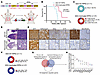 Figure 1
EED inactivation promotes a shift from SCLC toward LUAD histology in an RPR2 GEMM in vivo. (A) Schematic of the adenovirus used for intratracheal injection into the lungs of LSL-Cas9 mice to generate EED-inactivated or EED-WT autochthonous SCLC tumors. RPR2: sgRb1, sgTrp53, sgRbl2; sg“T”: sgEed or sgControl (nontargeting sgRNA). (B) Kaplan-Meier survival estimate of LSL-Cas9 mice intratracheally injected with adenoviruses as indicated. Median overall survival: EED-WT RPR2, 44.93 weeks; EED-inactivated RPR2, 65.00 weeks. n = 6 EED-WT RPR2 mice, n = 9 EED-inactivated RPR2 mice. Log-rank (Mantel-Cox) test was used to calculate P values. (C) Pie chart of lung cancer histology in EED-inactivated RPR2 tumors (n = 37 tumors from 33 independent mice) and EED-WT RPR2 tumors (n = 11 tumors from 8 independent mice). OS: osteosarcoma. (D and E) Representative H&E (D) and IHC (E) staining for GFP, H3K27me3, EED, synaptophysin (SYP), ASCL1, and NEUROD1 in EED-WT RPR2 SCLC tumors (n = 11, 2, 2, 2, 2, 2, and 10, respectively) and EED-inactivated RPR2 LUAD tumors (n = 36, 6, 4, 5, 3, 3, and 10, respectively). Scale bars: 1 mm (D), 100 μm (E). Insets are ×9 magnification of the corresponding main image. (F) Pie chart of liver metastases from EED-inactivated RPR2 (n = 27 independent mice) and EED-WT RPR2 (n = 11 independent mice) mice. (G) Venn diagram of differentially upregulated genes in transformed SCLC versus pretransformed LUAD from Oh et al. (64) and Quintanal-Villalonga et al. (5) overlapped with 120 epigenetic regulator genes (see Supplemental Table 1, tab 3). (H) Dot plot of log2 fold change of RNA-seq expression of transformed SCLC versus pretransformed LUAD from G. See also Supplemental Figure 1.
Consistent with prior studies nominating EZH2 as a therapeutic target in SCLC (23, 24), EED inactivation significantly delayed tumorigenesis, although tumors eventually formed in most mice injected with EED-inactivated RPR2 adenovirus (Figure 1, B–D). Strikingly, approximately 65% of EED-inactivated RPR2 mice developed tumors with pure LUAD histology and no evidence of SCLC (n = 24 of 37), whereas 27% developed independent LUAD and SCLC tumors within the same animal (n = 10 of 37), and only 1 mouse developed pure SCLC (Figure 1, C and D, Supplemental Figure 1B, and Supplemental Table 1, tab 1). IHC confirmed that all tumor cells were GFP-positive and had complete loss of EED and the PRC2 functional mark H3K27me3, while only SCLC tumors, and not LUAD tumors, expressed the neuroendocrine marker synaptophysin (Figure 1E, Supplemental Figure 1C, and Supplemental Table 1, tab 2). Consistent with prior work (20), EED-WT RPR2 SCLC tumors highly and homogeneously expressed ASCL1 and lacked NEUROD1 expression, whereas EED-inactivated RPR2 LUAD tumors lost ASCL1 expression (Figure 1E). CRISPR amplicon sequencing confirmed indels in Eed and Rb1 in select tumors (Supplemental Figure 1, D and E). Whereas most EED-WT RPR2 SCLC tumors metastasized to the liver, consistent with prior RPR2 studies (29, 34) and the highly metastatic nature of SCLC (1), EED-inactivated RPR2 LUAD tumors were confined to the lung without evidence of distant metastasis (Figure 1F). Together, these findings demonstrate strong selective pressure for a phenotypic shift from SCLC to LUAD following EED loss. However, because EED was inactivated at tumor initiation and tumors were analyzed at endpoint, these data do not distinguish when this selection toward LUAD occurred during tumorigenesis.
Given these findings, together with prior studies showing that SCLC transformation is associated with high EZH2 expression and increased PRC2 activity–associated gene signatures (5), we next asked whether PRC2 complex expression in SCLC is unique among epigenetic regulators. We analyzed publicly available RNA-seq data from 2 independent cohorts comparing transformed SCLC with pretransformed LUAD (5, 35) across 120 epigenetic modifiers (Figure 1G, Supplemental Figure 1F, and Supplemental Table 1, tab 3). Only 10 epigenetic modifiers were consistently upregulated in SCLC across both datasets, with EZH2 emerging as the most highly differentially expressed epigenetic regulator (Figure 1H and Supplemental Figure 1, G and H). Together, these data support a model in which PRC2 activity acts to constrain the SCLC neuroendocrine phenotype, with its loss promoting a histology shift toward LUAD.
EED inactivation Epigenetically Restores LUAD Oncogenic Signatures In Vivo. To investigate the molecular consequences of EED loss in the RPR2 GEMM, we performed RNA-seq on primary EED-inactivated RPR2 LUAD tumors and EED-WT RPR2 SCLC tumors (Supplemental Table 2, tab 1). Gene set enrichment using MSigDB (35), ENCODE (36), and ChEA (37) showed that genes upregulated in EED-inactivated tumors were enriched for targets normally bound and repressed by PRC2 members, including EZH2 and SUZ12 (Figure 2, A and B), consistent with functional PRC2 loss. EED-inactivated RPR2 LUAD tumors lost canonical neuroendocrine markers, including Chga, Insm1, and Syp (Figure 2C), and gained expression of RTK, KRAS, innate immune, and antigen processing and presentation pathways (Figure 2D). Flow cytometry confirmed restored MHC class I antigen presentation and increased immune and stromal infiltration (Supplemental Figure 2, A–D). Transcriptomic analysis showed that EED-inactivated RPR2 LUAD tumors correlated with pure and pretransformed human LUAD tumors, whereas EED-WT RPR2 SCLC tumors correlated with de novo and transformed SCLC tumors (Supplemental Figure 2E and Supplemental Table 2, tab 5), supporting the relevance of this model to human lung cancer (5).
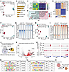 Figure 2
EED inactivation epigenetically restores LUAD oncogenic signatures in vivo. (A) Principal component analysis (PCA) of RNA-seq from 4 independent EED-WT RPR2 SCLC tumors (blue) and 4 independent EED-inactivated RPR2 LUAD tumors (red). (B) ENCODE/ChEA consensus transcription factor (TF) enrichment from RNA-seq in A showing top TF targets enriched in EED-inactivated RPR2 LUAD versus EED-WT RPR2 SCLC tumors. TFs with adjusted P value < 0.05 are shown. (C) Heatmap of neuroendocrine marker expression z scores from RNA-seq in A. Dark blue, low; yellow, high. (D) GSEA dot plot from RNA-seq in A. Dot color indicates FDR q value. Red, low; blue, high. Dot size indicates percentage of leading-edge genes. NES: normalized enrichment score. (E) Heatmap of top 500 differentially upregulated and downregulated genes ranked by z score. Red, high; blue, low. (F and H) PCA of ATAC-seq (F) and H3K27ac ChIP-seq (H) from 3 independent EED-WT RPR2 SCLC tumors (blue) and 3 independent EED-inactivated RPR2 LUAD tumors (red). (G and I) Heatmaps of differential ATAC-seq peaks (G) and H3K27ac ChIP-seq peaks (I). (J and K) Schematic (J) and volcano plot (K) showing differentially upregulated genes that gained both accessibility and H3K27ac in EED-inactivated RPR2 LUAD versus EED-WT RPR2 SCLC. log2FC: log2 fold change. (L) Averaged RNA-seq, ATAC-seq, and H3K27ac ChIP-seq tracks at Hnf4a, Met, and Cdx2. (M and N) HOMER motif analysis from ATAC-seq (M) and H3K27ac ChIP-seq (N). (O) Schematic showing escape from EED inactivation through SCLC-to-LUAD histological switching. Figure created with BioRender. See also Supplemental Figure 2.
Histologic analysis revealed that EED-inactivated LUAD tumors displayed mucinous differentiation (Supplemental Figure 2F), a feature observed in subsets of human LUAD associated with KRAS mutations, NKX2-1 loss, and gastrointestinal lineage gene expression (38–41). Correspondingly, EED-inactivated LUAD tumors upregulated gastrointestinal lineage markers including Cdx2, Hnf4a, and Pdx1, were enriched for gastrointestinal development signatures (Figure 2E and Supplemental Figure 2G), exhibited features of mucinous LUAD tumors (Supplemental Figure 2H), and lost NKX2-1 protein expression (Supplemental Figure 2I).
To define the chromatin-level changes underlying this reprogramming, we performed ATAC-seq (Assay for Transposase-Accessible Chromatin using sequencing) and H3K27ac ChIP-seq on EED-inactivated LUAD tumors and EED-WT SCLC tumors (Figure 2, F–I, Supplemental Figure 2, J and K, and Supplemental Table 2, tabs 3 and 4). Integrated analysis revealed that approximately 80% of upregulated genes gained both chromatin accessibility and H3K27 acetylation (Figure 2J), indicating widespread epigenetic activation. These included gastrointestinal differentiation factors Hnf4a and Cdx2 and the LUAD oncogene Met (Figure 2, K and L). HNF4A motifs were also enriched in regions with increased chromatin accessibility and H3K27ac in EED-inactivated RPR2 LUAD tumors (Figure 2, M and N). Together, these findings indicate that EED loss ultimately promotes LUAD histology in vivo associated with epigenetic activation of LUAD oncogenic programs and gastrointestinal differentiation factors (Figure 2O).
EED inactivation promotes rare NEUROD1-positive SCLC with early features of LUAD oncogenic signaling. Although EED-inactivated RPR2 mice predominantly developed LUAD tumors, a small subset formed tumors with SCLC histology (Figure 1C). These rare EED-inactivated RPR2 SCLC tumors showed complete loss of EED by CRISPR amplicon sequencing and IHC as well as complete loss of H3K27me3 by IHC (Supplemental Figure 1, C and D). Compared with EED-WT RPR2 SCLC tumors, EED-inactivated RPR2 SCLC tumors exhibited distinct morphology with giant cells and enlarged nuclei and were transcriptionally enriched for EMT, inflammatory response, and pancreatic β cell signatures (Figure 3, A–C, and Supplemental Table 3, tabs 1 and 2). A defining marker of the pancreatic β cell signature was Neurod1, which marks the NEUROD1-high SCLC-N subtype of human SCLC (11, 16, 17). These tumors showed high Neurod1 and NEUROD1-correlated gene expression with reduced Ascl1 and ASCL1-correlated gene expression (Figure 3, D and E), suggesting that EED inactivation promotes ASCL1-to-NEUROD1 subtype switching in vivo. FPKM values for key neuroendocrine and LUAD genes are shown in Supplemental Table 3, tab 3.
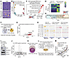 Figure 3
EED inactivation promotes rare NEUROD1-positive SCLC with early features of LUAD oncogenic signaling. (A) Representative H&Es of EED-WT (n = 11) and EED-inactivated (n = 2) RPR2 SCLC tumors. Yellow arrows indicate giant-cell features. Scale bars: 100 μm. (B) PCA of RNA-seq from EED-WT and EED-inactivated RPR2 SCLC and LUAD tumors. (C) Hallmark GSEA dot plot comparing EED-inactivated versus EED-WT RPR2 SCLC tumors. (D) Heatmap of neuroendocrine marker expression from z scored RNA-seq in B. Dark blue, low; yellow, high. (E) GSEA dot plot of indicated gene sets from RNA-seq in B. For C and E, dot color indicates FDR q value. Red, low; blue, high. Dot size indicates percentage of leading-edge genes. NES: normalized enrichment score. (F and G) Schematic (F) and volcano plot (G) showing upregulated genes that gained both accessibility and H3K27ac in EED-inactivated versus EED-WT RPR2 SCLC. log2FC, log2 fold change. (H) Averaged RNA-seq, ATAC-seq, and H3K27ac ChIP-seq tracks at Neurod1, Isl1, Met, and Fgfr1. (I and J) Representative immunoblot from 3 experiments (I) and RNA-seq volcano plot (J) of 1014 RPR2 EED-WT SCLC cells transduced with sgRNAs targeting Eed or sgControl. In J, vertical lines indicate absolute fold change of 2; horizontal line indicates adjusted P value = 0.05. (K) Schematic identifying direct EED targets by overlap of EED and H3K27me3 ChIP-seq in EED-WT RPR2 SCLC tumors. (L) Schematic integrating RNA-seq, ATAC-seq, and H3K27ac ChIP-seq to identify direct functional EED targets in EED-inactivated SCLC. (M) Rank plot of RNA-seq log2 fold change of functional EED targets in EED-inactivated SCLC. EMT: epithelial-to-mesenchymal transition. See also Supplemental Figure 3.
ATAC-seq and H3K27ac ChIP-seq similarly showed that NEUROD1/NEUROG3 and ISL1/2 motifs were among the top motifs with increased chromatin accessibility in EED-inactivated RPR2 SCLC tumors (Supplemental Figure 3, A–I), with Neurod1 and Isl1 gaining both chromatin accessibility and H3K27ac (Figure 3, F–H, and Supplemental Table 3, tabs 4 and 5). LUAD-associated RTK signaling genes, including Met and Fgfr1, were also epigenetically activated in EED-inactivated RPR2 SCLC tumors relative to EED-WT RPR2 SCLC tumors (Figure 3, F–H). Genetic EED inactivation in the ASCL1-positive murine SCLC cell line 1014 increased Neurod1, Met, and Fgfr1 mRNA expression, as well as NEUROD1 protein levels (Figure 3, I and J, and Supplemental Table 3, tab 6). Similarly, acute treatment with clinical-grade EZH1/2 or EED inhibitors increased NEUROD1 protein expression in vitro (Supplemental Figure 3J). In contrast to EED-inactivated RPR2 SCLC tumors in vivo, genetic or pharmacologic EED inactivation did not reduce ASCL1 protein expression in 1014 cells in vitro (Figure 3I and Supplemental Figure 3J), suggesting that ASCL1 reduction in vivo is indirect and requires tumor microenvironmental influences and/or evolutionary selection. RNA-seq also identified an EED-inactivated RPR2 tumor positive for POU2F3, which marks a non-neuroendocrine SCLC subtype (Supplemental Figure 4, A–C) (17). However, EED inactivation in 1014 cells did not induce POU2F3 expression (Supplemental Figure 4D), suggesting that emergence of POU2F3-positive SCLC after EED loss is likely indirect.
Finally, ChIP-seq for EED and H3K27me3 identified 6,072 direct EED target genes, 289 of which were epigenetically activated and expressed in EED-inactivated RPR2 SCLC tumors (Figure 3, K and L, Supplemental Figure 3K, and Supplemental Table 4, tabs 1 and 2), including Neurod1, Isl1, and Pdx1 (Figure 3M and Supplemental Figure 3L). EED and H3K27me3 were highly enriched at the Neurod1 locus in EED-WT RPR2 SCLC tumors, without corresponding H3K27ac enrichment or chromatin accessibility, consistent with direct PRC2-mediated repression. In contrast, EED and H3K27me3 were not strongly enriched at the Ascl1 locus, which showed prominent H3K27ac enrichment and chromatin accessibility, consistent with active gene expression (Supplemental Figure 3, M and N). Together, these findings demonstrate that EED directly represses NEUROD1 and that EED inactivation derepresses NEUROD1, promoting rare NEUROD1-positive SCLC tumors with partial activation of LUAD-associated genes.
EED Inactivation Promotes LUAD Histology Likely Through a NEUROD1-Positive Intermediate State. Although EED loss promoted NEUROD1 expression in vitro and generated occasional NEUROD1-positive SCLC tumors in vivo, most tumors in EED-inactivated RPR2 mice were LUAD tumors (Figure 1, C and D), suggesting that sustained SCLC histology is selected against and/or that LUAD fate is favored after EED loss. In line with this possibility, histological examination of LUAD-bearing lungs from EED-inactivated RPR2 mice revealed rare early neuroendocrine-positive lesions expressing SCLC subtype transcription factors, including ASCL1, NEUROD1, and POU2F3 (Supplemental Figure 4E and Supplemental Table 5, tab 1). To better define the relationship of NEUROD1 to lung cancer histological subtypes formed after EED inactivation in vivo, we performed IHC for NEUROD1 and ASCL1 (Figure 4A). EED-inactivated RPR2 SCLC tumors expressed NEUROD1, whereas EED-WT RPR2 SCLC tumors expressed ASCL1 (Figure 4A). Surprisingly, EED-inactivated RPR2 tumors with pure LUAD histology retained sporadic NEUROD1-positive cells intermixed within tumors (Figure 4, A and B). This was unique to EED-inactivated RPR2 LUAD tumors, as NEUROD1-positive cells were not detected in a de novo LUAD from an EGFR-mutant RPR2 GEMM or in EED-WT RPR2 SCLC tumors (Figure 4, A and B, and Supplemental Table 5, tab 2). Neurod1 mRNA was also elevated in EED-inactivated RPR2 LUAD tumors relative to EED-WT RPR2 SCLC tumors (Figure 4C), suggesting that EED-inactivated RPR2 LUAD tumors may have evolved from NEUROD1-positive SCLC cells.
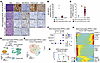 Figure 4
EED inactivation promotes LUAD histology likely through a NEUROD1-positive intermediate state. (A) Representative H&E and IHC for ASCL1, NEUROD1, H3K27me3, and EED in EED-WT RPR2 SCLC (n = 11, 2, 4, 2, and 2, respectively), EED-inactivated RPR2 SCLC (n = 2, 2, 2, 2, and 2, respectively), EED-inactivated RPR2 LUAD (n = 24, 3, 10, 4, and 5, respectively), and de novo LUAD tumors (n = 2, 2, and 2, respectively). Arrows indicate rare scattered NEUROD1-positive cells. Scale bars: 100 μm. Insets, ×9 magnification. (B) Quantitation of NEUROD1-positive cells from A. n = 4 EED-WT RPR2 SCLC, n = 2 EED-inactivated RPR2 SCLC, n = 10 EED-inactivated RPR2 LUAD, and n = 2 de novo LUAD tumors. (C) Normalized Neurod1 mRNA expression by RT-qPCR in indicated GEMM tumors. n = 2 EED-WT RPR2 SCLC, n = 2 EED-inactivated RPR2 SCLC, n = 39 EED-inactivated RPR2 LUAD, and n = 2 de novo LUAD tumors. (D and E) UMAP of all cells (D) and tumor cells (E) from snRNA-seq of 3 EED-inactivated RPR2 LUAD tumors from independent mice. n = 27,117 total cells; n = 16,590 tumor cells. (F) Dot plot showing average expression of indicated genes in each tumor cell cluster. Dot size indicates percentage of expressing cells; color indicates expression level. Blue, high; white, low. (G) Phylogenetic tree of tumor cell clusters from EED-inactivated RPR2 LUAD tumor 1350 from E using maximum parsimony method. (H) Heatmap of normalized expression of top 300 trajectory-defining genes along the EED-inactivated RPR2 LUAD tumor cell trajectory. Blue, low; red, high. See also Supplemental Figures 4 and 5.
To investigate this possibility, we performed single-nucleus RNA-seq (snRNA-seq) on 3 independent EED-inactivated RPR2 LUAD tumors. Unsupervised clustering identified 4 tumor cell clusters (Figure 4, D and E). Cluster 3 expressed neuroendocrine markers, including Neurod1, Chga, and Ncam1, and represented approximately 4% of tumor cells, whereas other clusters expressed LUAD genes such as Met (Figure 4F). InferCNV revealed distinct CNV landscapes across tumors (Supplemental Figure 5, A–C), but Neurod1-positive cluster 3 cells shared copy number alterations with other tumor clusters from the same sample, suggesting derivation from a common clone (Supplemental Figure 5, A–C) (42). CNV-based phylogenetic reconstruction showed that Neurod1-positive cluster 3 cells had fewer CNV events than other tumor clusters and could represent an ancestral population from which other clusters arose (Figure 4G and Supplemental Figure 5, D–F). Pseudotime analysis similarly showed early expression of neuroendocrine genes, followed by persistent Neurod1 and Ncam1 expression, eventual loss of neuroendocrine genes, and upregulation of LUAD and gastrointestinal differentiation genes, including Met, Hnf4a, and Nr1h4 (Figure 4H and Supplemental Figure 5, G and H). EMT genes, including Cdh6, Clu, and Itga2, were also elevated during the persistent Neurod1- and Ncam1-positive intermediate state.
To validate this trajectory, we performed scRNA-seq on EED-inactivated RPR2 LUAD tumors (n = 4) and SCLC tumors (n = 2) and compared them with published scRNA-seq data from EED-WT RPR2 SCLC tumors (n = 3) (Figure 5A and Supplemental Figure 6A) (20). As expected, EED-inactivated SCLC tumors expressed Neurod1, Isl1, and other neuroendocrine markers, whereas EED-inactivated LUAD tumors lost neuroendocrine features and expressed Hnf4a and Muc13 (Supplemental Figure 6B). Unsupervised ordering predicted a single trajectory from EED-WT RPR2 SCLC (Ascl1-positive) to EED-inactivated RPR2 SCLC (Neurod1-positive) to EED-inactivated RPR2 LUAD (Hnf4a-positive), supporting Neurod1-positive SCLC as an intermediate state before LUAD formation (Supplemental Figure 6, C–E).
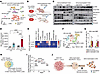 Figure 5
LUAD histology after EED inactivation requires cues from the tumor immune microenvironment. (A) UMAP of scRNA-seq from 3 independent EED-WT SCLC (n = 7,889 cells), 2 EED-inactivated SCLC (n = 2,064 cells), and 4 EED-inactivated LUAD (n = 2,013 cells) tumors; each tumor was from an independent mouse. Mϕ, macrophage. (B–D) Schematic (B), immunoblot (C), and RT-qPCR (D) of 1014 RPR2 EED-WT SCLC cells transduced with sgEed or sgControl and implanted subcutaneously in immunodeficient NCr nude or immunocompetent BL6J mice. For D, 1-way ANOVA with Tukey’s test was used to calculate 2-sided P values adjusted for multiple comparisons. **P < 0.01, ***P <0.001, ****P < 0.0001. (E) Heatmap of indicated tumor and immune cell types from scRNA-seq in A comparing EED-inactivated LUAD, EED-inactivated SCLC, and EED-WT SCLC tumors. (F and G) UMAP (F) and quantification (G) of 2 M2-like macrophage clusters as percentages of total cells in each sample. (H and I) Schematic (H) and rank plot (I) of scaled receptor expression fold changes in EED-inactivated LUAD tumor cells for receptor-ligand interactions enriched in EED-inactivated LUAD versus EED-WT SCLC tumors and receptors upregulated after CRISPR-mediated EED inactivation in 1014 cells in vitro. For Met, the corresponding ligand gene, Hgf, and tumor microenvironment component with higher expression, macrophages, are indicated. (J) Schematic showing LUAD histology arising through a NEUROD1-positive transient intermediate state requiring cues from the tumor immune microenvironment. Figure created with BioRender. See also Supplemental Figures 6 and 7.
LUAD histology after EED inactivation requires cues from the tumor immune microenvironment. Despite upregulating Neurod1 and Met, EED inactivation in 1014 cells in vitro did not induce LUAD lineage markers or a full LUAD phenotypic shift, suggesting that additional tumor microenvironmental factors and/or evolutionary selection are required for the complete shift to LUAD in vivo (Figure 3J). To test this, we transplanted EED-isogenic 1014 cells into immunocompetent BL6J and immunodeficient NCr nude mice (Figure 5B). In immunodeficient hosts, EED-inactivated tumors retained high NEUROD1 and failed to activate MAPK signaling. In contrast, EED-inactivated tumors in immunocompetent mice had lower NEUROD1 and increased MAPK signaling and Met, suggesting that the tumor immune microenvironment promotes LUAD-associated signaling after EED loss (Figure 5, C and D, and Supplemental Figure 7A). However, EED-inactivated 1014 tumors did not fully acquire LUAD histology or induce HNF4A (Supplemental Figure 7, A–D), likely reflecting differences between the transplantation model and the autochthonous GEMM. In the autochthonous GEMM, EED was inactivated at tumor initiation in individual lung cells, so selection against EED loss manifested as delayed tumorigenesis and eventual escape as LUAD. In contrast, in the transplantation model, EED was CRISPR-inactivated in a preestablished polyclonal population of 1014 SCLC cells before implantation, allowing cells retaining intact EED through incomplete editing or in-frame edits to outgrow. Consistent with this, endpoint tumors showed variable restoration of EED protein across tumor cells, suggesting that EED loss was selected against, particularly in immunocompetent hosts (Figure 5C, Supplemental Figure 7B, and Supplemental Table 5, tab 3).
To identify immune-derived cues that might promote LUAD histology, we analyzed the tumor immune microenvironment (Figure 5A). EED-WT RPR2 SCLC tumors contained relatively few immune cells, whereas EED-inactivated RPR2 LUAD tumors showed increased neutrophil and macrophage infiltration, and EED-inactivated RPR2 SCLC tumors showed increased macrophages (Figure 5E). M2-like macrophages expressing Msr1, Arg1, and Tgfb1 were enriched in both EED-inactivated LUAD and SCLC tumors (Figure 5, F and G, Supplemental Figure 6, F and G, and Supplemental Table 5, tab 4). Cell–cell interaction analysis comparing TIME ligands and tumor cell receptors in EED-inactivated LUAD tumors versus EED-WT SCLC tumors identified 58 enriched receptor-ligand pairs (Figure 5, H and I) (43). Cross-referencing with EED-inactivated 1014 RNA-seq (Figure 3I) identified 14 upregulated tumor-expressed receptors with corresponding TIME ligands, including LUAD RTK oncogenes Met, Ret, and Fgfr2, all converging on RAS signaling (Figure 5, H and I, and Supplemental Table 5, tab 5). Notably, Met was the second-highest expressed receptor, with its ligand Hgf highly expressed by macrophages in EED-inactivated RPR2 LUAD tumors. Together, these findings suggest that NEUROD1-positive tumor cells represent a transitional state toward LUAD following EED loss and that the tumor immune microenvironment promotes LUAD RTK oncogenic signaling to complete this process in vivo (Figure 5J).
EED Directly Binds and Represses RAS, PI3K, and MAPK Pathway LUAD Oncogenic Signaling Genes. To mechanistically understand how EED maintains the ASCL1-positive SCLC neuroendocrine state and why its loss promotes LUAD histology, we performed ChIP-seq for EED and H3K27me3, identifying 6,072 direct EED target genes (Figure 3K). Of these, 831 were epigenetically activated and expressed in EED-inactivated RPR2 LUAD tumors (Figure 6A and Supplemental Table 4, tab 3), including LUAD oncogenes such as Met and gastrointestinal lineage genes including Hnf4a and Cdx2 (Figure 6B). Activated EED targets were enriched for EMT, gastric cancer signatures, and RAS, PI3K, and MAPK signaling (Figure 6C and Supplemental Figure 8A). Consistent with these epigenomic data, immunoblot and IHC analyses demonstrated elevated phospho-ERK and phospho-AKT, providing protein-level evidence of MAPK and PI3K pathway activation in EED-inactivated RPR2 LUAD tumors (Figure 6D and Supplemental Figure 8, B and C). In contrast, EED-inactivated SCLC tumors showed increased phospho-AKT, but not phospho-ERK, consistent with an intermediate state. Only 151 EED target genes were commonly activated in both EED-inactivated RPR2 LUAD and SCLC tumors (Figure 6E), yet this shared set was also enriched for RAS and PI3K/AKT pathway genes (Figure 6F and Supplemental Figure 8D), including the RAS guanine nucleotide exchange factor Rasgrp1 (Figure 6G). Whole-exome sequencing of 6 EED-inactivated RPR2 LUAD tumors did not reveal recurrent copy number alterations or somatic mutations in canonical LUAD oncogenic drivers, with only 1 tumor harboring KRAS G12D and another Kras amplification (Supplemental Figure 9A). Together, these data support a dominant epigenetic mechanism driving RAS/PI3K/MAPK activation after EED loss.
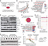 Figure 6
EED directly binds and represses RAS, PI3K, and MAPK pathway LUAD oncogenic signaling genes to support the SCLC neuroendocrine identity. (A) Schematic integrating direct EED targets with genes reactivated upon EED loss using RNA-seq, ATAC-seq, and H3K27ac ChIP-seq to identify direct functional EED targets in EED-inactivated LUAD tumors. (B) Rank plots of RNA-seq log2 fold change of functional EED targets in EED-inactivated LUAD tumors. (C) KEGG pathway enrichment of direct functional EED targets in EED-inactivated LUAD tumors. Dot color indicates Benjamini-Hochberg–adjusted (BH-adjusted) P value. Red, low; blue, high. Dot size indicates enriched gene counts. (D) Immunoblot of EED-WT SCLC, EED-inactivated SCLC, and EED-inactivated LUAD RPR2 GEMM tumors. (E and F) Schematic (E) and KEGG pathway enrichment analysis (F) of overlapping direct functional EED targets in EED-inactivated SCLC and LUAD. Color indicates BH-adjusted P value. (G) Averaged RNA-seq, ATAC-seq, and H3K27ac ChIP-seq tracks from EED-WT SCLC, EED-inactivated SCLC, and EED-inactivated LUAD tumors, and EED and H3K27me3 ChIP-seq tracks from EED-WT SCLC at Rasgrp1. (H) Immunoblot of cell lines from Supplemental Figure 9E treated overnight with 1 μM trametinib. (I) Normalized dose–response curves of cell lines from H treated with trametinib for 6 days, with EC50 values shown at right. If 50% killing was not reached at 1,000 nM, 1,000 nM was used for average EC50 calculation. Arrow indicates DMSO-treated sample used for normalization. n = 3 independent experiments. See also Supplemental Figures 8–10.
RAS pathway activation is a conserved hallmark of oncogenic signaling downstream of nearly all oncogenic RTKs in LUAD and antagonizes the neuroendocrine phenotype in murine SCLC (44), a finding we validated in human ASCL1-positive SCLC cell lines overexpressing WT or mutant KRAS G12C (Supplemental Figure 9, B and C). To test whether EED loss creates RAS pathway dependence, we derived 5 cell lines from EED-inactivated RPR2 LUAD tumors — 1350, 1339, 1343, 1344, and 1345 — which grew as adherent monolayers, similar to LUAD lines and unlike SCLC lines (Supplemental Figure 9, D and E). These lines exhibited elevated phospho-ERK and were highly sensitive to the pan-RAS inhibitor daraxonrasib (RMC-6236) (45) and MEK inhibitor trametinib relative to EED-WT RPR2 SCLC cells (Figure 6, H and I, and Supplemental Figure 9, F and G). Consistent with a direct on-target effect, reexpression of WT EED, but not a catalytically inactive EED mutant, in 2 EED-inactivated LUAD lines downregulated MAPK and RAP1 signaling pathways and PRC2 target genes (Supplemental Figure 10, A–G, and Supplemental Table 4, tabs 4 and 5). However, EED reexpression was insufficient to restore neuroendocrine gene expression (Supplemental Table 4, tabs 4 and 5), consistent with EED loss initiating a shift toward LUAD histology but requiring additional tumor microenvironmental cues to complete this process (Figure 5). Together, these data demonstrate that EED directly represses RAS signaling to maintain the SCLC neuroendocrine phenotype and that EED loss induces RAS oncogenic signaling to promote LUAD.
LUAD histology after PRC2 loss is associated with reactivation of bivalently marked RAS/PI3K/MAPK Pathway Genes and NEUROD1. Although 6,072 genes were marked by both EED and H3K27me3, relatively few were activated upon EED loss: 289 in SCLC and 831 in LUAD (Figure 3L and Figure 6A). Because bivalent genes, marked by both activating H3K4me3 and repressive H3K27me3, are selectively derepressed upon PRC2 inhibition during differentiation (46), we hypothesized that bivalent genes would be preferentially activated after EED loss and include regulators of SCLC-to-LUAD histological transformation (Figure 7A). To identify bivalent genes, we performed H3K4me3 ChIP-seq in EED-WT RPR2 SCLC tumors and overlaid these peaks with H3K27me3 peaks from the same samples, identifying 7,622 bivalent promoters (Figure 7B, Supplemental Figure 11, A and B, and Supplemental Table 6, tab 1). Consistent with prior studies (47, 48), GREAT and hallmark enrichment analyses showed that bivalent genes were enriched for MHC class I antigen presentation and EMT (Supplemental Figure 11, C–E).
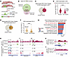 Figure 7
LUAD histology after PRC2 loss is associated with reactivation of bivalently marked RAS/PI3K/MAPK pathway genes and NEUROD1. (A) Schematic of genes with bivalent H3K4me3 and H3K27me3 marks poised for reactivation upon PRC2 loss. Figure created with BioRender. (B and C) Strategy to identify bivalent genes reactivated upon EED loss by overlapping H3K4me3 and H3K27me3 ChIP-seq peaks from EED-WT RPR2 SCLC tumors (B) with reactivated EED direct target genes in EED-inactivated RPR2 LUAD tumors (C). (D) Expression of reactivated EED direct targets that are bivalent or nonbivalent in EED-inactivated LUAD. (E) Identification of bivalent genes reactivated upon EED loss in EED-inactivated RPR2 SCLC using strategy from B. (F) Expression of reactivated EED direct targets that are bivalent or nonbivalent in EED-inactivated SCLC. (G) KEGG pathway enrichment of reactivated bivalent EED direct targets in EED-inactivated LUAD. Bar color indicates BH-adjusted P value. Red, low; blue, high. Count: enriched gene count. (H–J) Averaged H3K27me3, H3K4me3 ChIP-seq, and RNA-seq tracks at Akt1 (H), Mapkapk3 (I), and Erbb2 (J), showing bivalent peaks and RNA-seq activation upon EED loss. (K and L) Averaged H3K27me3 and H3K4me3 ChIP-seq tracks at Neurod1 (K) and Hnf4a (L), showing that Neurod1, but not Hnf4a, is bivalently marked. ****P < 0.0001. See also Supplemental Figure 11.
Consistent with our hypothesis, 417 of 831 genes activated in LUAD (~50%) and 118 of 289 genes activated upon EED loss in SCLC (41%) had bivalent promoters (Figure 7, C and E, and Supplemental Table 6, tab 2). Bivalent EED direct targets reactivated in both EED-inactivated SCLC and LUAD had higher expression than nonbivalent reactivated targets (Figure 7, D and F), consistent with bivalency poising genes for activation upon H3K27me3 loss (46). Bivalent genes activated in LUAD tumors after EED loss were enriched for PI3K/AKT and MAPK signaling, including Akt1, Mapkapk3, and upstream growth factor receptors such as Erbb2, with Akt1 also induced in EED-inactivated RPR2 SCLC (Figure 7, G–J). These findings indicate that EED directly represses bivalent RAS/PI3K/MAPK pathway genes poised for activation upon PRC2 loss.
Neurod1 was also a bivalent target reactivated in EED-inactivated RPR2 SCLC and among the most highly expressed genes (Figure 7, E, F, and K). In contrast, some reactivated EED direct targets, including gastrointestinal lineage genes Hnf4a, Cdx1, and Cdx2, were not bivalent (Figure 7, D and L), suggesting their activation requires further selection in vivo, consistent with their lack of induction after EED inactivation in 1014 cells in vitro (Figure 3J). Together, these findings suggest that H3K27me3/H3K4me3 bivalency poises RAS/PI3K/MAPK pathway genes and Neurod1 for robust activation after EED loss.
EED inactivation at tumor initiation prevents emergence of SCLC histology in an EGFR-mutant GEMM with Rb1 and Trp53 inactivation after EGFR withdrawal. Our data showed that EED supports ASCL1-positive SCLC histology in the RPR2 GEMM, with EED inactivation promoting LUAD histology (Figures 1–7). We therefore hypothesized that EED/PRC2 activity may also be important for acquisition of SCLC histology during LUAD-to-SCLC transformation, a resistance mechanism observed in EGFR-mutant LUAD tumors harboring RB1 and TP53 alterations (13). To test this, we developed an EGFR-mutant LUAD GEMM capable of histological transformation under selective pressure from EGFR suppression. We used a doxycycline-inducible (Dox-inducible) EGFR L858R transgenic model (49, 50) crossed with Trp53fl/fl, LSL rtTA, and LSL-Cas9-P2A-GFP mice (Figure 8A). We then generated adenoviruses encoding CMV-Cre to activate Cas9 and rtTA expression, sgRNAs targeting Rb1, and either an sgRNA targeting Eed or a nontargeting control sgRNA, generating EGFR-mutant LUAD GEMMs with concurrent Rb1 and Trp53 inactivation that were either EED-inactivated (EGFR-mutant ERP) or EED-WT (EGFR-mutant CRP). Immunoblotting validated Cre expression and efficient RB1 and EED inactivation (Supplemental Figure 12A).
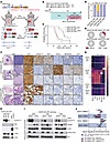 Figure 8
EED inactivation at tumor initiation prevents emergence of SCLC histology in an EGFR-mutant GEMM with Rb1 and Trp53 inactivation after EGFR withdrawal. (A) Schematic of CRISPR-based EGFR-mutant LUAD GEMM with Rb1 and Trp53 inactivation made isogenic for Eed using sgEed to generate ERP tumors or nontargeting sgRNA to generate CRP tumors. (B) Timeline of adenovirus injection, Dox induction, and Dox withdrawal. (C) Kaplan-Meier survival estimate of mice injected with indicated adenoviruses and treated as in B. P values calculated by log-rank test. (D) Percentage of mice with metastases. (E and F) Tumor histology pie charts (E) and representative H&E and IHC (F) for GFP, EGFR L858R, EED, ASCL1, NEUROD1, and SYP from CRP EGFR ON tumors (n = 14, 2, 2, 2, 2, 2, and 2, respectively), CRP EGFR OFF tumors (n = 12, 4, 3, 4, 4, 4, and 3, respectively), ERP EGFR ON tumors (n = 14, 2, 2, 2, 2, 2, and 2, respectively), and ERP EGFR OFF tumors (n = 12, 3, 3, 3, 3, 3, and 3, respectively). Scale bars: 1 mm (first column), 100 μm (remaining columns). Insets, ×9 magnification. For C–E, n = 14 EGFR ON CRP, n = 12 EGFR OFF CRP, and n = 14 EGFR ON ERP mice. For C, n = 16 EGFR OFF ERP mice; for D and E, n = 12 EGFR OFF ERP mice because 4 did not develop tumors. (G) Heatmap of z scored SCLC neuroendocrine and non-neuroendocrine marker expression from RNA-seq. Each row is 1 tumor. Black, low; yellow, high. n = 3 EGFR ON CRP, n = 9 EGFR OFF CRP, n = 2 EGFR ON ERP, n = 3 EGFR OFF ERP tumors. (H) Immunoblot of EGFR ON CRP cell lines cultured in 1 μg/mL Dox. (I) Immunoblot of cell lines in H cultured with or without Dox and treated with DMSO or MAK-683, 2 μM, for 6 days. (J) Gene signature enrichment scores from RNA-seq of EGFR CRP 1525 cells under conditions in I. For I, representative immunoblots from > 3 biological replicates. For J, n = 2 biological replicates. CRP, sgControl; sgRb1, Trp53−/−; ERP, sgEed; sgRb1, Trp53−/−; EGFR ON, Dox ON; EGFR OFF, Dox withdrawal. ***P < 0.001, ****P < 0.0001. See also Supplemental Figures 12 and 13.
Adenoviruses were intratracheally injected, and 1 week later mice were started on Dox chow to induce EGFR L858R expression. Pilot experiments showed that after 20 weeks on Dox, mice developed dyspnea with lung pathology consistent with EGFR-mutant LUAD, matching the expected latency of this model (Supplemental Figure 12, B and C) (49, 50). Therefore, after approximately 20 weeks, EGFR-mutant CRP and ERP mice were randomized to remain on Dox (Dox ON) or switch to regular chow (Dox OFF) to suppress EGFR L858R expression and were followed to endpoint (Figure 8B). Consistent with EGFR L858R acting as a potent oncogene, EGFR-mutant CRP and ERP Dox ON mice succumbed more rapidly, with median overall survival of 30.3 and 31.0 weeks, respectively. Survival was prolonged in Dox OFF mice, with median overall survival of 57.9 and 57.1 weeks for EGFR-mutant CRP and ERP cohorts, respectively (Figure 8C).
EGFR-mutant CRP Dox OFF and ERP Dox OFF mice developed distinct recurrent disease phenotypes. Tumors recurred in EGFR-mutant CRP Dox OFF mice, with 7 of 12 developing spontaneous metastases to liver, kidney, and brain, whereas no EGFR-mutant ERP Dox OFF mice developed distant metastases (Figure 8D, Supplemental Figure 12D, and Supplemental Table 7, tab 1). This was consistent with the absence of distant metastases when EED was inactivated at tumor initiation in the RPR2 GEMM (Figure 1F). Despite this difference, median overall survival was similar between EGFR-mutant CRP Dox OFF and ERP Dox OFF mice (Figure 8C), consistent with primary lung tumor burden and respiratory compromise driving endpoint in both groups (Supplemental Figure 12, E and F, and Supplemental Table 7, tab 1). Because analyses were performed at endpoint, we cannot distinguish whether EED loss suppresses early dissemination versus later metastatic outgrowth. However, recurrent tumors still developed in EGFR-mutant ERP Dox OFF mice, indicating that the reduction in metastasis does not simply reflect absence of recurrent disease.
IHC validated Dox-dependent EGFR L858R expression, GFP expression in tumor cells, and complete EED loss only in EGFR-mutant ERP tumors (Supplemental Figure 12G). DNA sequencing and immunoblotting confirmed expected genetic inactivation patterns in EGFR-mutant CRP and ERP tumors (Supplemental Figure 12, C, H, and I). Consistent with RB1 and TP53 alterations increasing SCLC transformation risk after EGFR inhibition in human EGFR-mutant LUAD (13), tumors in 3 of 12 EGFR-mutant CRP Dox OFF mice recurred as SCLC, whereas no EGFR-mutant ERP Dox OFF mice developed SCLC histology (0 of 12) (Figure 8E). IHC showed that transformed SCLC tumors in EGFR-mutant CRP Dox OFF mice either expressed ASCL1 alone or heterogeneously expressed ASCL1 and NEUROD1, whereas EGFR-mutant ERP counterparts were negative (Figure 8F and Supplemental Table 7, tab 2). This resembles human EGFR-mutant transformed SCLC, which shows heterogeneous transcription factor expression and enrichment for variant subtypes, including NEUROD1 (51), and supports transformation as a mechanism of escape from EGFR L858R suppression rather than de novo SCLC formation, as NEUROD1 expression is not observed in the RPR2 SCLC GEMM (19, 20, 31, 52). Consistent with the EED-inactivated RPR2 model, EGFR-mutant ERP Dox ON and Dox OFF tumors also showed LUAD with mucinous features (Figure 8F).
Bulk RNA-seq confirmed increased neuroendocrine markers, including Syp, Insm1, and Ncam1, as well as Ascl1, heterogeneous Neurod1, and enrichment for SCLC-related signatures, including MYC, G2M, and pancreas β cell programs, in EGFR-mutant CRP Dox OFF transformed SCLC tumors relative to EGFR-mutant CRP Dox ON, EGFR-mutant CRP Dox OFF LUAD, and EGFR-mutant ERP tumors (Figure 8G, Supplemental Figure 13, A–C, and Supplemental Table 7, tab 3). Transformed SCLC tumors also had lower MAPK target gene expression (Supplemental Figure 13D). EGFR-mutant CRP Dox OFF LUAD tumors were higher-grade tumors than EGFR-mutant CRP Dox ON LUAD tumors and were enriched for EMT signatures, consistent with poorly differentiated histology and increased metastatic propensity (Supplemental Figure 13E and Supplemental Table 7, tab 1). In line with the EED-inactivated RPR2 model, mucinous LUAD, tumor-associated macrophages, and neutrophil signatures were enriched in EGFR-mutant ERP Dox ON and Dox OFF tumors relative to EGFR-mutant CRP counterparts (Supplemental Figure 13, A, F, and G, and Supplemental Table 7, tab 4). Together, these data demonstrate that EGFR-mutant LUAD tumors with Rb1 and Trp53 loss can recur after EGFR oncogene withdrawal through distinct trajectories, including SCLC transformation or aggressive metastatic LUAD recurrence. EED inactivation prevents emergence of SCLC histology and reduces spontaneous metastasis, but does not prevent recurrence, instead redirecting tumor evolution toward LUAD with mucinous differentiation (Supplemental Figure 13H).
Although these studies demonstrate that EED inactivation at tumor initiation prevents SCLC histology after EGFR oncogene withdrawal, they do not determine whether pharmacologic EED inhibition suppresses SCLC transformation during tumor maintenance. Because therapeutic studies in the EGFR ON/OFF GEMM would require prolonged treatment in a long-latency autochthonous model, we tested whether pharmacologic EED inhibition attenuates early neuroendocrine changes after EGFR withdrawal in vitro. We generated 3 independent cell lines — 1525, 1533, 1544 — from EGFR-mutant CRP tumors maintained on Dox, each with EGFR L858R expression and Rb1 and Trp53 inactivation (Figure 8H). EGFR withdrawal reduced proliferation in 2 of 3 lines (Supplemental Figure 13I). In all 3 lines, EGFR withdrawal induced the neuroendocrine marker INSM1 (53), which was attenuated by the clinical-grade EED inhibitor MAK683 (54), while MAK683 completely suppressed H3K27me3, confirming on-target PRC2 inhibition (Figure 8I). RNA-seq in 1525 cells showed that EGFR withdrawal enriched gene signatures associated with SCLC, including neuroendocrine, E2F target, G2M checkpoint, and MYC-associated signatures (15), which were attenuated by MAK683 (Figure 8J). Thus, pharmacologic EED inhibition attenuates INSM1 induction and selected SCLC- and proliferation-associated programs following EGFR withdrawal in vitro. However, these experiments do not establish that pharmacologic EED inhibition suppresses SCLC transformation in established tumors in vivo, which requires future study.
H3K27me3 represses LUAD oncogenic programs in human SCLC through bivalent silencing of RAS, PI3K, and MAPK pathway genes. Having identified that PRC2 represses LUAD oncogenic programs in murine SCLC models, we next asked whether similar bivalent repression patterns occur in human lung cancer. We analyzed H3K27me3 and H3K4me3 ChIP-seq data from 5 de novo neuroendocrine SCLC patient-derived xenografts (PDXs) and 4 de novo EGFR-mutant LUAD PDXs (Figure 9, A, B, and E) (55). ChEA and ENCODE analyses of H3K27me3 peaks identified SUZ12 and EZH2 as dominant regulators in both histologies, validating the dataset (Supplemental Figure 14, A and B). Consistent with our murine data and prior studies (47, 48), Gene Ontology analysis showed that MHC class I antigen presentation genes were enriched for H3K27me3 in SCLC, whereas axon guidance pathways were enriched for H3K27me3 in LUAD (Supplemental Figure 14, C and D), indicating that H3K27me3 repression patterns distinguish key features of SCLC and LUAD. De novo neuroendocrine SCLC had more numerous and stronger H3K27me3 peaks than LUAD, with top SCLC-gained H3K27me3 peaks including PI3K and MAPK pathway genes (Figure 9, C and D, and Supplemental Table 6, tab 3), mirroring our GEMM findings. Consistently, bulk H3K27me3 was elevated in EGFR-mutant CRP Dox OFF tumors that transformed to SCLC relative to EGFR-mutant CRP Dox ON LUAD tumors (Supplemental Figure 14, E and F).
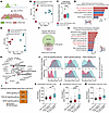 Figure 9
H3K27me3 represses LUAD oncogenic programs in human SCLC through bivalent silencing of RAS, PI3K, and MAPK pathway genes. (A) Schematic showing PRC2-mediated repression of genes with bivalent H3K4me3 and H3K27me3 marks. Figure created with BioRender. (B and E) PCA of H3K27me3 ChIP-seq (B) and H3K4me3 ChIP-seq (E) from 5 independent human de novo neuroendocrine (NE) SCLC PDX tumors and 4 independent human EGFR-mutant LUAD PDX tumors. (C) Box-and-whisker plots of normalized H3K27me3 ChIP-seq read counts in the indicated groups. Mann-Whitney U test was used to calculate 2-sided P value. (D) KEGG pathway enrichment of genes mapped to differential H3K27me3 peaks gained in neuroendocrine SCLC versus EGFR-mutant LUAD PDX tumors. (F) Schematic identifying bivalent genes marked by H3K4me3 and H3K27me3 in human neuroendocrine SCLC PDX tumors. (G) KEGG pathway enrichment of bivalent genes in human neuroendocrine SCLC PDX tumors. For D and G, bar color indicates BH-adjusted P value. Red, low; blue, high. Count: enriched gene count. (H) Gene network analysis of enriched RAS, PI3K-AKT, and MAPK pathways from G. Dot size indicates enriched gene count. (I) Bar plots showing the percentage of H3K4me3-marked genes in the indicated pathways that gained H3K27me3 in neuroendocrine SCLC PDX tumors and are therefore bivalent. (J) Density plots of normalized H3K4me3 and H3K27me3 ChIP-seq read counts mapped to genes in KRAS signaling UP, PI3K/AKT, and MAPK KEGG pathways in human neuroendocrine SCLC and EGFR-mutant LUAD PDX tumors. (K–M) RNA-seq batch-normalized expression of EED (K), EZH2 (L), and SUZ12 (M) in human EGFR-mutant LUAD tumors with indicated RB1 and TP53 alterations from The Cancer Genome Atlas (65). The box-and-whisker plots depict the 10th and 90th percentile (whiskers), the upper and lower quartiles, and the median. RB1/TP53 WT, n = 22; TP53 mutation or deep deletion, n = 34; RB1/TP53 mutation or deep deletion, n = 10 tumors. One-way ANOVA with Tukey’s test was used to calculate 2-sided P values adjusted for multiple comparisons. **P < 0.01, ***P < 0.001, ****P < 0.0001. See also Supplemental Figure 14.
We next identified bivalent promoters comarked by H3K27me3 and H3K4me3 in these human PDXs. SCLC PDXs contained 548 bivalent genes compared with 202 in LUAD (Figure 9F, Supplemental Figure 14G, and Supplemental Table 6, tab 4). Bivalent genes in SCLC were enriched for RAS, PI3K/AKT, and MAPK signaling, including RASGRP1 (Figure 9, G and H), and were largely distinct from those in LUAD (Supplemental Figure 14, H and I). Notably, approximately 90% of RAS/PI3K/AKT/MAPK pathway genes that gained H3K27me3 in SCLC were bivalent (Figure 9I), and the genome-wide H3K27me3 signal was higher across these pathways in SCLC than in LUAD (Figure 9J). Finally, because RB1 and TP53 loss are nearly universal in SCLC (10), common in LUAD tumors that transform to SCLC (13), and RB1 loss promotes E2F activity that drives PRC2 component expression (56), we asked whether RB1/TP53 inactivation was associated with elevated PRC2 expression in human LUAD. Indeed, RB1/TP53-mutant LUAD tumors expressed higher levels of EZH2, EED, and SUZ12 than RB1/TP53-WT LUAD tumors (Figure 9, K–M, and Supplemental Figure 14, J–L). Together, these data provide evidence in human tumors that H3K27me3 represses LUAD-associated oncogenic signaling programs through bivalent chromatin in SCLC, supporting a mechanism through which the PRC2 complex promotes the neuroendocrine phenotype.


Copyright © 2026 American Society for Clinical Investigation
ISSN: 0021-9738 (print), 1558-8238 (online)