
Advertisement
Research ArticleDevelopmentGeneticsNeuroscience
Open Access |  10.1172/JCI198696
Epilepsy-associated digenic variants affecting an actin/mitochondria/glutamate pathway promote seizure susceptibility
Shenzhao Lu,1,2 Mengqi Ma,1,2 Shabab B. Hannan,2,3 Mingxi Deng,1,2 Hu Chen,2,4 Zhijian Yu,2,4 Lindsey D. Goodman,1,2 Haein Kim,2,4 Yun Zhao,2,3 Sandeep Kumar Dubey,3 Wen-Wen Lin,1,2 Xueyang Pan,1,2 Debdeep Dutta,1,2 Vishnu Anand Cuddapah,2,4 Jill A. Rosenfeld,1,5 Xi Luo,1,5 Zhandong Liu,2,4 Joshua M. Shulman,1,2,3,6 and Hugo J. Bellen1,2,6
1Department of Molecular and Human Genetics, Baylor College of Medicine, Houston, Texas, USA.
2Jan and Dan Duncan Neurological Research Institute, Texas Children’s Hospital, Houston, Texas, USA.
3Department of Neurology, Baylor College of Medicine, Houston, Texas, USA.
4Department of Pediatrics, Baylor College of Medicine, Houston, Texas, USA.
5Baylor Genetics Laboratories, Houston, Texas, USA.
6Department of Neuroscience, Baylor College of Medicine, Houston, Texas, USA.
Address correspondence to: Hugo J. Bellen, Department of Molecular and Human Genetics, Baylor College of Medicine, 1250 Moursund St., Room N1165.08, Houston, Texas 77030, USA. Phone: 713.798.5272; Email: hbellen@bcm.edu.
Find articles by
Lu, S.
in:
PubMed
|
Google Scholar
|

1Department of Molecular and Human Genetics, Baylor College of Medicine, Houston, Texas, USA.
2Jan and Dan Duncan Neurological Research Institute, Texas Children’s Hospital, Houston, Texas, USA.
3Department of Neurology, Baylor College of Medicine, Houston, Texas, USA.
4Department of Pediatrics, Baylor College of Medicine, Houston, Texas, USA.
5Baylor Genetics Laboratories, Houston, Texas, USA.
6Department of Neuroscience, Baylor College of Medicine, Houston, Texas, USA.
Address correspondence to: Hugo J. Bellen, Department of Molecular and Human Genetics, Baylor College of Medicine, 1250 Moursund St., Room N1165.08, Houston, Texas 77030, USA. Phone: 713.798.5272; Email: hbellen@bcm.edu.
Find articles by Ma, M. in: PubMed | Google Scholar
1Department of Molecular and Human Genetics, Baylor College of Medicine, Houston, Texas, USA.
2Jan and Dan Duncan Neurological Research Institute, Texas Children’s Hospital, Houston, Texas, USA.
3Department of Neurology, Baylor College of Medicine, Houston, Texas, USA.
4Department of Pediatrics, Baylor College of Medicine, Houston, Texas, USA.
5Baylor Genetics Laboratories, Houston, Texas, USA.
6Department of Neuroscience, Baylor College of Medicine, Houston, Texas, USA.
Address correspondence to: Hugo J. Bellen, Department of Molecular and Human Genetics, Baylor College of Medicine, 1250 Moursund St., Room N1165.08, Houston, Texas 77030, USA. Phone: 713.798.5272; Email: hbellen@bcm.edu.
Find articles by Hannan, S. in: PubMed | Google Scholar
1Department of Molecular and Human Genetics, Baylor College of Medicine, Houston, Texas, USA.
2Jan and Dan Duncan Neurological Research Institute, Texas Children’s Hospital, Houston, Texas, USA.
3Department of Neurology, Baylor College of Medicine, Houston, Texas, USA.
4Department of Pediatrics, Baylor College of Medicine, Houston, Texas, USA.
5Baylor Genetics Laboratories, Houston, Texas, USA.
6Department of Neuroscience, Baylor College of Medicine, Houston, Texas, USA.
Address correspondence to: Hugo J. Bellen, Department of Molecular and Human Genetics, Baylor College of Medicine, 1250 Moursund St., Room N1165.08, Houston, Texas 77030, USA. Phone: 713.798.5272; Email: hbellen@bcm.edu.
Find articles by Deng, M. in: PubMed | Google Scholar
1Department of Molecular and Human Genetics, Baylor College of Medicine, Houston, Texas, USA.
2Jan and Dan Duncan Neurological Research Institute, Texas Children’s Hospital, Houston, Texas, USA.
3Department of Neurology, Baylor College of Medicine, Houston, Texas, USA.
4Department of Pediatrics, Baylor College of Medicine, Houston, Texas, USA.
5Baylor Genetics Laboratories, Houston, Texas, USA.
6Department of Neuroscience, Baylor College of Medicine, Houston, Texas, USA.
Address correspondence to: Hugo J. Bellen, Department of Molecular and Human Genetics, Baylor College of Medicine, 1250 Moursund St., Room N1165.08, Houston, Texas 77030, USA. Phone: 713.798.5272; Email: hbellen@bcm.edu.
Find articles by Chen, H. in: PubMed | Google Scholar
1Department of Molecular and Human Genetics, Baylor College of Medicine, Houston, Texas, USA.
2Jan and Dan Duncan Neurological Research Institute, Texas Children’s Hospital, Houston, Texas, USA.
3Department of Neurology, Baylor College of Medicine, Houston, Texas, USA.
4Department of Pediatrics, Baylor College of Medicine, Houston, Texas, USA.
5Baylor Genetics Laboratories, Houston, Texas, USA.
6Department of Neuroscience, Baylor College of Medicine, Houston, Texas, USA.
Address correspondence to: Hugo J. Bellen, Department of Molecular and Human Genetics, Baylor College of Medicine, 1250 Moursund St., Room N1165.08, Houston, Texas 77030, USA. Phone: 713.798.5272; Email: hbellen@bcm.edu.
Find articles by Yu, Z. in: PubMed | Google Scholar
1Department of Molecular and Human Genetics, Baylor College of Medicine, Houston, Texas, USA.
2Jan and Dan Duncan Neurological Research Institute, Texas Children’s Hospital, Houston, Texas, USA.
3Department of Neurology, Baylor College of Medicine, Houston, Texas, USA.
4Department of Pediatrics, Baylor College of Medicine, Houston, Texas, USA.
5Baylor Genetics Laboratories, Houston, Texas, USA.
6Department of Neuroscience, Baylor College of Medicine, Houston, Texas, USA.
Address correspondence to: Hugo J. Bellen, Department of Molecular and Human Genetics, Baylor College of Medicine, 1250 Moursund St., Room N1165.08, Houston, Texas 77030, USA. Phone: 713.798.5272; Email: hbellen@bcm.edu.
Find articles by Goodman, L. in: PubMed | Google Scholar
1Department of Molecular and Human Genetics, Baylor College of Medicine, Houston, Texas, USA.
2Jan and Dan Duncan Neurological Research Institute, Texas Children’s Hospital, Houston, Texas, USA.
3Department of Neurology, Baylor College of Medicine, Houston, Texas, USA.
4Department of Pediatrics, Baylor College of Medicine, Houston, Texas, USA.
5Baylor Genetics Laboratories, Houston, Texas, USA.
6Department of Neuroscience, Baylor College of Medicine, Houston, Texas, USA.
Address correspondence to: Hugo J. Bellen, Department of Molecular and Human Genetics, Baylor College of Medicine, 1250 Moursund St., Room N1165.08, Houston, Texas 77030, USA. Phone: 713.798.5272; Email: hbellen@bcm.edu.
Find articles by Kim, H. in: PubMed | Google Scholar
1Department of Molecular and Human Genetics, Baylor College of Medicine, Houston, Texas, USA.
2Jan and Dan Duncan Neurological Research Institute, Texas Children’s Hospital, Houston, Texas, USA.
3Department of Neurology, Baylor College of Medicine, Houston, Texas, USA.
4Department of Pediatrics, Baylor College of Medicine, Houston, Texas, USA.
5Baylor Genetics Laboratories, Houston, Texas, USA.
6Department of Neuroscience, Baylor College of Medicine, Houston, Texas, USA.
Address correspondence to: Hugo J. Bellen, Department of Molecular and Human Genetics, Baylor College of Medicine, 1250 Moursund St., Room N1165.08, Houston, Texas 77030, USA. Phone: 713.798.5272; Email: hbellen@bcm.edu.
Find articles by Zhao, Y. in: PubMed | Google Scholar
1Department of Molecular and Human Genetics, Baylor College of Medicine, Houston, Texas, USA.
2Jan and Dan Duncan Neurological Research Institute, Texas Children’s Hospital, Houston, Texas, USA.
3Department of Neurology, Baylor College of Medicine, Houston, Texas, USA.
4Department of Pediatrics, Baylor College of Medicine, Houston, Texas, USA.
5Baylor Genetics Laboratories, Houston, Texas, USA.
6Department of Neuroscience, Baylor College of Medicine, Houston, Texas, USA.
Address correspondence to: Hugo J. Bellen, Department of Molecular and Human Genetics, Baylor College of Medicine, 1250 Moursund St., Room N1165.08, Houston, Texas 77030, USA. Phone: 713.798.5272; Email: hbellen@bcm.edu.
Find articles by Dubey, S. in: PubMed | Google Scholar
1Department of Molecular and Human Genetics, Baylor College of Medicine, Houston, Texas, USA.
2Jan and Dan Duncan Neurological Research Institute, Texas Children’s Hospital, Houston, Texas, USA.
3Department of Neurology, Baylor College of Medicine, Houston, Texas, USA.
4Department of Pediatrics, Baylor College of Medicine, Houston, Texas, USA.
5Baylor Genetics Laboratories, Houston, Texas, USA.
6Department of Neuroscience, Baylor College of Medicine, Houston, Texas, USA.
Address correspondence to: Hugo J. Bellen, Department of Molecular and Human Genetics, Baylor College of Medicine, 1250 Moursund St., Room N1165.08, Houston, Texas 77030, USA. Phone: 713.798.5272; Email: hbellen@bcm.edu.
Find articles by Lin, W. in: PubMed | Google Scholar
1Department of Molecular and Human Genetics, Baylor College of Medicine, Houston, Texas, USA.
2Jan and Dan Duncan Neurological Research Institute, Texas Children’s Hospital, Houston, Texas, USA.
3Department of Neurology, Baylor College of Medicine, Houston, Texas, USA.
4Department of Pediatrics, Baylor College of Medicine, Houston, Texas, USA.
5Baylor Genetics Laboratories, Houston, Texas, USA.
6Department of Neuroscience, Baylor College of Medicine, Houston, Texas, USA.
Address correspondence to: Hugo J. Bellen, Department of Molecular and Human Genetics, Baylor College of Medicine, 1250 Moursund St., Room N1165.08, Houston, Texas 77030, USA. Phone: 713.798.5272; Email: hbellen@bcm.edu.
Find articles by Pan, X. in: PubMed | Google Scholar
1Department of Molecular and Human Genetics, Baylor College of Medicine, Houston, Texas, USA.
2Jan and Dan Duncan Neurological Research Institute, Texas Children’s Hospital, Houston, Texas, USA.
3Department of Neurology, Baylor College of Medicine, Houston, Texas, USA.
4Department of Pediatrics, Baylor College of Medicine, Houston, Texas, USA.
5Baylor Genetics Laboratories, Houston, Texas, USA.
6Department of Neuroscience, Baylor College of Medicine, Houston, Texas, USA.
Address correspondence to: Hugo J. Bellen, Department of Molecular and Human Genetics, Baylor College of Medicine, 1250 Moursund St., Room N1165.08, Houston, Texas 77030, USA. Phone: 713.798.5272; Email: hbellen@bcm.edu.
Find articles by Dutta, D. in: PubMed | Google Scholar
1Department of Molecular and Human Genetics, Baylor College of Medicine, Houston, Texas, USA.
2Jan and Dan Duncan Neurological Research Institute, Texas Children’s Hospital, Houston, Texas, USA.
3Department of Neurology, Baylor College of Medicine, Houston, Texas, USA.
4Department of Pediatrics, Baylor College of Medicine, Houston, Texas, USA.
5Baylor Genetics Laboratories, Houston, Texas, USA.
6Department of Neuroscience, Baylor College of Medicine, Houston, Texas, USA.
Address correspondence to: Hugo J. Bellen, Department of Molecular and Human Genetics, Baylor College of Medicine, 1250 Moursund St., Room N1165.08, Houston, Texas 77030, USA. Phone: 713.798.5272; Email: hbellen@bcm.edu.
Find articles by Cuddapah, V. in: PubMed | Google Scholar
1Department of Molecular and Human Genetics, Baylor College of Medicine, Houston, Texas, USA.
2Jan and Dan Duncan Neurological Research Institute, Texas Children’s Hospital, Houston, Texas, USA.
3Department of Neurology, Baylor College of Medicine, Houston, Texas, USA.
4Department of Pediatrics, Baylor College of Medicine, Houston, Texas, USA.
5Baylor Genetics Laboratories, Houston, Texas, USA.
6Department of Neuroscience, Baylor College of Medicine, Houston, Texas, USA.
Address correspondence to: Hugo J. Bellen, Department of Molecular and Human Genetics, Baylor College of Medicine, 1250 Moursund St., Room N1165.08, Houston, Texas 77030, USA. Phone: 713.798.5272; Email: hbellen@bcm.edu.
Find articles by
Rosenfeld, J.
in:
PubMed
|
Google Scholar
|
1Department of Molecular and Human Genetics, Baylor College of Medicine, Houston, Texas, USA.
2Jan and Dan Duncan Neurological Research Institute, Texas Children’s Hospital, Houston, Texas, USA.
3Department of Neurology, Baylor College of Medicine, Houston, Texas, USA.
4Department of Pediatrics, Baylor College of Medicine, Houston, Texas, USA.
5Baylor Genetics Laboratories, Houston, Texas, USA.
6Department of Neuroscience, Baylor College of Medicine, Houston, Texas, USA.
Address correspondence to: Hugo J. Bellen, Department of Molecular and Human Genetics, Baylor College of Medicine, 1250 Moursund St., Room N1165.08, Houston, Texas 77030, USA. Phone: 713.798.5272; Email: hbellen@bcm.edu.
Find articles by Luo, X. in: PubMed | Google Scholar
1Department of Molecular and Human Genetics, Baylor College of Medicine, Houston, Texas, USA.
2Jan and Dan Duncan Neurological Research Institute, Texas Children’s Hospital, Houston, Texas, USA.
3Department of Neurology, Baylor College of Medicine, Houston, Texas, USA.
4Department of Pediatrics, Baylor College of Medicine, Houston, Texas, USA.
5Baylor Genetics Laboratories, Houston, Texas, USA.
6Department of Neuroscience, Baylor College of Medicine, Houston, Texas, USA.
Address correspondence to: Hugo J. Bellen, Department of Molecular and Human Genetics, Baylor College of Medicine, 1250 Moursund St., Room N1165.08, Houston, Texas 77030, USA. Phone: 713.798.5272; Email: hbellen@bcm.edu.
Find articles by Liu, Z. in: PubMed | Google Scholar
1Department of Molecular and Human Genetics, Baylor College of Medicine, Houston, Texas, USA.
2Jan and Dan Duncan Neurological Research Institute, Texas Children’s Hospital, Houston, Texas, USA.
3Department of Neurology, Baylor College of Medicine, Houston, Texas, USA.
4Department of Pediatrics, Baylor College of Medicine, Houston, Texas, USA.
5Baylor Genetics Laboratories, Houston, Texas, USA.
6Department of Neuroscience, Baylor College of Medicine, Houston, Texas, USA.
Address correspondence to: Hugo J. Bellen, Department of Molecular and Human Genetics, Baylor College of Medicine, 1250 Moursund St., Room N1165.08, Houston, Texas 77030, USA. Phone: 713.798.5272; Email: hbellen@bcm.edu.
Find articles by Shulman, J. in: PubMed | Google Scholar
1Department of Molecular and Human Genetics, Baylor College of Medicine, Houston, Texas, USA.
2Jan and Dan Duncan Neurological Research Institute, Texas Children’s Hospital, Houston, Texas, USA.
3Department of Neurology, Baylor College of Medicine, Houston, Texas, USA.
4Department of Pediatrics, Baylor College of Medicine, Houston, Texas, USA.
5Baylor Genetics Laboratories, Houston, Texas, USA.
6Department of Neuroscience, Baylor College of Medicine, Houston, Texas, USA.
Address correspondence to: Hugo J. Bellen, Department of Molecular and Human Genetics, Baylor College of Medicine, 1250 Moursund St., Room N1165.08, Houston, Texas 77030, USA. Phone: 713.798.5272; Email: hbellen@bcm.edu.
Find articles by Bellen, H. in: PubMed | Google Scholar
Published July 16, 2026 - More info
J Clin Invest. 2026;136(17):e198696. https://doi.org/10.1172/JCI198696.
© 2026 Lu et al. This work is licensed under the Creative Commons Attribution 4.0 International License. To view a copy of this license, visit http://creativecommons.org/licenses/by/4.0/.
Received: August 8, 2025; Accepted: July 9, 2026

Related video:
Seizure susceptibility is promoted by pathways affected by epilepsy-associated digenic variants
In this episode, Hugo Bellen explains how these findings establish the AMG pathway as a mechanistic framework for identifying digenic etiologies in epilepsy and highlight potential therapeutic targets.

-
Results
Behavioral defects in sif LoF mutants are mediated by glutamatergic and cholinergic neurons. By querying the literature (1, 17) and the Online Mendelian Inheritance in Man (OMIM) database (18), we identified approximately 50 human genes that are involved in actin regulation and epilepsy among approximately 1,000 genes associated with epilepsy (Supplemental Figure 1A and Supplemental Table 1; supplemental material available online with this article; https://doi.org/10.1172/JCI198696DS1). Many of these correspond to downstream effectors of Rho GEFs and actin-binding proteins.
Flies can be used to study the mechanisms underlying epilepsy. Mutations in fly orthologs of human genes that are associated with seizures often cause behavioral phenotypes consistent with human seizures, including mechanical stress–induced hyperactivation and paralysis (bang sensitivity) and/or heat stress–induced seizures and paralysis (heat sensitivity) (19–21). This hyperactivation of neurons has been confirmed, using electrophysiological studies, in some fly mutants that have seizures (22, 23). Furthermore, antiepileptic drugs used in humans often ameliorate these seizure-like phenotypes in flies (24–26). We previously identified individuals with variants in TIAM1 who exhibited developmental delay and epilepsy (16) and showed that lack of sif (still life, the fly TIAM1 ortholog) in neurons leads to seizure-like phenotypes, including bang and heat sensitivity (16). Since TIAM1 plays an important role in actin dynamics (15, 27) (Supplemental Figure 1B), we explored the phenotypes associated with loss of sif in fly neurons. We validated the notion that ubiquitous expression of a UAS-sif RNA interference (RNAi) (~88% knockdown efficiency) can induce severe phenotypes (Figure 1, A–C) that are very similar to those observed in sif-mutant animals (16). We next determined which neuronal cell populations require sif by selectively inducing expression of the UAS-sif RNAi in specific neuronal cell subtypes using established Galactose-responsive transcription factor (GAL4) drivers (Table 1). Interestingly, the sif RNAi in cholinergic neurons caused the most severe climbing defects (Figure 1A). However, sif RNAi in glutamatergic neurons caused the most severe bang sensitivity and heat sensitivity, but milder climbing defects when compared with the cholinergic sif RNAi (Figure 1, A–C). In contrast, knockdown of sif in either GABAergic neurons or dopaminergic neurons did not induce these phenotypes. These data indicate that sif was required in glutamatergic neurons and cholinergic neurons for proper neuronal activity. Given that glutamatergic and cholinergic transmissions can be excitatory in both humans and Drosophila, whereas GABAergic transmission is primarily inhibitory (28, 29), the above observations indicate that sif loss may lead to an increase in excitatory synaptic transmission.
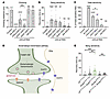 Figure 1
Behavioral defects in sif LoF mutants are mediated by glutamatergic and cholinergic neurons. (A–C) sif RNAi in glutamatergic neurons and cholinergic neurons led to climbing defects (A), bang sensitivity (B), and heat sensitivity (C) when compared with the control (n = 24–50 for A and B; n = 4–6 for C). Control: da-GAL4>luciferase RNAi. (D) Schematic of glutamatergic transmission (created with BioRender). NMDAR is a glutamate receptor. EAAT2 is a glutamate transporter that recycles glutamate from the synaptic cleft. (E) Genetic interactions of LoF alleles show that the transheterozygous combination of the sifT2A-GAL4 allele with Eaat2T2A-GAL4 allele causes a bang-sensitive phenotype (n = 32–59). Flies were tested at 3–5 days post eclosion (dpe). Data are presented as the mean ± SEM. *P < 0.05, **P < 0.01, ***P < 0.001, and ****P < 0.0001, by unpaired 2 tailed t test with Welch’s correction for unequal variances (A–C, and E).
Since glutamatergic sif RNAi causes the most severe seizure-like phenotypes, we further assessed the sensitivity of glutamatergic neurons to sif loss. We first considered whether loss of sif could genetically interact with genes required in the glutamatergic network — Eaat2 and Nmdar1 (Figure 1D). Loss of EAAT2, a glutamate transporter that is required for glutamate clearance, causes developmental epilepsy due to an accumulation of glutamate in the synaptic cleft (30). Interestingly, transheterozygous (GeneALoF/+ GeneBLoF/+) flies carrying 1 copy of a strong LoF allele for sif and Eaat2 exhibit more severe bang sensitivity when compared with the single heterozygous flies (Figure 1E, detailed genotypes are provided in Supplemental Table 2). Hence, sif loss enhanced the seizure phenotype caused by Eaat2 loss, probably by promoting glutamatergic transmission. Loss of Nmdar1, which encodes a glutamate receptor, should decrease synaptic transmission (31) and hence may not interact with sif. Indeed, we found that transheterozygous flies with a single LoF allele of Nmdar1 and sif did not exhibit bang sensitivity (Figure 1E). In summary, these data indicate that glutamatergic transmission is at the root of the bang sensitivity associated with sif loss.
sif loss causes hyperexcitability of glutamatergic neurons. To assess whether the activity of glutamatergic neurons is increased upon sif loss, we generated VGlut-GAL4>CaLexA flies. CaLexA induces a calcium-dependent nuclear import of LexA, which is stimulated by Ca2+-regulated dephosphorylation and nuclear translocation of nuclear factor of activated T cells (NFAT) (32). It includes 2 components: UAS-mLexA-VP16-NFAT and lexAop-rCD2-GFP. In the presence of VGlut-GAL4 and increased Ca2+ activities, GFP will be expressed and localized to the membrane of glutamatergic neurons. As a result, sif RNAi flies showed a significant increase in GFP intensity in the CNS, including the brain lobes and the ventral nerve cord (VNC), when compared with the control RNAi (Supplemental Figure 2, A and B). These data indicate that sif loss in glutamatergic neurons led to increased neuronal activity.
Given that synaptic transmission at the fly neuromuscular junction (NMJ) is mediated by glutamate and an increase in synaptic transmission at larval NMJs has previously been shown to cause crawling defects (22, 23, 33), we evaluated crawling behavior of sif-mutant larvae. Control third instar larvae crawled approximately 3 cm in a minute (Figure 2A). In contrast, sif-mutant third instar larvae only crawled approximately 0.8 cm in a minute. The sifT2A-GAL4 is a severe LoF allele (16) and produces GAL4 under the endogenous sif regulatory elements, which allows expression of the UAS cDNA in the proper spatial and temporal pattern of sif to assess the rescue effect (16). We observed that the crawling defects were fully rescued by expressing WT sif cDNA in the mutant background (Figure 2A, sif rescue), demonstrating that this phenotype was due to sif loss. These data suggest a defect in either muscle function, synaptic transmission, or both during larval development.
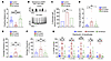 Figure 2
sif mutants display hyperexcitability at neuromuscular junctions. (A) The L3 larva crawling assay. Larvae were placed on an agar plate and allowed to crawl for 1 minute, and the crawling distance was measured (n = 15–34). (B) An electrode was placed on L3 larval muscles 6/7 in the abdominal segment, and spontaneous EPSPs were recorded for 2 minutes. Representative traces for spontaneous EPSPs with an intact CNS are shown. (C) The percentage of muscles with active events (>5 mV) were calculated. Fisher’s exact test. (D) Average amplitude of spontaneous EPSP events. (E) Average frequency of spontaneous EPSP events. (F) Percentage of events of greater than 1.5 mV for each muscle. (G) Distribution of EPSP amplitudes. Recordings were performed in 1 mM Ca2+ for 2 minutes. n = 12–15 cells recorded for each genotype. Genotypes: control (w1118), sif mutant (w; UAS-lacZ/+; sifT2A-GAL4/Df), sif rescue (w; UAS-sif/+; sifT2A-GAL4/Df). Data are presented as the mean ± SEM. *P < 0.05, **P < 0.01, ***P < 0.001, and ****P < 0.0001, by unpaired 2 tailed t test with Welch’s correction for unequal variances (A, and D–G).
To determine if there are morphological defects at the larval NMJ, we stained the NMJs with HRP, a neuronal membrane marker (34), and syt EGFP, a synaptic vesicle marker (35). We did not observe obvious changes in fluorescence of HRP or syt EGFP when compared with controls (Supplemental Figure 3, A–E). Hence, sif did not obviously affect the neuronal morphology or distribution of synaptic vesicles in third instar larvae.
To assess the electrophysiological properties of NMJs, we performed intracellular recordings of third instar muscles. Consistent with a previous study (36), we did not observe obvious differences in the average amplitude and frequency of miniature excitatory postsynaptic potentials (mEPSPs) in sif mutants when the axons were severed (Supplemental Figure 4, A–C). We therefore recorded without severing the axons of glutamatergic motor neurons from the CNS. In control animals, approximately 30% of the muscles showed patterned EPSPs in a 2-minute recording period (Figure 2, B and C). These periodic contractions were induced by the CNS pattern generator, which drives muscle contractions (37, 38). In contrast, 80% of the sif-mutant larvae exhibited large, nonperiodic bursts of EPSPs in a 2-minute recording (Figure 2, B and C). Hence, the frequency and amplitude of EPSPs were significantly increased in sif-mutant muscles when compared with controls (Figure 2, D–G). Note that the increase in glutamatergic transmission was significantly rescued by expression of sif cDNA (Figure 2, D–G). In summary, our data indicate that loss of sif increased glutamatergic transmission without altering NMJ morphology.
Sif loss promotes seizures by affecting actin polymerization. To determine whether the seizure-like phenotypes of sif mutants are due to a disruption in actin dynamics, we assessed dominant interactions between sif and other actin regulatory genes. As a GEF, TIAM1/Sif regulates actin remodeling by activating the Rho GTPase RAC1. The downstream effectors of Rho GTPases regulate actin filaments together with the actin-binding proteins that promote actin polymerization, including profilin (fly Chickadee, Chic) and actin related protein 2/3 complex (ARP2/3) (Figure 3A). We assessed chic01320/+ (a LoF allele) flies and observed that they had mild bang sensitivity, whereas Arp3EP3640/+ (a LoF allele) single-heterozygous flies did not show obvious bang sensitivity (Figure 3B). However, transheterozygous animals that carried a single LoF allele of sif and chic exhibited a severe bang-sensitive phenotype (Figure 3B). Similarly, sif and Arp3 transheterozygous flies were also bang sensitive (Figure 3B). These phenotypes were also observed using another LoF of the chic221 allele and the Arp3515FC allele (Supplemental Figure 5A). We also tested cofilin (fly Twinstar, Tsr), a protein that disassembles actin filaments (13) (Figure 3A). Neither tsrT2A-GAL4/+ (a LoF allele) flies nor the sifT2A-GAL4/+ tsrT2A-GAL4/+ transheterozygous flies displayed obvious bang sensitivity (Figure 3B). These data indicate that the loss of sif affected bang sensitivity by reducing actin polymerization and that other proteins required for proper actin polymerization also play a critical role in seizures.
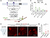 Figure 3
Actin polymerization defects are associated with seizures. (A) Graphical illustration of actin regulatory pathways in neurons (created with BioRender). Profilin (chic) promotes actin polymerization. The ARP2/3 complex nucleates actin filaments into branched networks. Cofilin (Tsr) mediates actin depolymerization. (B) Transheterozygous flies with combined LoF alleles of sif and chic displayed more severe bang sensitivity when compared with single-heterozygous flies (n = 32–56). (C) Glutamatergic motor neurons of the VNC from sif RNAi and chic RNAi animals. Actin filaments were visualized by LifeAct-RFP. Created with BioRender. Scale bar: 10 μm. (D) Quantification of the density of actin dots in neuronal cell body in the VNC (n = 12–23). Flies were tested at 3–5 dpe. Data are presented as the mean ± SEM. *P < 0.05, **P < 0.01, and ****P < 0.0001, by unpaired 2 tailed t test with Welch’s correction for unequal variances (B and D).
We next aimed to visualize the defects in actin filaments in neurons using an established reporter, Lifeact-RFP (39, 40). Lifeact is a 17 amino acid peptide that binds to 2 consecutive actin subunits on the F-actin filament in cells and tissues (40), and its intensity is proportional to the amount and length of F-actin present (41). Knockdown of sif in motor neurons leads to an accumulation of small “actin patches” in cell bodies. These resemble actin patches in yeast (42) and correspond to short actin filaments. Interestingly, we also observed this phenotype when we knocked down chic (Figure 3, C and D). Since profilin (Chic) is known to promote actin polymerization, these actin patches likely reflect a reduction in actin polymerization levels. Hence, these data provide compelling evidence that loss of sif affects actin polymerization as previously shown in mammalian cells (27).
Actin polymerization defects promote seizures in flies. Numerous genes encoding proteins that regulate actin dynamics are associated with developmental epilepsy. GEF proteins regulate the actin cytoskeleton by activating Rho GTPases like RAC1 and RAC3, and variants in these genes have been associated with seizures in humans (43, 44). In addition, although mice that lack Rac1 or Rac3 do not exhibit seizures, double-homozygous-KO mice exhibit spontaneous seizures (45). We therefore performed genetic interaction studies using 2 fly Rac alleles: Rac1J11 (a LoF allele) and Rac2KG05681 (a LoF allele), and 1 LoF chic allele. Both Rac1J11 and Rac2KG05681 synergized with chic01320, and the transheterozygous flies were bang sensitive (Supplemental Figure 5B, Supplemental Video 1). These data are consistent with seizures resulting from a disruption to a GEF/RAC GTPase pathway that reduces actin polymerization.
We next assessed whether loss of 1 copy of 2 different genes that promote actin polymerization can similarly affect seizure sensitivity. WASHC4 (fly SWIP) encodes a subunit of the WASH complex that promotes actin polymerization (46), and FLNA (fly cher) encodes an actin cross-linking protein filamin A, which stabilizes polymerized actin filaments (Figure 3A). Pathogenic LoF variants in both WASHC4 and FLNA have been associated with seizures in humans (1, 46, 47). Both SWIPCR70302/+ chic01320/+ and cherMiMIC/+ chic01320/+ flies exhibit increased bang sensitivity when compared with chic01320/+ flies (Supplemental Figure 5B and Supplemental Videos 2 and 3). In contrast, single-heterozygous loss of SWIP or cher alone does not cause obvious bang sensitivity. These data further indicate that disruptions in actin polymerization are associated with increased bang sensitivity and that digenic loss of genes regulating actin polymerization increases seizure susceptibility.
To determine if the actin regulatory proteins cause seizures by affecting glutamatergic transmission, similar to loss of TIAM1/sif, we performed genetic interaction studies with genes encoding proteins that are known to affect glutamate uptake at the fly NMJs. The previously tested mutations — Rac1J11, Rac2KG05681b, SWIPCR70302, and cherMIMIC alleles — all caused an obvious increase in bang sensitivity when combined with a single Eaat2T2A-GAL4 LoF allele (Supplemental Figure 5C). Together, these data provide evidence that seizures can be caused by reduced actin polymerization levels in glutamatergic neurons.
sif loss leads to an accumulation of small mitochondria in glutamatergic neurons. Next, we asked how actin polymerization defects affect neuronal activity. Since actin dynamics are important for neuronal migration and axonal formation, we tested whether sif loss leads to neural wiring phenotypes. Our previous work has shown that sif is enriched in the antennal lobe during the developmental stage (16). We therefore examined whether the neural wiring of olfactory projection neurons (PNs) was affected (48, 49). As a result, loss of sif in the PNs did not cause obvious defects in neural wiring (Supplemental Figure 6, A–E). We also assessed the neural wiring pattern in the mushroom body using OK107-GAL4–driven GFP (50). Likewise, sif RNAi in the mushroom body also did not cause any significant changes in neural wiring (Supplemental Figure 6, F and G). These data suggest that sif loss may not obviously alter the neuronal projections and synapse formation during development.
In addition to neuronal morphology and neural circuit regulation, the actin cytoskeleton participates in a variety of other cellular activities, including the regulation of mitochondrial function. Experimental paradigms have documented how cytoskeletal dynamics affect mitochondrial function by affecting their motility, fission, and mitophagy (51–55). We therefore assessed mitochondrial distribution using mito-GFP (56) in flies with sif loss. Interestingly, we observed an increase in the mito-GFP signal in both the soma and axons of glutamatergic motor neurons in sif RNAi when compared with the control RNAi (Figure 4, A–D). We also performed transmission electron microscopy (TEM) analysis of cross-sections of the VNC of sif mutants and observed a significant increase in the number and a decrease in the size of mitochondria when compared with the WT control (Figure 4, E–H). The mitochondrial phenotypes observed in sif mutants were rescued by expression of the sif cDNA (sif rescue) (Figure 4, E–H). These data indicate that sif loss perturbed normal mitochondrial dynamics.
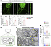 Figure 4
sif loss leads to an accumulation of mitochondria in glutamatergic neurons. (A) Representative images of third instar larval VNC cell bodies and axons of glutamatergic motor neurons from control (luciferase) RNAi and sif RNAi animals. Mitochondria are visualized by mito-GFP. (B) Quantification of normalized mito-GFP intensity in neuronal cell bodies (n = 13). Scale bars: 10 μm and 50 μm. (C and D) Quantification of normalized signal intensity (C) and percentage of area (D) of mito-GFP in neuronal axons (n = 8–10). (E) Diagram of the CNS of third instar larva. The CNS was dissected, and a transverse section was made for TEM. (F) Representative VNC neuronal cell body TEM images of sif-mutant and sif rescue neuronal cell bodies. Yellow arrowheads indicate mitochondria. Scale bar: 5 μm. Original magnification, ×1.85. (G and H) Quantification of mitochondria numbers (G) (n = 29 cells) and size (H) (n = 7–8 cells) in the VNC for WT, sif rescue, and sif-mutant animals (n = 3 animals for each genotype). Genotypes: Canton S (WT); w UAS-sif/+ sifT2A-GAL4/Df (sif-mutant); and w UAS-lacZ/+ sifT2A-GAL4/Df (sif rescue) Data are presented as the mean ± SEM. **P < 0.01, ***P < 0.001, and ****P < 0.0001, by unpaired 2 tailed t test with Welch’s correction for unequal variances (B–D, and G and H).
Next, we assessed mitochondrial membrane potential using tetramethylrhodamine, methyl ester (TMRM) staining of sif-mutant brains and did not observe a significant change in TMRM intensity, indicating that these mitochondria maintained their membrane potential (Supplemental Figure 7, A and B). To further evaluate mitochondrial function, we measured ATP production. ATP levels in the adult fly head of sif mutants showed an approximately 1.5-fold increase when compared with levels in sif rescue flies (Supplemental Figure 7C). These data suggest that the mitochondrial function was not overtly compromised.
Increased number and decreased size of mitochondria are associated with elevated Drp1 levels. During mitochondrial fission, the ER forms a ring around the fission site. Upon calcium release from the ER, G-actin is recruited to this site, where it forms short filaments. These short filaments recruit and bind the mitochondrial fission regulator dynamin-related protein 1 (DRP1), promoting its oligomerization and GTPase activity (51–55, 57) (Figure 5A). However, the relationship between actin dynamics, mitochondrial fission, and epilepsy has not been established.
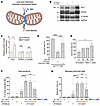 Figure 5
Inhibition of mitochondrial fission significantly rescues sif LoF phenotypes. (A) Graphical illustration of actin filaments mediated mitochondrial fission via recruitment of DRP1 (created with BioRender). (B) Protein levels in sif rescue and sif-mutant animals were determined by Western blotting. Third instar larvae were collected for protein extraction and analysis. (C) Quantification of protein levels. Data points represent the relative protein levels per independent repeat (n = 5). (D) Survival rate of sif-mutant animals with the expression of UAS-Drp1 RNAi. The survival rate was measured using the expected Mendelian ratio by counting flies that eclosed and were viable after 24 hours. (n = 3–9, with over 300 progenies counted for each genotype). (E) Survival rate of sif-mutant animals fed different doses of Mdivi-1 (0, 20, 50, and 100 μM in food) (n = 4 for 0 μM and 100 μM groups, n = 1 for 20 μM and 50 μM groups). (F and G) Quantification of heat sensitivity for control (w1118), sif heterozygote (sifT2A-GAL4/+), sif-mutant (w UAS-lacZ/+ sifT2A-GAL4/Df), and sif-mutant flies fed different doses of Mdivi-1. The flies were tested at 3–5 dpe. Data points in F represent replicates (n = 5–8), and each replicate contains 5–10 animals; data points in G represent the recovery time for each animal (n = 24–54). Data are presented as the mean ± SEM. *P < 0.05, **P < 0.01, ***P < 0.001, and ****P < 0.0001, by unpaired 2 tailed t test with Welch’s correction for unequal variances (C–G).
Given that the mitochondrial number was increased but mitochondria size was decreased in flies lacking sif, we speculated that mitochondrial fission may be increased by increased Drp1 levels. We first assessed Drp1 protein levels and found that they were indeed upregulated in sif mutants (Figure 5, B and C). To assess whether this elevation contributes to the phenotypes, we reduced Drp1 levels in sif mutants. Drp1 RNAi significantly rescued the viability of sif mutants at 1 day post eclosion (dpe) from approximately 30% to 80% (Figure 5D), but it did not significantly improve the climbing defects or bang sensitivity (Supplemental Figure 8, A and B). However, we have previously documented that loss-of-function mutations in DNM1L (also known as Drp1) cause severe seizures in both humans (58, 59) and flies (60) (see Discussion). As actin polymerization is also required for targeting damaged mitochondria to the autophagosome (51, 61), we next considered whether other genes that affect mitophagy might play a role. We found that expression of Pink1 or parkin, genes that promote mitophagy (62, 63), did not affect the survival rate of sif mutants (Supplemental Figure 9). These data argue that the increase in the number of mitochondria and the decrease in their size in sif mutants were, at least in part, associated with elevated mitochondrial fission driven by increased Drp1 levels.
Given that the Drp1 RNAi improves sif-mutant survival but does not suppress seizures, we next assessed whether feeding sif mutants a small compound, mitochondrial division inhibitor 1 (Mdivi-1), was effective. Mdivi-1 decreases the GTPase activity of DRP1 without significantly reducing total DRP1 protein levels (64, 65). Here, Mdivi-1 treatment not only significantly improved the survival rate of sif mutants in a dose-dependent manner (Figure 5E) but also suppressed the heat sensitivity in surviving sif-mutant adults at the 100 μM dose (Figure 5, F and G). Together, these data indicate a pharmacological benefit of Mdivi-1 in our fly model for TIAM1-associated epilepsy.
Elevated ROS causes seizure-like behaviors in flies. Since increased mitochondrial fission leads to elevated ROS (66, 67), and high ROS is associated with hyperexcitability and toxicity (23), we assessed if there is increased ROS production in sif mutants (Figure 6A). We measured the enzymatic activity of the citric acid cycle protein mitochondrial aconitase (m-aconitase). Both m-aconitase and cytosolic aconitase (c-aconitase) are highly sensitive to ROS-induced damage and have reduced activity in high ROS environments (68, 69). Indeed, the enzymatic activity of m-aconitase in sif mutants was decreased by approximately 86% when compared with control animals (Figure 6B), reflecting increased mitochondrial ROS. Furthermore, we observed that c-aconitase activity was also reduced by 20% (Figure 6C), indicating a mild increase in cytoplasmic ROS levels. Importantly, these reductions in aconitase activity were rescued by expressing a sif cDNA in the sif-mutant flies (Figure 6, B and C). To address whether ROS are elevated using another assay, we performed MitoSOX staining of fly brains. We found that sif-mutant brains displayed a significantly increased MitoSOX signal, indicating elevated ROS (Supplemental Figure 10, A and B). Finally, we used a third ROS indicator, UAS-mito-roGFP2-Orp1 (70, 71), and tested for ROS in glutamatergic neurons and GABAergic neurons. VGlut-GAL4>sif RNAi flies exhibited an increase in mitochondrial oxidation in the brain (Supplemental Figure 10, C and D). However, Gad1-GAL4>sif RNAi flies did not show altered mitochondrial oxidation in the brain (Supplemental Figure 10, E and F), consistent with their lack of seizure phenotypes when sif was reduced in GABAergic neurons (Figure 1, C and D).
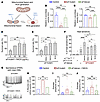 Figure 6
Inhibition of mitochondrial ROS using NACA significantly rescues sif LoF phenotypes. (A) Graphical illustration of mitochondrial fission and ROS generation (created with BioRender). (B and C) An aconitase activity assay was used to reveal high ROS in sif mutants. This high ROS was rescuable by expression of sif cDNA in these mutants (n = 9). (D and E) Survival rate of sif LoF mutants fed with different doses of NACA (0, 40, 80, and 160 μg/mL in food) (D) or Mdivi-1 and NACA (M+N) (Mdivi-1: 100 μM; NACA: 80 μg/mL) (E). n ≥3, except for NACA: 40 μg/mL group (n = 1), with over 200 progenies counted for each group. (F) Quantification of heat sensitivity for control (w1118), sif heterozygote (sifT2A-GAL4/+), sif-mutant (w UAS-lacZ/+ sifT2A-GAL4/Df), and sif-mutant flies fed Mdivi-1 (100 μM), NACA (80 μg/mL), or their combination (M+N). Flies were tested at 3–5 dpe. (G) Representative traces of spontaneous EPSPs from muscles with intact CNS of sif mutants fed normal food (top) or NACA (bottom). (H) Percentage of muscle cells with active events (events >5 mV). Fisher’s exact test. (I) Average amplitude of spontaneous EPSP events (n = 13–17). (J) Average frequency of spontaneous EPSP events (n = 13–17). Data are presented as the mean ± SEM. *P < 0.05, **P < 0.01, ***P < 0.001, and ****P < 0.0001, by unpaired 2 tailed t test with Welch’s correction for unequal variances (B–F, I, and J).
It has been reported that oxidative stress in neurons leads to upregulation of the glial innate immune response (IIR) genes, which in turn contributes to the progression of epilepsy (70). We therefore evaluated the expression of IIR genes. Consistent with previous findings, we observed that many IIR genes were significantly upregulated in the head of sif mutants when compared with the sif rescue group (Supplemental Figure 11). In summary, sif loss significantly increased the expression of IIR genes.
To further determine the effect of ROS on sif loss–induced seizures, we genetically increased ROS by reducing the levels of superoxide dismutase 1 (SOD1), an enzyme that breaks down superoxide radicals (72), using a sod1T2A-GAL4 LoF allele. sifT2A-GAL4/+ sod1T2A-GAL4/+ transheterozygous flies were more bang sensitive than sod1T2A-GAL4/+ flies (Supplemental Figure 12A). Together, these data argue that elevated ROS production indeed contributed to seizures in sif mutants.
To assess whether ROS may also underlie seizure phenotypes in other epilepsy-associated actin regulatory genes, we performed genetic interaction tests between the sod1T2A-GAL4 allele and additional actin regulatory genes. Transheterozygous flies carrying the sod1T2A-GAL4 allele and the Rac1J11, Rac2KG05681b, SWIPCR70302, or cherMiMIC alleles showed bang sensitivity (Supplemental Figure 12A). Given that these genes also dominantly interact with Profilin (chic) and EAAT2 (Eaat2) (Supplemental Figure 12B), they seem to share a pathway defined by an AMG pathway in epilepsy.
Inhibition of ROS by NACA significantly rescues the phenotypes of sif mutants. We next determined whether the phenotypes of sif mutants can be ameliorated by pharmacological inhibition of ROS. NACA is a potent antioxidant that crosses the blood-brain barrier (BBB) and reduces ROS (73). We raised sif mutants on NACA-containing food or control food. We observed that NACA treatment significantly improved the survival rate of sif mutants in a dose-dependent manner, with 80 μg/mL NACA having the highest rescue ability: it improved the survival rate from approximately 30% to approximately 90% (Figure 6D). Combining Mdivi-1 and NACA did not further improve the survival rate (Figure 6E). We also tested the behavioral rescue and found that NACA treatment significantly ameliorated the heat sensitivity phenotype (Figure 6F). Mdivi-1 combined with NACA did not further improve the heat sensitivity phenotype (Figure 6F). These data argue that mitochondrial fission defects and ROS affect the same pathway.
To test whether NACA ameliorates seizures by reducing hyperexcitability, we performed NMJ electrophysiological recordings. The sif mutants were raised on NACA food (80 μg/mL), and the third instar larvae with an intact CNS were used for the recordings. Spontaneous EPSPs were significantly inhibited in the NACA-treated group, and the percentage of muscles with events above 5 mV were significantly decreased (Figure 6, G and H). The amplitude and frequency of EPSPs were significantly reduced in the NACA-treated group (Figure 6, I and J). Hence, reducing ROS via NACA significantly reduced glutamatergic transmission in sif mutants.
Key features of the AMG pathway are observed in mammalian cells with TIAM1 siRNA. To determine if the AMG pathway is conserved in mammals, we performed TIAM1 RNAi in the human Daoy neuronal cell line (74) using a previously validated TIAM1-targeting siRNA (75). We found that this siRNA downregulated TIAM1 protein expression to less than 10% in Daoy cells (Supplemental Figure 13, A and B). Staining for F-actin using phalloidin revealed a significant decrease in long actin filaments and an increase in short actin filaments when compared with cells transfected with a control siRNA (Figure 7, A and B). We also assessed mitochondrial morphology using ATP5A immunostaining. TIAM1 siRNA significantly increased the number and decreased the length of mitochondria in Daoy cells (Figure 7, C–F), consistent with what we observed in fly neurons. In addition, we also observed a significant increase in total DRP1 protein levels upon TIAM1 knockdown (Figure 7, G and H), as well as a robust increase in phosphorylated DRP1 at Ser616 (p-DRP1 S616) protein levels (Figure 7, G–I). Note that DRP1 (S616) phosphorylation promotes mitochondrial fission (76). The elevation of p-DRP1 S616 was significantly rescued by treating the cells with NACA (Figure 7, G–I) or Mdivi-1 (Supplemental Figure 13, A–C). These data are consistent with our findings in flies, indicating a conserved AMG pathway in flies and humans. Overall, the data reveal an AMG pathway that may be disrupted in some individuals with seizures.
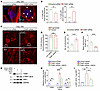 Figure 7
Knockdown of TIAM1 in mammalian cells leads to reduced actin polymerization and increased mitochondrial fission. (A) Representative image of actin filaments in Daoy cells transfected with either a negative control siRNA or an siRNA targeting TIAM1 (TIAM1 siRNA). Red, phalloidin; blue, DAPI. (B) The percentage of cells with long and short actin filaments was quantified (n = 3 with 190–192 cells for each group). (C) Representative images of the ATP5A-labeled mitochondria in Daoy cells. (D) Quantitative analysis of the mitochondrial phenotype in each cell (n = 15–21). Fisher’s exact test. (E and F) Quantitative analysis of the number of mitochondria (E) and mean mitochondria length (F) in the region of interest (ROI) of each Daoy cell (n = 15). (G) Western blots of Daoy cells transfected with control siRNA or TIAM1 siRNA and treated with or without NACA (750 μM, 24 hours). (H) Quantifications of protein levels indicate a significant increase in p-DRP1 (S616) and total DRP1 protein levels in the TIAM1 siRNA group (n = 4–7). (I) The elevated p-DRP1 (S616)/total DRP1 ratio in TIAM1 siRNA cells was significantly rescued by NACA treatment (n = 4). Data are presented as the mean ± SEM. *P < 0.05, **P < 0.01, ***P < 0.001, and ****P < 0.0001, by unpaired 2 tailed t test with Welch’s correction for unequal variances (B, E, F, H, and I).
Higher frequency of heterozygous LoF variants in AMG pathway genes in the seizure cohort. Besides the approximately 50 actin regulatory genes associated with epilepsy, there are approximately 140 genes associated with epilepsy that are involved in the regulation of mitochondrial function, and approximately 30 genes that affect glutamate synthesis and transmission based on the literature (1, 17, 18) (Supplemental Table 1). Hence, variants in more than 200 genes may affect the AMG pathway. Since our findings indicated that seizures can be caused by 2 heterozygous LoF variants in different genes affecting the AMG pathway, it is possible that digenic pathogenic variants that affect actin polymerization, mitochondrial ROS, and/or glutamatergic transmission cause a subset of seizures. We therefore gathered an undiagnosed cohort of 433 individuals with seizures and 2,167 individuals without seizures from Baylor Genetics and analyzed their sequencing data by ranking the top risk variants using AI-MARRVEL, a comprehensive tool to prioritize possible disease-causing variants (77) (Figure 8A). Among the top-10 variants of each individual, the percentage of individuals with 2 or more LoF variants from AMG genes was significantly higher in the seizure cohort (5.5%) when compared with the nonseizure cohort (1.2%) (Figure 8B). We also downloaded and analyzed the sequencing data for 774 unaffected individuals as nonpatient controls from the 1000 Genomes Project dataset (78). The percentage of control individuals with 2 or more LoF variants in the AMG pathway genes was extremely low (<0.3%) (Supplemental Figure 14).
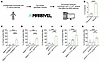 Figure 8
Increased burden of heterozygous LoF variants in AMG pathway genes among individuals with undiagnosed epilepsy. (A) Sequencing data of undiagnosed individuals with or without epilepsy were analyzed and the variants were ranked by AI-MARRVEL (v.1.1.0rc). The top 10 variants were used for the enrichment analysis. Created with BioRender. (B) The percentage of individuals with 2 or more LoF variants from the AMG gene set were counted. Fisher’s exact test. (C–G) Heat sensitivity of adult flies. The combinations of the LoF alleles show synergistic or additive effects for heat sensitivity (n = 5–14). Flies were tested at 6–9 dpe. Heat-sensitive flies are counted after heating at 42°C for 40 seconds (C), 50 seconds (D–F), or 60 seconds (G). Data are presented as the mean ± SEM. **P < 0.01, ***P < 0.001, and ****P < 0.0001, by unpaired 2 tailed t test with Welch’s correction for unequal variances (C–G).
We selected 16 individuals with digenic LoF variants in genes known to regulate either actin polymerization or ROS (Supplemental Table 3). We evaluated the effects of the affected orthologous genes in flies for 7 of these individuals (Table 2 and Supplemental Figure 15) and observed that 5 of the 7 transheterozygous mutants had synergistic or additive effects with respect to heat sensitivity, suggesting that combining these alleles had a pathological consequence (Figure 8, C–G). In the remaining 2 digenic combinations, we did not observe obvious synergistic or additive effects (Supplemental Figure 15, A–D).
To assess whether digenic LoF variants cause spontaneous seizures, we recorded the behaviors of the flies for 4 days using a video-tracking system (79). As a negative control, we used w1118 flies, as they do not exhibit seizures. As expected, we observed spontaneous seizures in VGlut>sif RNAi flies (Supplemental Figure 16, A and B). Next, we video-tracked 9 combinations of transheterozygous mutant flies including the 5 that modeled the individuals in Figure 8. As a result, we detected seizure-like events in 7 of the 10 genotypes at 25°C (Supplemental Figure 16A). We then recorded 4 of the genotypes at 29°C and observed spontaneous seizures (Supplemental Figure 16B and Supplemental Videos 4–7). These data show that digenic variants in genes that regulate the AMG pathway can promote seizure susceptibility.


Copyright © 2026 American Society for Clinical Investigation
ISSN: 0021-9738 (print), 1558-8238 (online)