
Advertisement
Research ArticleGeneticsNeuroscience
Open Access |  10.1172/JCI169297
Mouse model of atypical DAT deficiency syndrome uncovers dopamine dysfunction associated with parkinsonism and ADHD
Freja Herborg,1 Lisa K. Konrad,1 Søren H. Jørgensen,1 Jamila H. Lilja,1 Benoît Delignat-Lavaud,2 Leonie P. Posselt,1 Ciara F. Pugh,1 Sofie A. Bach,1,3 Cecilia F. Ratner,4 Nora Awadallah,5 Jose A. Pino,6 Frida Berlin,1 Aske L. Ejdrup,1 Mikkel V. Olesen,3 Mattias Rickhag,1,7 Birgitte Holst,4 Susana Aznar,3 Felix P. Mayer,1 David Woldbye,1 Gonzalo Torres,5,8 Louis-Eric Trudeau,2 and Ulrik Gether1
1Molecular Neuropharmacology and Genetics Laboratory, Department of Neuroscience, Faculty of Health and Medical Sciences, University of Copenhagen, Copenhagen, Denmark.
2CNS Research Group, Department of Pharmacology and Physiology, Department of Neurosciences, Faculty of Medicine, Université de Montréal, Montréal, Quebec, Canada.
3Centre for Neuroscience and Stereology, Copenhagen University Hospital Bispebjerg-Frederiksberg, Copenhagen, Denmark.
4Department of Biomedical Sciences, University of Copenhagen, Copenhagen, Denmark.
5Department of Molecular, Cellular, and Biomedical Sciences, City University of New York School of Medicine, New York, New York, USA.
6Laboratorio de Bioquímica y Farmacología Molecular, Escuela de Ciencias, Facultad de Ciencias de la Vida, Universidad Viña del Mar, Valparaíso, Chile.
7Danish Research Centre for Magnetic Resonance (DRCMR), Department of Radiology and Nuclear Medicine, Copenhagen University Hospital-Amager and Hvidovre, Copenhagen, Denmark.
8Department of Molecular Pharmacology and Neuroscience, Stritch School of Medicine, Loyola University, Chicago, USA.
Address correspondence to: Freja Herborg or Ulrik Gether, Department of Neuroscience, Maersk Tower 7.5, University of Copenhagen, Blegdamsvej 3B, DK-2200 N, Copenhagen, Denmark. Phone: 45.5360.9699; Email: frejahh@sund.ku.dk (FH). Phone: 45.2875.7548; Email: gether@sund.ku.dk (UG).
Find articles by
Herborg, F.
in:
PubMed
|
Google Scholar
|

1Molecular Neuropharmacology and Genetics Laboratory, Department of Neuroscience, Faculty of Health and Medical Sciences, University of Copenhagen, Copenhagen, Denmark.
2CNS Research Group, Department of Pharmacology and Physiology, Department of Neurosciences, Faculty of Medicine, Université de Montréal, Montréal, Quebec, Canada.
3Centre for Neuroscience and Stereology, Copenhagen University Hospital Bispebjerg-Frederiksberg, Copenhagen, Denmark.
4Department of Biomedical Sciences, University of Copenhagen, Copenhagen, Denmark.
5Department of Molecular, Cellular, and Biomedical Sciences, City University of New York School of Medicine, New York, New York, USA.
6Laboratorio de Bioquímica y Farmacología Molecular, Escuela de Ciencias, Facultad de Ciencias de la Vida, Universidad Viña del Mar, Valparaíso, Chile.
7Danish Research Centre for Magnetic Resonance (DRCMR), Department of Radiology and Nuclear Medicine, Copenhagen University Hospital-Amager and Hvidovre, Copenhagen, Denmark.
8Department of Molecular Pharmacology and Neuroscience, Stritch School of Medicine, Loyola University, Chicago, USA.
Address correspondence to: Freja Herborg or Ulrik Gether, Department of Neuroscience, Maersk Tower 7.5, University of Copenhagen, Blegdamsvej 3B, DK-2200 N, Copenhagen, Denmark. Phone: 45.5360.9699; Email: frejahh@sund.ku.dk (FH). Phone: 45.2875.7548; Email: gether@sund.ku.dk (UG).
Find articles by
Konrad, L.
in:
PubMed
|
Google Scholar
|
1Molecular Neuropharmacology and Genetics Laboratory, Department of Neuroscience, Faculty of Health and Medical Sciences, University of Copenhagen, Copenhagen, Denmark.
2CNS Research Group, Department of Pharmacology and Physiology, Department of Neurosciences, Faculty of Medicine, Université de Montréal, Montréal, Quebec, Canada.
3Centre for Neuroscience and Stereology, Copenhagen University Hospital Bispebjerg-Frederiksberg, Copenhagen, Denmark.
4Department of Biomedical Sciences, University of Copenhagen, Copenhagen, Denmark.
5Department of Molecular, Cellular, and Biomedical Sciences, City University of New York School of Medicine, New York, New York, USA.
6Laboratorio de Bioquímica y Farmacología Molecular, Escuela de Ciencias, Facultad de Ciencias de la Vida, Universidad Viña del Mar, Valparaíso, Chile.
7Danish Research Centre for Magnetic Resonance (DRCMR), Department of Radiology and Nuclear Medicine, Copenhagen University Hospital-Amager and Hvidovre, Copenhagen, Denmark.
8Department of Molecular Pharmacology and Neuroscience, Stritch School of Medicine, Loyola University, Chicago, USA.
Address correspondence to: Freja Herborg or Ulrik Gether, Department of Neuroscience, Maersk Tower 7.5, University of Copenhagen, Blegdamsvej 3B, DK-2200 N, Copenhagen, Denmark. Phone: 45.5360.9699; Email: frejahh@sund.ku.dk (FH). Phone: 45.2875.7548; Email: gether@sund.ku.dk (UG).
Find articles by Jørgensen, S. in: PubMed | Google Scholar
1Molecular Neuropharmacology and Genetics Laboratory, Department of Neuroscience, Faculty of Health and Medical Sciences, University of Copenhagen, Copenhagen, Denmark.
2CNS Research Group, Department of Pharmacology and Physiology, Department of Neurosciences, Faculty of Medicine, Université de Montréal, Montréal, Quebec, Canada.
3Centre for Neuroscience and Stereology, Copenhagen University Hospital Bispebjerg-Frederiksberg, Copenhagen, Denmark.
4Department of Biomedical Sciences, University of Copenhagen, Copenhagen, Denmark.
5Department of Molecular, Cellular, and Biomedical Sciences, City University of New York School of Medicine, New York, New York, USA.
6Laboratorio de Bioquímica y Farmacología Molecular, Escuela de Ciencias, Facultad de Ciencias de la Vida, Universidad Viña del Mar, Valparaíso, Chile.
7Danish Research Centre for Magnetic Resonance (DRCMR), Department of Radiology and Nuclear Medicine, Copenhagen University Hospital-Amager and Hvidovre, Copenhagen, Denmark.
8Department of Molecular Pharmacology and Neuroscience, Stritch School of Medicine, Loyola University, Chicago, USA.
Address correspondence to: Freja Herborg or Ulrik Gether, Department of Neuroscience, Maersk Tower 7.5, University of Copenhagen, Blegdamsvej 3B, DK-2200 N, Copenhagen, Denmark. Phone: 45.5360.9699; Email: frejahh@sund.ku.dk (FH). Phone: 45.2875.7548; Email: gether@sund.ku.dk (UG).
Find articles by Lilja, J. in: PubMed | Google Scholar
1Molecular Neuropharmacology and Genetics Laboratory, Department of Neuroscience, Faculty of Health and Medical Sciences, University of Copenhagen, Copenhagen, Denmark.
2CNS Research Group, Department of Pharmacology and Physiology, Department of Neurosciences, Faculty of Medicine, Université de Montréal, Montréal, Quebec, Canada.
3Centre for Neuroscience and Stereology, Copenhagen University Hospital Bispebjerg-Frederiksberg, Copenhagen, Denmark.
4Department of Biomedical Sciences, University of Copenhagen, Copenhagen, Denmark.
5Department of Molecular, Cellular, and Biomedical Sciences, City University of New York School of Medicine, New York, New York, USA.
6Laboratorio de Bioquímica y Farmacología Molecular, Escuela de Ciencias, Facultad de Ciencias de la Vida, Universidad Viña del Mar, Valparaíso, Chile.
7Danish Research Centre for Magnetic Resonance (DRCMR), Department of Radiology and Nuclear Medicine, Copenhagen University Hospital-Amager and Hvidovre, Copenhagen, Denmark.
8Department of Molecular Pharmacology and Neuroscience, Stritch School of Medicine, Loyola University, Chicago, USA.
Address correspondence to: Freja Herborg or Ulrik Gether, Department of Neuroscience, Maersk Tower 7.5, University of Copenhagen, Blegdamsvej 3B, DK-2200 N, Copenhagen, Denmark. Phone: 45.5360.9699; Email: frejahh@sund.ku.dk (FH). Phone: 45.2875.7548; Email: gether@sund.ku.dk (UG).
Find articles by Delignat-Lavaud, B. in: PubMed | Google Scholar
1Molecular Neuropharmacology and Genetics Laboratory, Department of Neuroscience, Faculty of Health and Medical Sciences, University of Copenhagen, Copenhagen, Denmark.
2CNS Research Group, Department of Pharmacology and Physiology, Department of Neurosciences, Faculty of Medicine, Université de Montréal, Montréal, Quebec, Canada.
3Centre for Neuroscience and Stereology, Copenhagen University Hospital Bispebjerg-Frederiksberg, Copenhagen, Denmark.
4Department of Biomedical Sciences, University of Copenhagen, Copenhagen, Denmark.
5Department of Molecular, Cellular, and Biomedical Sciences, City University of New York School of Medicine, New York, New York, USA.
6Laboratorio de Bioquímica y Farmacología Molecular, Escuela de Ciencias, Facultad de Ciencias de la Vida, Universidad Viña del Mar, Valparaíso, Chile.
7Danish Research Centre for Magnetic Resonance (DRCMR), Department of Radiology and Nuclear Medicine, Copenhagen University Hospital-Amager and Hvidovre, Copenhagen, Denmark.
8Department of Molecular Pharmacology and Neuroscience, Stritch School of Medicine, Loyola University, Chicago, USA.
Address correspondence to: Freja Herborg or Ulrik Gether, Department of Neuroscience, Maersk Tower 7.5, University of Copenhagen, Blegdamsvej 3B, DK-2200 N, Copenhagen, Denmark. Phone: 45.5360.9699; Email: frejahh@sund.ku.dk (FH). Phone: 45.2875.7548; Email: gether@sund.ku.dk (UG).
Find articles by
Posselt, L.
in:
PubMed
|
Google Scholar
|
1Molecular Neuropharmacology and Genetics Laboratory, Department of Neuroscience, Faculty of Health and Medical Sciences, University of Copenhagen, Copenhagen, Denmark.
2CNS Research Group, Department of Pharmacology and Physiology, Department of Neurosciences, Faculty of Medicine, Université de Montréal, Montréal, Quebec, Canada.
3Centre for Neuroscience and Stereology, Copenhagen University Hospital Bispebjerg-Frederiksberg, Copenhagen, Denmark.
4Department of Biomedical Sciences, University of Copenhagen, Copenhagen, Denmark.
5Department of Molecular, Cellular, and Biomedical Sciences, City University of New York School of Medicine, New York, New York, USA.
6Laboratorio de Bioquímica y Farmacología Molecular, Escuela de Ciencias, Facultad de Ciencias de la Vida, Universidad Viña del Mar, Valparaíso, Chile.
7Danish Research Centre for Magnetic Resonance (DRCMR), Department of Radiology and Nuclear Medicine, Copenhagen University Hospital-Amager and Hvidovre, Copenhagen, Denmark.
8Department of Molecular Pharmacology and Neuroscience, Stritch School of Medicine, Loyola University, Chicago, USA.
Address correspondence to: Freja Herborg or Ulrik Gether, Department of Neuroscience, Maersk Tower 7.5, University of Copenhagen, Blegdamsvej 3B, DK-2200 N, Copenhagen, Denmark. Phone: 45.5360.9699; Email: frejahh@sund.ku.dk (FH). Phone: 45.2875.7548; Email: gether@sund.ku.dk (UG).
Find articles by
Pugh, C.
in:
PubMed
|
Google Scholar
|
1Molecular Neuropharmacology and Genetics Laboratory, Department of Neuroscience, Faculty of Health and Medical Sciences, University of Copenhagen, Copenhagen, Denmark.
2CNS Research Group, Department of Pharmacology and Physiology, Department of Neurosciences, Faculty of Medicine, Université de Montréal, Montréal, Quebec, Canada.
3Centre for Neuroscience and Stereology, Copenhagen University Hospital Bispebjerg-Frederiksberg, Copenhagen, Denmark.
4Department of Biomedical Sciences, University of Copenhagen, Copenhagen, Denmark.
5Department of Molecular, Cellular, and Biomedical Sciences, City University of New York School of Medicine, New York, New York, USA.
6Laboratorio de Bioquímica y Farmacología Molecular, Escuela de Ciencias, Facultad de Ciencias de la Vida, Universidad Viña del Mar, Valparaíso, Chile.
7Danish Research Centre for Magnetic Resonance (DRCMR), Department of Radiology and Nuclear Medicine, Copenhagen University Hospital-Amager and Hvidovre, Copenhagen, Denmark.
8Department of Molecular Pharmacology and Neuroscience, Stritch School of Medicine, Loyola University, Chicago, USA.
Address correspondence to: Freja Herborg or Ulrik Gether, Department of Neuroscience, Maersk Tower 7.5, University of Copenhagen, Blegdamsvej 3B, DK-2200 N, Copenhagen, Denmark. Phone: 45.5360.9699; Email: frejahh@sund.ku.dk (FH). Phone: 45.2875.7548; Email: gether@sund.ku.dk (UG).
Find articles by Bach, S. in: PubMed | Google Scholar
1Molecular Neuropharmacology and Genetics Laboratory, Department of Neuroscience, Faculty of Health and Medical Sciences, University of Copenhagen, Copenhagen, Denmark.
2CNS Research Group, Department of Pharmacology and Physiology, Department of Neurosciences, Faculty of Medicine, Université de Montréal, Montréal, Quebec, Canada.
3Centre for Neuroscience and Stereology, Copenhagen University Hospital Bispebjerg-Frederiksberg, Copenhagen, Denmark.
4Department of Biomedical Sciences, University of Copenhagen, Copenhagen, Denmark.
5Department of Molecular, Cellular, and Biomedical Sciences, City University of New York School of Medicine, New York, New York, USA.
6Laboratorio de Bioquímica y Farmacología Molecular, Escuela de Ciencias, Facultad de Ciencias de la Vida, Universidad Viña del Mar, Valparaíso, Chile.
7Danish Research Centre for Magnetic Resonance (DRCMR), Department of Radiology and Nuclear Medicine, Copenhagen University Hospital-Amager and Hvidovre, Copenhagen, Denmark.
8Department of Molecular Pharmacology and Neuroscience, Stritch School of Medicine, Loyola University, Chicago, USA.
Address correspondence to: Freja Herborg or Ulrik Gether, Department of Neuroscience, Maersk Tower 7.5, University of Copenhagen, Blegdamsvej 3B, DK-2200 N, Copenhagen, Denmark. Phone: 45.5360.9699; Email: frejahh@sund.ku.dk (FH). Phone: 45.2875.7548; Email: gether@sund.ku.dk (UG).
Find articles by Ratner, C. in: PubMed | Google Scholar
1Molecular Neuropharmacology and Genetics Laboratory, Department of Neuroscience, Faculty of Health and Medical Sciences, University of Copenhagen, Copenhagen, Denmark.
2CNS Research Group, Department of Pharmacology and Physiology, Department of Neurosciences, Faculty of Medicine, Université de Montréal, Montréal, Quebec, Canada.
3Centre for Neuroscience and Stereology, Copenhagen University Hospital Bispebjerg-Frederiksberg, Copenhagen, Denmark.
4Department of Biomedical Sciences, University of Copenhagen, Copenhagen, Denmark.
5Department of Molecular, Cellular, and Biomedical Sciences, City University of New York School of Medicine, New York, New York, USA.
6Laboratorio de Bioquímica y Farmacología Molecular, Escuela de Ciencias, Facultad de Ciencias de la Vida, Universidad Viña del Mar, Valparaíso, Chile.
7Danish Research Centre for Magnetic Resonance (DRCMR), Department of Radiology and Nuclear Medicine, Copenhagen University Hospital-Amager and Hvidovre, Copenhagen, Denmark.
8Department of Molecular Pharmacology and Neuroscience, Stritch School of Medicine, Loyola University, Chicago, USA.
Address correspondence to: Freja Herborg or Ulrik Gether, Department of Neuroscience, Maersk Tower 7.5, University of Copenhagen, Blegdamsvej 3B, DK-2200 N, Copenhagen, Denmark. Phone: 45.5360.9699; Email: frejahh@sund.ku.dk (FH). Phone: 45.2875.7548; Email: gether@sund.ku.dk (UG).
Find articles by Awadallah, N. in: PubMed | Google Scholar
1Molecular Neuropharmacology and Genetics Laboratory, Department of Neuroscience, Faculty of Health and Medical Sciences, University of Copenhagen, Copenhagen, Denmark.
2CNS Research Group, Department of Pharmacology and Physiology, Department of Neurosciences, Faculty of Medicine, Université de Montréal, Montréal, Quebec, Canada.
3Centre for Neuroscience and Stereology, Copenhagen University Hospital Bispebjerg-Frederiksberg, Copenhagen, Denmark.
4Department of Biomedical Sciences, University of Copenhagen, Copenhagen, Denmark.
5Department of Molecular, Cellular, and Biomedical Sciences, City University of New York School of Medicine, New York, New York, USA.
6Laboratorio de Bioquímica y Farmacología Molecular, Escuela de Ciencias, Facultad de Ciencias de la Vida, Universidad Viña del Mar, Valparaíso, Chile.
7Danish Research Centre for Magnetic Resonance (DRCMR), Department of Radiology and Nuclear Medicine, Copenhagen University Hospital-Amager and Hvidovre, Copenhagen, Denmark.
8Department of Molecular Pharmacology and Neuroscience, Stritch School of Medicine, Loyola University, Chicago, USA.
Address correspondence to: Freja Herborg or Ulrik Gether, Department of Neuroscience, Maersk Tower 7.5, University of Copenhagen, Blegdamsvej 3B, DK-2200 N, Copenhagen, Denmark. Phone: 45.5360.9699; Email: frejahh@sund.ku.dk (FH). Phone: 45.2875.7548; Email: gether@sund.ku.dk (UG).
Find articles by
Pino, J.
in:
PubMed
|
Google Scholar
|
1Molecular Neuropharmacology and Genetics Laboratory, Department of Neuroscience, Faculty of Health and Medical Sciences, University of Copenhagen, Copenhagen, Denmark.
2CNS Research Group, Department of Pharmacology and Physiology, Department of Neurosciences, Faculty of Medicine, Université de Montréal, Montréal, Quebec, Canada.
3Centre for Neuroscience and Stereology, Copenhagen University Hospital Bispebjerg-Frederiksberg, Copenhagen, Denmark.
4Department of Biomedical Sciences, University of Copenhagen, Copenhagen, Denmark.
5Department of Molecular, Cellular, and Biomedical Sciences, City University of New York School of Medicine, New York, New York, USA.
6Laboratorio de Bioquímica y Farmacología Molecular, Escuela de Ciencias, Facultad de Ciencias de la Vida, Universidad Viña del Mar, Valparaíso, Chile.
7Danish Research Centre for Magnetic Resonance (DRCMR), Department of Radiology and Nuclear Medicine, Copenhagen University Hospital-Amager and Hvidovre, Copenhagen, Denmark.
8Department of Molecular Pharmacology and Neuroscience, Stritch School of Medicine, Loyola University, Chicago, USA.
Address correspondence to: Freja Herborg or Ulrik Gether, Department of Neuroscience, Maersk Tower 7.5, University of Copenhagen, Blegdamsvej 3B, DK-2200 N, Copenhagen, Denmark. Phone: 45.5360.9699; Email: frejahh@sund.ku.dk (FH). Phone: 45.2875.7548; Email: gether@sund.ku.dk (UG).
Find articles by
Berlin, F.
in:
PubMed
|
Google Scholar
|
1Molecular Neuropharmacology and Genetics Laboratory, Department of Neuroscience, Faculty of Health and Medical Sciences, University of Copenhagen, Copenhagen, Denmark.
2CNS Research Group, Department of Pharmacology and Physiology, Department of Neurosciences, Faculty of Medicine, Université de Montréal, Montréal, Quebec, Canada.
3Centre for Neuroscience and Stereology, Copenhagen University Hospital Bispebjerg-Frederiksberg, Copenhagen, Denmark.
4Department of Biomedical Sciences, University of Copenhagen, Copenhagen, Denmark.
5Department of Molecular, Cellular, and Biomedical Sciences, City University of New York School of Medicine, New York, New York, USA.
6Laboratorio de Bioquímica y Farmacología Molecular, Escuela de Ciencias, Facultad de Ciencias de la Vida, Universidad Viña del Mar, Valparaíso, Chile.
7Danish Research Centre for Magnetic Resonance (DRCMR), Department of Radiology and Nuclear Medicine, Copenhagen University Hospital-Amager and Hvidovre, Copenhagen, Denmark.
8Department of Molecular Pharmacology and Neuroscience, Stritch School of Medicine, Loyola University, Chicago, USA.
Address correspondence to: Freja Herborg or Ulrik Gether, Department of Neuroscience, Maersk Tower 7.5, University of Copenhagen, Blegdamsvej 3B, DK-2200 N, Copenhagen, Denmark. Phone: 45.5360.9699; Email: frejahh@sund.ku.dk (FH). Phone: 45.2875.7548; Email: gether@sund.ku.dk (UG).
Find articles by Ejdrup, A. in: PubMed | Google Scholar
1Molecular Neuropharmacology and Genetics Laboratory, Department of Neuroscience, Faculty of Health and Medical Sciences, University of Copenhagen, Copenhagen, Denmark.
2CNS Research Group, Department of Pharmacology and Physiology, Department of Neurosciences, Faculty of Medicine, Université de Montréal, Montréal, Quebec, Canada.
3Centre for Neuroscience and Stereology, Copenhagen University Hospital Bispebjerg-Frederiksberg, Copenhagen, Denmark.
4Department of Biomedical Sciences, University of Copenhagen, Copenhagen, Denmark.
5Department of Molecular, Cellular, and Biomedical Sciences, City University of New York School of Medicine, New York, New York, USA.
6Laboratorio de Bioquímica y Farmacología Molecular, Escuela de Ciencias, Facultad de Ciencias de la Vida, Universidad Viña del Mar, Valparaíso, Chile.
7Danish Research Centre for Magnetic Resonance (DRCMR), Department of Radiology and Nuclear Medicine, Copenhagen University Hospital-Amager and Hvidovre, Copenhagen, Denmark.
8Department of Molecular Pharmacology and Neuroscience, Stritch School of Medicine, Loyola University, Chicago, USA.
Address correspondence to: Freja Herborg or Ulrik Gether, Department of Neuroscience, Maersk Tower 7.5, University of Copenhagen, Blegdamsvej 3B, DK-2200 N, Copenhagen, Denmark. Phone: 45.5360.9699; Email: frejahh@sund.ku.dk (FH). Phone: 45.2875.7548; Email: gether@sund.ku.dk (UG).
Find articles by Olesen, M. in: PubMed | Google Scholar
1Molecular Neuropharmacology and Genetics Laboratory, Department of Neuroscience, Faculty of Health and Medical Sciences, University of Copenhagen, Copenhagen, Denmark.
2CNS Research Group, Department of Pharmacology and Physiology, Department of Neurosciences, Faculty of Medicine, Université de Montréal, Montréal, Quebec, Canada.
3Centre for Neuroscience and Stereology, Copenhagen University Hospital Bispebjerg-Frederiksberg, Copenhagen, Denmark.
4Department of Biomedical Sciences, University of Copenhagen, Copenhagen, Denmark.
5Department of Molecular, Cellular, and Biomedical Sciences, City University of New York School of Medicine, New York, New York, USA.
6Laboratorio de Bioquímica y Farmacología Molecular, Escuela de Ciencias, Facultad de Ciencias de la Vida, Universidad Viña del Mar, Valparaíso, Chile.
7Danish Research Centre for Magnetic Resonance (DRCMR), Department of Radiology and Nuclear Medicine, Copenhagen University Hospital-Amager and Hvidovre, Copenhagen, Denmark.
8Department of Molecular Pharmacology and Neuroscience, Stritch School of Medicine, Loyola University, Chicago, USA.
Address correspondence to: Freja Herborg or Ulrik Gether, Department of Neuroscience, Maersk Tower 7.5, University of Copenhagen, Blegdamsvej 3B, DK-2200 N, Copenhagen, Denmark. Phone: 45.5360.9699; Email: frejahh@sund.ku.dk (FH). Phone: 45.2875.7548; Email: gether@sund.ku.dk (UG).
Find articles by Rickhag, M. in: PubMed | Google Scholar
1Molecular Neuropharmacology and Genetics Laboratory, Department of Neuroscience, Faculty of Health and Medical Sciences, University of Copenhagen, Copenhagen, Denmark.
2CNS Research Group, Department of Pharmacology and Physiology, Department of Neurosciences, Faculty of Medicine, Université de Montréal, Montréal, Quebec, Canada.
3Centre for Neuroscience and Stereology, Copenhagen University Hospital Bispebjerg-Frederiksberg, Copenhagen, Denmark.
4Department of Biomedical Sciences, University of Copenhagen, Copenhagen, Denmark.
5Department of Molecular, Cellular, and Biomedical Sciences, City University of New York School of Medicine, New York, New York, USA.
6Laboratorio de Bioquímica y Farmacología Molecular, Escuela de Ciencias, Facultad de Ciencias de la Vida, Universidad Viña del Mar, Valparaíso, Chile.
7Danish Research Centre for Magnetic Resonance (DRCMR), Department of Radiology and Nuclear Medicine, Copenhagen University Hospital-Amager and Hvidovre, Copenhagen, Denmark.
8Department of Molecular Pharmacology and Neuroscience, Stritch School of Medicine, Loyola University, Chicago, USA.
Address correspondence to: Freja Herborg or Ulrik Gether, Department of Neuroscience, Maersk Tower 7.5, University of Copenhagen, Blegdamsvej 3B, DK-2200 N, Copenhagen, Denmark. Phone: 45.5360.9699; Email: frejahh@sund.ku.dk (FH). Phone: 45.2875.7548; Email: gether@sund.ku.dk (UG).
Find articles by Holst, B. in: PubMed | Google Scholar
1Molecular Neuropharmacology and Genetics Laboratory, Department of Neuroscience, Faculty of Health and Medical Sciences, University of Copenhagen, Copenhagen, Denmark.
2CNS Research Group, Department of Pharmacology and Physiology, Department of Neurosciences, Faculty of Medicine, Université de Montréal, Montréal, Quebec, Canada.
3Centre for Neuroscience and Stereology, Copenhagen University Hospital Bispebjerg-Frederiksberg, Copenhagen, Denmark.
4Department of Biomedical Sciences, University of Copenhagen, Copenhagen, Denmark.
5Department of Molecular, Cellular, and Biomedical Sciences, City University of New York School of Medicine, New York, New York, USA.
6Laboratorio de Bioquímica y Farmacología Molecular, Escuela de Ciencias, Facultad de Ciencias de la Vida, Universidad Viña del Mar, Valparaíso, Chile.
7Danish Research Centre for Magnetic Resonance (DRCMR), Department of Radiology and Nuclear Medicine, Copenhagen University Hospital-Amager and Hvidovre, Copenhagen, Denmark.
8Department of Molecular Pharmacology and Neuroscience, Stritch School of Medicine, Loyola University, Chicago, USA.
Address correspondence to: Freja Herborg or Ulrik Gether, Department of Neuroscience, Maersk Tower 7.5, University of Copenhagen, Blegdamsvej 3B, DK-2200 N, Copenhagen, Denmark. Phone: 45.5360.9699; Email: frejahh@sund.ku.dk (FH). Phone: 45.2875.7548; Email: gether@sund.ku.dk (UG).
Find articles by Aznar, S. in: PubMed | Google Scholar
1Molecular Neuropharmacology and Genetics Laboratory, Department of Neuroscience, Faculty of Health and Medical Sciences, University of Copenhagen, Copenhagen, Denmark.
2CNS Research Group, Department of Pharmacology and Physiology, Department of Neurosciences, Faculty of Medicine, Université de Montréal, Montréal, Quebec, Canada.
3Centre for Neuroscience and Stereology, Copenhagen University Hospital Bispebjerg-Frederiksberg, Copenhagen, Denmark.
4Department of Biomedical Sciences, University of Copenhagen, Copenhagen, Denmark.
5Department of Molecular, Cellular, and Biomedical Sciences, City University of New York School of Medicine, New York, New York, USA.
6Laboratorio de Bioquímica y Farmacología Molecular, Escuela de Ciencias, Facultad de Ciencias de la Vida, Universidad Viña del Mar, Valparaíso, Chile.
7Danish Research Centre for Magnetic Resonance (DRCMR), Department of Radiology and Nuclear Medicine, Copenhagen University Hospital-Amager and Hvidovre, Copenhagen, Denmark.
8Department of Molecular Pharmacology and Neuroscience, Stritch School of Medicine, Loyola University, Chicago, USA.
Address correspondence to: Freja Herborg or Ulrik Gether, Department of Neuroscience, Maersk Tower 7.5, University of Copenhagen, Blegdamsvej 3B, DK-2200 N, Copenhagen, Denmark. Phone: 45.5360.9699; Email: frejahh@sund.ku.dk (FH). Phone: 45.2875.7548; Email: gether@sund.ku.dk (UG).
Find articles by Mayer, F. in: PubMed | Google Scholar
1Molecular Neuropharmacology and Genetics Laboratory, Department of Neuroscience, Faculty of Health and Medical Sciences, University of Copenhagen, Copenhagen, Denmark.
2CNS Research Group, Department of Pharmacology and Physiology, Department of Neurosciences, Faculty of Medicine, Université de Montréal, Montréal, Quebec, Canada.
3Centre for Neuroscience and Stereology, Copenhagen University Hospital Bispebjerg-Frederiksberg, Copenhagen, Denmark.
4Department of Biomedical Sciences, University of Copenhagen, Copenhagen, Denmark.
5Department of Molecular, Cellular, and Biomedical Sciences, City University of New York School of Medicine, New York, New York, USA.
6Laboratorio de Bioquímica y Farmacología Molecular, Escuela de Ciencias, Facultad de Ciencias de la Vida, Universidad Viña del Mar, Valparaíso, Chile.
7Danish Research Centre for Magnetic Resonance (DRCMR), Department of Radiology and Nuclear Medicine, Copenhagen University Hospital-Amager and Hvidovre, Copenhagen, Denmark.
8Department of Molecular Pharmacology and Neuroscience, Stritch School of Medicine, Loyola University, Chicago, USA.
Address correspondence to: Freja Herborg or Ulrik Gether, Department of Neuroscience, Maersk Tower 7.5, University of Copenhagen, Blegdamsvej 3B, DK-2200 N, Copenhagen, Denmark. Phone: 45.5360.9699; Email: frejahh@sund.ku.dk (FH). Phone: 45.2875.7548; Email: gether@sund.ku.dk (UG).
Find articles by Woldbye, D. in: PubMed | Google Scholar
1Molecular Neuropharmacology and Genetics Laboratory, Department of Neuroscience, Faculty of Health and Medical Sciences, University of Copenhagen, Copenhagen, Denmark.
2CNS Research Group, Department of Pharmacology and Physiology, Department of Neurosciences, Faculty of Medicine, Université de Montréal, Montréal, Quebec, Canada.
3Centre for Neuroscience and Stereology, Copenhagen University Hospital Bispebjerg-Frederiksberg, Copenhagen, Denmark.
4Department of Biomedical Sciences, University of Copenhagen, Copenhagen, Denmark.
5Department of Molecular, Cellular, and Biomedical Sciences, City University of New York School of Medicine, New York, New York, USA.
6Laboratorio de Bioquímica y Farmacología Molecular, Escuela de Ciencias, Facultad de Ciencias de la Vida, Universidad Viña del Mar, Valparaíso, Chile.
7Danish Research Centre for Magnetic Resonance (DRCMR), Department of Radiology and Nuclear Medicine, Copenhagen University Hospital-Amager and Hvidovre, Copenhagen, Denmark.
8Department of Molecular Pharmacology and Neuroscience, Stritch School of Medicine, Loyola University, Chicago, USA.
Address correspondence to: Freja Herborg or Ulrik Gether, Department of Neuroscience, Maersk Tower 7.5, University of Copenhagen, Blegdamsvej 3B, DK-2200 N, Copenhagen, Denmark. Phone: 45.5360.9699; Email: frejahh@sund.ku.dk (FH). Phone: 45.2875.7548; Email: gether@sund.ku.dk (UG).
Find articles by Torres, G. in: PubMed | Google Scholar
1Molecular Neuropharmacology and Genetics Laboratory, Department of Neuroscience, Faculty of Health and Medical Sciences, University of Copenhagen, Copenhagen, Denmark.
2CNS Research Group, Department of Pharmacology and Physiology, Department of Neurosciences, Faculty of Medicine, Université de Montréal, Montréal, Quebec, Canada.
3Centre for Neuroscience and Stereology, Copenhagen University Hospital Bispebjerg-Frederiksberg, Copenhagen, Denmark.
4Department of Biomedical Sciences, University of Copenhagen, Copenhagen, Denmark.
5Department of Molecular, Cellular, and Biomedical Sciences, City University of New York School of Medicine, New York, New York, USA.
6Laboratorio de Bioquímica y Farmacología Molecular, Escuela de Ciencias, Facultad de Ciencias de la Vida, Universidad Viña del Mar, Valparaíso, Chile.
7Danish Research Centre for Magnetic Resonance (DRCMR), Department of Radiology and Nuclear Medicine, Copenhagen University Hospital-Amager and Hvidovre, Copenhagen, Denmark.
8Department of Molecular Pharmacology and Neuroscience, Stritch School of Medicine, Loyola University, Chicago, USA.
Address correspondence to: Freja Herborg or Ulrik Gether, Department of Neuroscience, Maersk Tower 7.5, University of Copenhagen, Blegdamsvej 3B, DK-2200 N, Copenhagen, Denmark. Phone: 45.5360.9699; Email: frejahh@sund.ku.dk (FH). Phone: 45.2875.7548; Email: gether@sund.ku.dk (UG).
Find articles by Trudeau, L. in: PubMed | Google Scholar
1Molecular Neuropharmacology and Genetics Laboratory, Department of Neuroscience, Faculty of Health and Medical Sciences, University of Copenhagen, Copenhagen, Denmark.
2CNS Research Group, Department of Pharmacology and Physiology, Department of Neurosciences, Faculty of Medicine, Université de Montréal, Montréal, Quebec, Canada.
3Centre for Neuroscience and Stereology, Copenhagen University Hospital Bispebjerg-Frederiksberg, Copenhagen, Denmark.
4Department of Biomedical Sciences, University of Copenhagen, Copenhagen, Denmark.
5Department of Molecular, Cellular, and Biomedical Sciences, City University of New York School of Medicine, New York, New York, USA.
6Laboratorio de Bioquímica y Farmacología Molecular, Escuela de Ciencias, Facultad de Ciencias de la Vida, Universidad Viña del Mar, Valparaíso, Chile.
7Danish Research Centre for Magnetic Resonance (DRCMR), Department of Radiology and Nuclear Medicine, Copenhagen University Hospital-Amager and Hvidovre, Copenhagen, Denmark.
8Department of Molecular Pharmacology and Neuroscience, Stritch School of Medicine, Loyola University, Chicago, USA.
Address correspondence to: Freja Herborg or Ulrik Gether, Department of Neuroscience, Maersk Tower 7.5, University of Copenhagen, Blegdamsvej 3B, DK-2200 N, Copenhagen, Denmark. Phone: 45.5360.9699; Email: frejahh@sund.ku.dk (FH). Phone: 45.2875.7548; Email: gether@sund.ku.dk (UG).
Find articles by
Gether, U.
in:
PubMed
|
Google Scholar
|
Published January 27, 2026 - More info
J Clin Invest. 2026;136(6):e169297. https://doi.org/10.1172/JCI169297.
© 2026 Herborg et al. This work is licensed under the Creative Commons Attribution 4.0 International License. To view a copy of this license, visit http://creativecommons.org/licenses/by/4.0/.
Received: February 2, 2023; Accepted: January 16, 2026


-
Results
DAT-I312F/D421N+/+ mice display changes in DA regulation. Generation of the DAT-I312F/D421N+/+ compound heterozygous mice was achieved by crossbreeding of DAT-I312F+/WT heterozygotes with DAT-D421N+/WT heterozygotes (Supplemental Figure 1; supplemental material available online with this article; https://doi.org/10.1172/JCI169297DS1). To directly measure if the mutations have a functional impact on DA uptake in vivo, we first measured 3H-DA uptake using striatal synaptosomes. Relative to WT mice, the maximal uptake capacity of DAT-I312F/D421N+/+ mice was reduced by approximately 75% (Figure 1, A and B), without significant changes in the apparent DA affinity (Km WT = 0.16 ± 0.09 μM versus Km DAT-I312F/D421N+/+ = 0.25 ± 0.1 μM, P > 0.05, Figure 1A), substantiating the disruptive properties of the disease-associated mutations previously described in vitro.
 Figure 1
DA dysregulation in DAT-I312F/D421N+/+ mice. (A) 3H-DA saturation uptake on striatal synaptosomes. Data show mean uptake curves from 4 WT and DAT-I312F/D421N+/+ (IF/DN) pairs, each normalized to the WT Vmax. (B) Relative Vmax for 3H-DA synaptosomal uptake in DAT-I312F/D421N+/+ vs. WT mice (P < 0.01, paired t test, N = 4 WT:IF/DN pairs; Km values: 0.16 ± 0.09 μM for WT vs. 0.25 ± 0.1 μM for I312F/D421N+/+, P > 0.05, unpaired t test). (C) HPLC DA tissue content in midbrain (N = 9 WT, 6 DAT-I312F/D421N+/+ mice, P < 0.05, unpaired t test) and total striatum (N = 8 WT, 6 DAT-I312F/D421N+/+ mice, P < 0.01, unpaired t test). (D) Striatal DA content by subregion (DS and VS) in DAT-I312F/D421N+/+ mice (N = 8 WT, 6 DAT-I312F/D421N+/+ mice, 2-way ANOVA with Holm-Šidák correction; DS, P < 0.001; VS, P > 0.05). (E) Regional penetrance of DAT-I312F/D421N+/+ genotype on DA content in DS and VS. For each DAT-I312F/D421N+/+ mouse, DA content in DS and VS was normalized to mean of WT controls’ corresponding regions (N = 6 DAT-I312F/D421N+/+ mice, P < 0.05, paired t test). (F) Example FSCV traces of evoked DA release from DS (top) and VS (bottom) in acute striatal slices from WT and DAT-I312F/D421N+/+ mice. (G) Peak DA concentrations, [DA0], averaged across all recording sites per slice (left, WT vs. DAT-I312F/D421N+/+, P < 0.001, unpaired t test) and analyzed by subregion (right, DS vs. VS, P < 0.0001, 2-way ANOVA with Holm-Šidák correction). (H) Regional penetrance of DAT-I312F/D421N+/+ genotype on DA release in DS and VS. Paired DS and VS recordings from DAT-I312F/D421N+/+ slices were normalized to the corresponding means from WT (P < 0.001, paired t test). Data for G and H are N = 9 WT slices (6 mice) and 8 DAT-I312F/D421N+/+ slices (5 mice). (I) Sulpiride-mediated disinhibition of DA release during train stimulation, expressed as percentage of baseline in DS (left) and VS (right) (P < 0.05, unpaired t test, N = 9 WT slices (6 mice) and 10 (DS) or 12 (VS) DAT-I312F/D421N+/+ mouse slices (6 mice). Data are mean ± SEM. *P < 0.05, **P < 0.01, ***P < 0.001, ****P < 0.0001.
Next, we measured the total DA tissue content in dorsal striatal (DS), ventral striatal (VS), and midbrain samples. In general, the DA tissue content was diminished by approximately 50% in DAT-I312F/D421N+/+ mice in both combined striatum (DS+VS) and midbrain (Figure 1C). Interestingly, however, when analyzing the DS and VS samples separately, we found that the DA tissue content was significantly reduced only in DS but not VS of DAT-I312F/D421N+/+ mice (Figure 1D). To directly compare the regional effects of the mutations in the striatum, we normalized the DA content in DS and VS of DAT-I312F/D421N+/+ mice to the DA content in corresponding regions of WT control mice. This confirmed that DS was significantly more affected than VS in DAT-I312F/D421N+/+ mice (Figure 1E), suggesting that penetrance of the DAT-I312F/D421N+/+ genotype is heterogeneous across striatum.
To further assess the impact of the 2 mutations on DA neurotransmission, we used fast-scan cyclic voltammetry (FSCV) to record stimulated DA release and reuptake in striatal slices. We carried out single-pulse stimulations with matched measurements of DA release and reuptake in DS and VS in each slice. Figure 1, F and G, show peak DA concentrations along with representative traces of the recorded DA current in WT mice and DAT-I312F/D421N+/+ mice. Across the striatum, evoked DA release was reduced by more than 80% in DAT-I312F/D421N+/+ mice compared with WT mice (Figure 1G). Analysis of DA kinetics showed clear increases in both time to peak (TTP; ~2.5 fold) and halftime of clearance (t1/2; ~5 fold) in DAT-I312F/D421N+/+ mice, consistent with a profound impairment in DAT function (Supplemental Table 1). Interestingly, when we compared the functional deficits from matched recordings in DS and VS from each DAT-I312F/D421N+/+ striatal slice to the corresponding regions in WT mice, DA release was significantly more compromised in DS than in VS (6.0% ± 2% of WT in DS versus 22% ± 3% of WT in VS, P < 0.001; Figure 1, G and H). Likewise, both TTP and the halftime of DA clearance were relatively longer in DS than in VS of DAT-I312F/D421N+/+ mice (Supplemental Table 1), further supporting a region-specific penetrance of the disease-associated DAT variants. Noting that the reduction in evoked DA release was more substantial than the decrease in total DA tissue content, we hypothesized that the disease-associated DAT variants would not only alter the distribution of DA between intracellular and extracellular compartments but also promote D2 autoreceptor-mediated inhibition of DA release via elevated extracellular DA tone (25). To address this, we conducted paired recordings of DA release during train stimulations in the absence and presence of the D2 receptor (DR2) antagonist, sulpiride, to unmask D2-mediated autoinhibition of DA release (26, 27) (Figure 1I). Intriguingly, sulpiride produced larger disinhibition of DA release in striatal slices from DAT-I312F/D421N+/+ mice compared with WT in both DS and VS (Figure 1I), supporting the hypothesis that increased tonic activation of DR2 in DAT-I312F/D421N+/+ mice contributes to the observed deficiencies in stimulated DA release.
DAT-I312F/D421N+/+ mice show synaptopathy with terminal loss and DA receptor downregulation. Impaired DA uptake in DAT-I312F/D421N+/+ mice could reflect either reduced striatal protein levels or direct impairment of transport activity. To assess the expression and striatal targeting of the disease-associated DAT variants, we visualized DAT in striatal and midbrain slices by immunohistochemistry. In striatal slices, we observed a clear DAT immunosignal in both WT and DAT-I312F/D421N+/+ slices without a significant difference in mean labeling intensity across striatum (Figure 2A). Likewise, staining of midbrain slices showed comparable DAT immunosignal from DAergic neurons in both genotypes (Figure 2B). These findings indicate, in agreement with previous in vitro studies (23), that the DAT-I312F and DAT-D421N mutations do not cause an overall folding deficiency with ER retention and loss of striatal targeting. As a further investigation of striatal DAT protein levels, we performed Western blotting on striatal synaptosomes. We observed approximately 23% decrease in total DAT protein levels in DAT-I312F/D421N+/+ mice (Figure 2C), which was not apparent from the immunohistochemistry images. This finding was further validated when we performed surface biotinylation experiments on slices from DS, which showed a reduction in total DAT levels in DAT-I312F/D421N+/+ mice, whereas the surface/total ratio was unchanged (Supplemental Figure 2). Interestingly, similar experiments on slices from VS did not show significant changes in total or surface DAT protein levels, further indicating that DA projections to DS appear particularly sensitive to the DAT-I312F/D421N mutations (Supplemental Figure 2). We also assessed the expression of tyrosine hydroxylase (TH), the rate-limiting enzyme in DA synthesis, and the vesicular monoamine transporter 2 (VMAT2), which mediates DA sequestration into synaptic vesicles. Here, we observed a reduction in the mean TH immunosignal in striatal slices, but no change in midbrain slices, from DAT-I312F/D421N+/+ mice compared with WT littermates (Figure 2, D and E). This finding was further confirmed by Western blotting of striatal synaptosomes and total protein lysates, which showed an approximately 45% decrease in striatal TH expression in DAT-I312F/D421N+/+ mice (Figure 2F and Supplemental Figure 2). As observed for DAT, widefield fluorescence imaging of VMAT2 in striatal and midbrain slices showed no apparent genotype differences (Figure 2, G and H), while Western blotting revealed a significant reduction in striatal VMAT2 protein levels of approximately 30% in I312F/D421N+/+ mice (Figure 2I). Of notice, we did not observe genotype differences in DAT, TH, and VMAT2 mRNA levels in midbrain lysates, indicating that reduced protein expression of DAT, TH, and VMAT2 in striatum was not driven by transcriptional changes (Supplemental Figure 2).
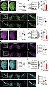 Figure 2
Expression of DAT, TH, and VMAT2 in midbrain and striatum of DAT-I312F/D421N+/+ mice. (A and B) Immunohistochemical visualization by wide-field imaging of DAT in coronal striatal (A) and midbrain sections (B) of WT and DAT-I312F/D421N+/+ (IF/DN) mice with quantification of mean fluorescence intensities (P > 0.05, ratio-paired t test. N = 6 WT:IF/DN pairs. Each WT:IF/DN pair was independently immunolabeled and imaged in parallel). (C) Quantification of striatal DAT protein levels by Western blotting of crude synaptosomal preparations from DAT-I312F/D421N+/+ mice relative to WT controls (P < 0.05, ratio-paired t test. N = 6 WT:IF/DN pairs. Each pair was processed and analyzed in parallel). (D and E) Immunohistochemical visualization by wide-field imaging of TH in coronal striatal (D) and midbrain sections (E) of WT and DAT-I312F/D421N+/+ mice with quantification of mean fluorescence intensities (P < 0.01 for striatum and P > 0.05 for midbrain, N = 6 WT:IF/DN pairs, ratio-paired t tests). (F) Quantification of striatal TH protein levels by Western blotting of crude synaptosomal preparations from DAT-I312F/D421N+/+ mice relative to WT controls (P < 0.0001, ratio-paired t test. N = 6 WT:IF/DN pairs). (G and H) Immunohistochemical visualization by wide-field imaging of VMAT2 in coronal striatal (G) and midbrain sections (H) of WT and DAT-I312F/D421N+/+ mice with quantification of mean fluorescence intensities (P > 0.05, ratio-paired t tests. N = 7 WT:IF/DN pairs for striatum. P > 0.05. N = 6 WT:IF/DN pairs for midbrain). (I) Quantification of striatal VMAT2 protein levels by Western blotting of crude synaptosomal preparations from DAT-I312F/D421N+/+ mice relative to WT controls (P < 0.01, ratio-paired t test. N = 6 WT:IF/DN pairs). Data are mean ± SEM. Scale bars: striatum overview: 100 μm; striatum zoom-in and midbrain images: 10 μm. *P < 0.05, **P < 0.01, ****P < 0.0001.
The parallel reduction in striatal protein levels of DAT, VMAT2, and TH in DAT-I312F/D421N+/+ mice found by Western blot analysis of whole striatal lysates led us to investigate if the DAT-I312F/D421N+/+ mice display structural changes in DA terminals, which may not be resolved in widefield fluorescence images. We therefore systematically acquired confocal images from striatal slices labeled for DAT and TH to visualize the DAergic arbor in more detail. As expected, the 2 DAergic markers showed almost complete overlap in both WT and DAT-I312F/D421N+/+ mice (Figure 3A). The higher resolution confocal images revealed an apparent reduction in the area density of DAergic terminals in images from DAT-I312F/D421N+/+ mice compared with WT mice. To directly quantify the area density of DAergic terminals, we applied a line-scan intensity analysis to count the terminals intersecting with a line grid in each image (28) (see Supplemental Methods). In agreement with the visual impression, image-level quantification showed reduced area density of both DAT- and TH-positive terminals in the DAT-I312F/D421N+/+ mice compared with WT mice (Figure 3, B and C). These data suggest that the parallel reduction in striatal levels of DAergic markers that we observed by Western blot analysis may be explained, at least in part, by axonal loss in DAT-I312F/D421N+/+ mice and, thus, that genetic injury to DAT can compromise the microscale integrity of DAergic axon terminals. To determine whether the observed deficit in fiber density arises during development, we examined the striatal terminal density in neonatal pups. Interestingly, across the sampled images, the area density of both TH- and DAT-positive striatal fibers was reduced in DAT-I312F/D421N+/+ pups (Supplemental Figure 3). As we noticed that these reductions were proportionally smaller than those observed in adult mutant mice, these data suggest that the reduced density of DAergic terminals reflects, at least in part, a developmental impairment in terminal formation or maintenance.
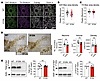 Figure 3
Reduced DAergic terminal density in DAT-I312F/D421N+/+ mice. (A) Representative confocal images of striatal DA projections immunolabeled for DAT and TH. (B and C) Quantification of area density of (B) DAT- and (C) TH-labeled fibers in DAT-I312F/D421N+/+ (IF/DN) mice compared with WT shown as SuperPlots (small points represent images; large points represent mouse means). Images were systematically sampled from striatal slices from 3 independent WT:IF/DN pairs, stained and imaged in parallel, and analyzed blinded to genotype. Image-level statistic shows significant reductions in both DAT and TH area density (P < 0.001 for DAT, N = 96 WT and 96 IF/DN; P < 0.01 for TH, unpaired t test, N = 95 WT and 90 IF/DN images). Mouse-level statistics: DAT P = 0.165; TH P = 0.37, N = 3, paired t test). (D) Stereological counting of TH-positive cells. Left, representative visualization of TH-positive cells by DAB staining. Right, quantification of neurons, volume, and density in the substantia nigra of WT and DAT-I312F/D421N+/+ mice (P > 0.05, unpaired t test. N = 9 WT and 8 IF/DN mice. See also Supplemental Tables 2 and 3). (E) Assessment of α-synuclein (α-Syn) levels in midbrain lysates from WT and DAT-I312F/D421N+/+ mice by Western blotting. Left, representative gel and quantification of total α-Syn soluble monomers (N = 8 WT:IF/DN pairs). Right, representative gel and quantification of phosphorylated α-Syn soluble monomers (pS129, N = 4 WT:IF/DN pairs) (P > 0.05, ratio-paired t test). Data are mean ± SEM.
Despite the deficit in axon fiber area density in striatal images, stereological counting of DAergic neurons in substantia nigra (SN) of the adult mice did not show any genotype differences in cell count, volume, or density (Figure 3D and Supplemental Tables 2 and 3).We also found comparable levels of both unphosphorylated and phosphorylated (p-S129) soluble α-synuclein monomers in midbrain lysates, indicating that the loss of striatal DA terminals in DAT-I312F/D421N+/+ mice is not associated with α-synuclein pathology or cell body degeneration (Figure 3E).
Changes in DA release and in DAergic axon terminal density in the striatum could lead to additional adaptations in the expression of DA D1 receptor (DR1) and DR2 in the striatum. We thus examined this using knockout-validated antibodies and qPCR (29, 30). Immunostaining of striatal slices showed decreased immunolabeling intensity of both DR1 and DR2 in DAT-I312F/D421N+/+ mice compared with WT mice (Figure 4, A and B). This reduction in striatal DR1 and DR2 protein levels was also observed upon Western blotting of lysates from DS (Figure 4, C and D). To determine if the changes in DR1 and DR2 protein levels involved transcriptional changes, we quantified mRNA levels in DS lysates by qPCR. Interestingly, we found a significant reduction in DR2 mRNA levels in DAT-I312F/D421N+/+ mice, whereas DR1 mRNA levels were comparable to WT mice, indicating that the reduction in DR2, but not DR1, protein levels involves transcriptional changes (Figure 4, E and F). As we did not find changes in DR2 mRNA levels in midbrain lysates, these changes in DS DR2 mRNA levels (Supplemental Figure 2A) presumably reflect postsynaptic adaptations.
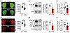 Figure 4
Synaptic adaptations in DAT-I312F/D421N+/+ mice. (A and B) Immunohistochemical visualization (representative images) of DA D1R (A) and D2R (B) by wide-field imaging on coronal striatal brain slices from pairs of WT and DAT-I312F/D421N+/+ mice (see refs. 29 and 30 for knockout validation of antibodies) with quantification of mean fluorescence intensity (P < 0.01, ratio-paired t test. N = 7 WT:IF/DN pairs that were independently labeled and imaged in parallel. (C and D) D1R and D2R Western blot analysis on DS homogenates from pairs of WT and DAT-I312F/D421N+/+ mice (left, representative blots; right, quantification; P < 0.05, ratio-paired t test. N = 4 WT:IF/DN pairs, each processed and analyzed in parallel). (E and F) D1R and D2R mRNA levels in DS lysates by quantitative real-time PCR shown as fold-change above the WT mean (2-ΔΔCt, P > 0.05 for D1R, P < 0.05 for D2R, N = 10 mice per genotype, multiple unpaired t test with Holm-Šidák correction). Data are mean ± SEM. Scale bar: 300 μm. *P < 0.05, **P < 0.01.
Taken together, our findings demonstrate that the genetic insult to DAT function causes a DA dysfunction that extends beyond DA reuptake and release to also affect DA synthesis, postsynaptic receptor levels, and the microscale integrity of DAergic axonal arborization.
DAT-I312F/D421N+/+ mice display persistent hyperactivity and increased explorative behavior. We next characterized the overall behavioral phenotype of the DAT-I312F/D421N+/+ mice. These analyses were performed on adult male and female mice during a 5-week period starting at age 15 ± 2 weeks. Of note, both male and female DAT-I312F/D421N+/+ mice were, by visual inspection, smaller than WT littermates. Compared with WT mice, the body weight was on average 4.6 g lower in DAT-I312F/D421N+/+ males (15.6%) and 3.6 g lower (15.2%) in females (Figure 5A).
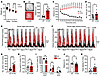 Figure 5
DAT-I312F/D421N+/+ mice display persistent hyperactivity and increased exploratory behavior. (A) Body weight for male and female WT and DAT-I312F/D421N+/+ (IF/DN) mice (P < 0.0001, 2-way ANOVA with Holm-Šidák’s correction, Nmales = 20 WT, 13 IF/DN. Nfemales = 14 WT, 20 IF/DN). (B) Locomotor activity during a 2-hour open-field test (OFT); left, mouse trajectory plots; right, total distance moved (P < 0.0001, Welch’s t test, N = 15 IF/DN and 14 WT). (C) Locomotor activity during the 2-hour OFT across 10-minute intervals (P < 0.001, multiple Welch’s t test with Holm-Šidák’s correction for multiple comparisons, N = 15 IF/DN and 14 WT). (D) Rearing during 10-minute cylinder test (P < 0.05, Welch’s t tests, N = 15 WT and 13 IF/DN mice). (E and F) Home cage activity recordings of total (E) and vertical (F) locomotor activity (dark phases in gray). Total activity in the home cage environment, quantified as AUC (P < 0.001, Welch’s t test, E: N = 7 WT and 7 IF/DN; F: N = 7 WT and 6 IF/DN mice). (G) Center zone entries during the 2-hour OFT (P < 0.0001, Welch’s t test, N = 14 WT and 15 IF/DN). (H) Time spent in center zone (%) during the 2-hour OFT (P > 0.05, Mann-Whitney U test, N = 13 WT and 15 IF/DN). (I) Elevated plus maze (EPM): % time spent in closed arms, center zone, and open arms for DAT-I312F/D421N+/+ versus WT mice (multiple Welch’s t test with Holm-Šidák’s correction, N = 7 WT and 10 IF/DN mice). (J) Distance traveled in the EPM (P > 0.05, Welch t test, N = 7 WT and 10 IF/DN mice). (K) Head-dipping in the EPM (P < 0.05, Welch’s t test, N = 7 WT and 10 IF/DN mice). All data are shown as mean ± SEM. *P < 0.05, **P < 0.01, ***P < 0.001, ****P < 0.0001.
Disturbances in DAergic neurotransmission have been widely implicated in ADHD, and the patient modeled by the DAT-I312F/D421N+/+ mice was diagnosed with ADHD in addition to atypical parkinsonism (23). A core symptom of ADHD is hyperactivity, and we therefore compared spontaneous locomotor activity of DAT-I312F/D421N+/+ mice with WT littermates in a 2-hour, novel open-field test. On average, the DAT-I312F/D421N+/+ mice traveled more than 3 times the distance of their WT littermates (Figure 5B). This hyperlocomotive behavior of DAT-I312F/D421N+/+ mice was evident throughout the entire 2-hour test period, but became more pronounced over time, as the WT mice habituated to the novel environment during 2 hours of recording while the DAT-I312F/D421N+/+ mice showed persistently high locomotor activity (Figure 5C and Supplemental Videos 1–4). We compared the rearing behavior of WT and DAT-I312F/D421N+/+ mice in a novel environment by counting the rears after placing the mice in a glass cylinder for 10 minutes. The DAT-I312F/D421N+/+ mice displayed nearly twice as many rears as the WT mice (Figure 5D), collectively demonstrating that the DAT-I312F/D421N+/+ mice have both increased horizontal and vertical activity in novel environments.
To assess if the observed hyperactivity was novelty dependent, we performed extended activity recordings of DAT-I312F/D421N+/+ and WT mice in activity cages. During 8.5 days of monitoring, the DAT-I312F/D421N+/+ mice displayed persistently higher activity than WT mice both in total activity (Figure 5E) and in vertical activity (Figure 5F), showing that the hyperlocomotive phenotype of DAT-I312F/D421N+/+ mice is present even in the absence of novelty.
We next examined the effect of genotype on explorative versus anxiety-related phenotypes by analyzing the number of center zone entries and time spent in the center zone in the open-field test. The DAT-I312F/D421N+/+ mice showed a 2.6-fold increase in the number of center zone entries (Figure 5G), but no difference in percentage time spent in center zone (Figure 5H), indicating no anxiety-related center zone aversion. We further tested the mice for their propensity to explore versus their tendency to seek protected areas in an elevated plus maze (EPM) paradigm. We found that the DAT-I312F/D421N+/+ mice spent more time in the center zone and open arms than WT mice (Figure 5I). Of notice, this increase was not caused by an overall increase in locomotion, as the distance traveled by DAT-I312F/D421N+/+ mice during the test was not increased compared with WT mice (Figure 5J). Consistent with an increased explorative drive, the DAT-I312F/D421N+/+ mice also spent almost 3-fold more time engaged in head dipping over the sides of the open arms (Figure 5K), which is an additional behavioral correlate of explorative activity (31).
In summary, the DA imbalance imposed by the disease-associated mutations in DAT drives an ADHD-relevant phenotype characterized by hyperactivity and an enhanced exploratory drive.
DAT-I312F/D421N+/+ mice show increased hind limb clasping and kyphosis. Transgenic mouse models expressing genetic variants associated with hereditable forms of PD display notoriously few, if any, motor deficits (32–35). To assess the motor function of DAT-I312F/D421N+/+ mice, we performed a battery of motor tests comprising rotarod, pole test, hanging wire test, and hind limb clasping during tail suspension. Most prominently, we observed a clear motor abnormality in the DAT-I312F/D421N+/+ mice when assessing hind limb clasping. While DAT-WT mice showed practically no clasping behavior when lifted by their tail for 30 seconds, the DAT-I312F/D421N+/+ mice displayed clear, but varying, degrees of clasping, seen as partial or full retraction of hind limbs toward the midline in a dystonic fashion (Figure 6A and Supplemental Videos 5 and 6). This clasping phenotype of DAT-I312F/D421N+/+ mice was not accompanied by reduced motor performance on an accelerating rotarod, in the pole test, or in the hanging wire test (Figure 6, B–D), suggesting that DAT-I312F/D421N+/+ mice can acquire and execute motor tasks to a similar level of performance as WT mice. Nonetheless, in addition to receiving higher clasping scores, the DAT-I312F/D421N+/+ mice also received higher scores when visually inspected for dorsal kyphosis (Figure 6E), a phenotype that has been linked to premature aging and movement disorders (36–38).
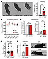 Figure 6
DAT-I312F/D421N+/+ mice exhibit clasping behavior and kyphosis. (A) Representative images of hind limb clasping phenotypes in DAT-I312F/D421N+/+ mice during tail suspension with quantification of clasping score (see Methods; P < 0.0001, Mann-Whitney U test. N = 14 WT and 12 IF/DN mice). (B) Accelerating rotarod test (P > 0.05 for all test days by 2-way ANOVA with Holm-Šidák multiple comparisons test. N = 13 WT and 12 IF/DN mice). (C) Hanging wire test with escape strategy scores, left (P > 0.05, Mann-Whitney U test. N = 12 WT and 11 IF/DN mice); and escape time to platform, right (P > 0.05, Mann-Whitney U test. N = 12 WT and 10 IF/DN mice). (D) Pole test descent time (P > 0.05, Mann-Whitney U test. N = 11 WT and 11 IF/DN mice). (E) DAT-I312F/D421N+/+ received higher scores in visual inspection for dorsal kyphosis than WT mice (P < 0.01, Mann-Whitney U test. N = 22 WT and 22 IF/DN mice). All data are shown as mean ± SEM. **P < 0.01, ****P < 0.0001.
Hyperactivity in DAT-I312F/D421N+/+ mice is ameliorated by amphetamine and orphenadrine. Hyperactivity is a core symptom of ADHD and recapitulated in the DAT-I312F/D421N+/+ mouse. Since amphetamine (AMPH) is frequently used to treat ADHD in humans, we next evaluated the effect of AMPH on spontaneous locomotion in an open-field arena. Following a 1-hour habituation period, DAT-I312F/D421N+/+ mice and WT littermates were first treated with either a single dose of AMPH (2 mg/kg) or a saline injection, after which activity was recorded for an additional 1 hour. For AMPH-treated WT mice, we observed, as expected, a significant AMPH-induced increase in the distance traveled compared with saline controls (Figure 7, A and B). In striking contrast, the hyperactive behavior of DAT-I312F/D421N+/+ mice was ameliorated by AMPH treatment, showing a reduction in traveled distance (Figure 7, A and B).
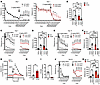 Figure 7
AMPH and orphenadrine alleviate hyperactivity in DAT-I312F/D421N+/+ mice. (A) Open-field locomotion before and after AMPH (2 mg/kg, i.p.) in WT and DAT-I312F/D421N+/+ (IF/DN) mice (RM 2-way ANOVA with Holm-Šidák correction). (B) Total distance traveled after saline or AMPH (2 mg/kg) injections (2-way ANOVA with Holm-Šidák correction, N = 9 WTsaline, 8 WTAMPH, 10 IF/DNsaline, and 10 IF/DNAMPH). (C–F) Reexposure to saline or 5 mg/kg AMPH (C and D) and saline or 10 mg/kg AMPH (E and F) after 7 days’ washout. Locomotion is shown in 10-minute bins (C and E; RM 2-way ANOVA with Holm-Šidák correction) and as total distance over 1 hour after injections (D and F, 2-way ANOVA with Holm-Šidák correction, N = 9 WTsaline, 10 IF/DNsaline, 8 WT5 mg/kg, 10 IF/DN5 mg/kg, 8 WT10 mg/kg, 7 IF/DN10 mg/kg). (G) Slow-flow microdialysis of extracellular striatal DA in anesthetized WT and DAT-I312F/D421N+/+ mice before and after AMPH (2 mg/kg, i.p.). Data are expressed as percentage of WT mean baseline (mixed-effect model with Holm-Šidák correction, N = 7 WT and 6 IF/DN mice). (H) Relative baseline DA levels in percentage of WT mean (P < 0.01, unpaired t test, N = 7 WT and 6 IF/DN mice). (I) Peak AMPH-induced DA levels in WT and DAT-I312F/D421N+/+ mice (P < 0.001, unpaired t test, N = 7 WT and 6 IF/DN mice). (J) Open-field locomotion in 10-minute time bins after ORPH (30 mg/kg, i.p.) administration to WT and DAT-I312F/D421N+/+ mice (RM 2-way ANOVA with Holm-Šidák correction). (K) Total distance traveled after saline or ORPH (30 mg/kg) (2-way ANOVA with Holm-Šidák correction). N for J and K = 9 WTsaline, 8 WTORPH, 10 IF/DNsaline, 9 IF/DNORPH. All data are shown as mean ± SEM. *P < 0.05, **P < 0.01, ***P < 0.001, ****P < 0.0001.
We previously showed that both the DAT-D421N and the DAT-I312F mutations alter DAT’s substrate and inhibitor binding properties (23, 39). To exclude the possibility that higher doses of AMPH were required to elicit AMPH-induced hyperlocomotion in DAT-I312F/D421N+/+ mice, we reexposed the mice to 5 mg/kg AMPH after a 1-week washout period and again to 10 mg/kg AMPH after a second 1-week washout period. Whereas WT littermates showed even more robust increases in locomotion in response to 5 mg/kg and 10 mg/kg AMPH (Figure 7, C–F), the DAT-I312F/D421N+/+ mice still demonstrated a pronounced decrease in activity at both 5 mg/kg and 10 mg/kg AMPH (Figure 7, C–F), further consolidating the attenuating effect of AMPH on the hyperactive DAT-I312F/D421N+/+ phenotype.
To gain further insights into the effect of AMPH on DAT-I312F/D421N+/+ mice, we carried out slow-flow microdialysis experiments on anesthetized mice to compare baseline DA levels and AMPH-induced DA release in the DS. Baseline DA levels were measured over a 40-minute time window before i.p. administration of 2 mg/kg AMPH (Figure 7G). Importantly, baseline DA measurements revealed that extracellular DA levels in DAT-I312F/D421N+/+ mice were 2.8- ± 0.4-fold higher than in WT mice, demonstrating that the DAT-I312F/D421N+/+ genotype causes a chronic increase in extracellular DAergic tone (Figure 7H). Administration of AMPH (2 mg/kg) elicited, as expected, a marked increase in extracellular DA in WT mice (Figure 7, G and I). More surprisingly, AMPH also evoked an increase in extracellular DA levels in DAT-I312F/D421N+/+ mice (Figure 7, G and I). However, peak DA levels increased 17- ± 2.8-fold above baseline in WT mice whereas only a 1.6- ± 0.13-fold increase above baseline was recorded in DAT-I312F/D421N+/+ mice (Figure 7I). Still, these findings suggest that the seemingly calming effect of AMPH on DAT-I312F/D421N+/+ mice is not established by a paradoxical decrease in extracellular DA levels leading to a reduced exploratory drive. Rather, the AMPH-treated DAT-I312F/D421N+/+ mice are less active despite increased extracellular DA levels.
The index patient with atypical DTDS was treated with the anticholinergic drug, ORPH, to alleviate parkinsonian symptoms. To uncover potential disease-relevant alterations in cholinergic responses (40, 41), the effect of ORPH was evaluated in the open-field test comparing WT and DAT-I312F/D421N+/+ mice. Interestingly, analysis of locomotion in 10-minute bins revealed an immediate, but short-lasting (10 minutes), increase in locomotion in ORPH-treated (30 mg/kg) WT mice relative to saline-treated WT mice (Figure 7J). This initial hyperlocomotive response to ORPH was also seen, though less pronounced, in DAT-I312F/D421N+/+ mice. However, while ORPH-treated WT mice rapidly resumed activity levels similar to saline-treated mice, DAT-I312F/D421N+/+ mice treated with ORPH showed a significant reduction in locomotor activity compared with saline control mice, an effect that persisted throughout the remaining test period (Figure 7J). Thus, during 1-hour recordings, the total distance traveled by ORPH-treated WT mice did not differ significantly from saline-treated mice (Figure 7K), while ORPH alleviated the hyperlocomotive phenotype of DAT-I312F/D421N+/+ mice (Figure 7K).
These data further support the predictive validity of the DAT-I312F/D421N+/+ mice to identify and validate potential new treatments while pointing to a potentially important cholinergic impairment that likely arises from the primary DA-related dysfunction.
DAT-I312F/D421N+/+ mice show loss of fast DA dynamics and region-specific changes in amphetamine responses. Recent work has substantiated that temporally precise DA fluctuations are critical for encoding information and shaping striatal output (42–44). We therefore turned to fiber photometry with the genetically encoded DA sensor dLight1.3b (45) to examine how the patient-derived DAT mutations affect the temporal structure and regional coordination of DA signaling and to interrogate the paradoxical response of DAT-I312F/D421N+/+ mice to AMPH in further detail. To this end, dLight1.3b was virally expressed in the right DS and left VS of WT and DAT-I312F/D421N+/+ mice, and fluorescence signals were recorded more than 3 weeks later during open-field exploration, followed by vehicle or AMPH injections. The DA signal in WT mice during baseline (self-paced exploratory) activity confirmed previous findings, with continuous rapid DA fluctuations in the DS and slower fluctuations in the VS, yet with distinct DA peaks (transients) (42, 46) (Figure 8A). In DAT-I312F/D421N+/+ mice, however, the fast DA fluctuations were essentially absent in both DS and VS (Figure 8B). In the DS, the characteristic swift DA peaks were replaced by long, irregular waves overlaid with smaller oscillations, while in the VS, DA signals showed even more prominent, enduring slow waves without the sharp peaks observed WT mice (Figure 8B). Spectral energy density plots across the entire recording period of vehicle-injected mice confirmed, for both DS and VS, a clear reduction in high-frequency DA fluctuations, coupled with the presence of slower, longer lasting DA signals in DAT-I312F/D421N+/+ mice (Figure 8C). These results reveal the detrimental effect of the patient-derived mutations on maintaining temporally precise DA signaling.
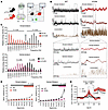 Figure 8
Temporally disrupted and low-complexity striatal DA dynamics in DAT-I312F/D421N+/+ mice. (A) Fiber photometry with dLight1.3b in dorsal striatum (DS) and ventral striatum (VS) of WT and DAT-I312F/D421N+/+ mice during 1-hour, self-paced open-field activity. (B) Representative DS and VS DA traces. WT mice show rapid DA fluctuations in DS and transient peaks in VS, whereas DAT-I312F/D421N+/+ mice show longer, irregular waves of DA without rapid transients. (C) Spectral energy density plots demonstrating decreased high-frequency DA activity and increased low-frequency components in both DS and VS of DAT-I312F/D421N+/+ mice (multiple unpaired t test with Holm-Šidák correction. N = 9 WT and 6 DS or 8 VS IF/DN mice). (D) Variance of the first derivative of DA signals across deciles of signal intensity (normalized to the lowest decile) indicates reduced structured, nonrandom DA signals in DAT-I312F/D421N+/+ mice (RM2-way ANOVA, followed by within-genotype simple-effect comparisons of deciles relative to baseline, with Benjamini-Hochberg correction. N = 9 WT and 6 DS or 8 VS IF/DN mice). (E) DS–VS cross-correlation analysis shows a sharp asymmetric peak at approximately 0.35 second lag in WT mice (DS leading), consistent with temporally structured coordination. DAT-I312F/D421N+/+ mice exhibit a broader and less defined correlation (N = 8 WT and 5 IF/DN), indicating disrupted temporal coupling. Data in C–E are shown as mean ± SEM.
To evaluate how the changes in DA signals in DAT-I312F/D421N+/+ mice affect the potential informational content of the DA fluctuations, we examined the degree to which the signals differed from random variations arising from a homogeneous Poisson process. We previously showed that the DA signals in both DS and VS of WT mice are not derived from random variations but are heterogeneous, supporting that the signals may encode behaviorally relevant information (42). We demonstrated this by computing the variance of the first derivative of the DA signal intensity, as this should be constant across signal intensities (binned into deciles) in a homogeneous process (42). In agreement with these previous data, and consistent with a heterogeneous release pattern, the variance of the first derivative in WT mice was significantly larger for all deciles compared with the bottom 10th percentile in DS and for the upper half of the signal in VS (Figure 8D). In contrast, DA signals in DAT-I312F/D421N+/+ mice were homogeneous across a wider range of signal intensities than in WT mice and showed reduced variability even in the bins with significant heterogeneous variation. This supports that the DAT mutations impair the dynamic complexity of striatal DA release patterns, that, in turn, may limit its informational value. We also performed a cross-correlation analysis to examine the temporal coordination of DA signals between DS and VS. In agreement with our earlier observations in WT mice (42), we observed a sharp, asymmetric peak with the DS signal preceding the VS DA signal by about 0.35 seconds, supporting a highly temporally structured and directional relationship between these regions. DAT-I312F/D421N+/+ mice, however, exhibited a markedly different temporal coordination, evident as a prolonged and less defined correlation pattern (although a peak still was seen at ~0.35 seconds). Thus, the precise temporal coupling between DS and VS DA release events appeared lost and replaced by more diffuse waves of DA that unfolded over longer times.
Upon AMPH administration, we observed an expected increased locomotor activity in the WT mice, which was accompanied by a large increase in striatal DA (Figure 9, A–E). Notably, the magnitude of the DA response was substantially greater in the VS than in the DS (~35 times SD of baseline in VS versus ~10 times SD of baseline in DS; Figure 9). In the DAT-I312F/D421N+/+ mice, locomotor activity was again markedly reduced by AMPH. Strikingly, this hypolocomotion was not accompanied by a significant change in the overall AMPH-induced DA response in DS compared with WT mice (P = 0.065, quantified by the AUC; Figure 9, B and C). In VS, however, AMPH-evoked DA elevation was profoundly impaired (Figure 9, D and E). Finally, we evaluated AMPH’s effect on fast DA dynamics. Visual inspection of representative 30-second traces before and after AMPH administration revealed a marked blunting of rapid DA dynamics in DS of WT mice with reduced signal amplitude (Figure 9, F and G), whereas signal amplitudes in the VS were increased (Figure 9, H and I). These observations were supported by isolating the fast (0.01–10 Hz) frequency components of the DA signals across the entire 90-minute recording periods and quantifying mean amplitudes before and after AMPH administration (Figure 9, G and I). In contrast, DAT-I312F/D421N+/+ mice showed no apparent change in VS dynamics and only a modest, yet significant, effect in DS (Figure 9, F–I).
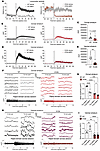 Figure 9
Region-specific disruption of AMPH-evoked DA release and fast DA dynamics in DAT-I312F/D421N+/+ mice. (A) Locomotor activity in 1-second time bins for WT and DAT-I312F/D421N+/+ mice during in vivo recording of DA signals before and after AMPH (2 mg/kg, i.p.) administration. (B–E) Average saline- and AMPH-induced DA responses in WT (left) and IF/DN (right) mice measured in DS (B and C) and VS (D and E). Area under the curve (AUC) following AMPH administration shows no significant genotype difference in DS (C, P = 0.065, unpaired t test. NAMPH = 6 per genotype) but a marked reduction in AMPH-evoked DA release in VS of IF/DN mice (E, P < 0.01, unpaired t test. NAMPH = 6 WT and 6 IF/DN mice). (F–I) Analysis of AMPH’s impact on fast DA dynamics. F and H show representative 30-second DA traces from WT and IF/DN mice in DS and VS, respectively, alongside representative examples of the rapid signal component across an entire recording session (band-pass filtering for rapid fluctuations: 0.01–10 Hz). G and I show the quantification of mean signal amplitude of the rapid DA signal component before and after AMPH injection. In WT mice, AMPH reduces rapid signal amplitude in DS (F and G) but increases the mean amplitude in VS (H and I), whereas in IF/DN mice AMPH produces only a modest increase in DS and no significant change in VS (2-way ANOVA with Holm-Šidák correction. N = 6 WT and 6 DS or 7 VS IF/DN mice). Data are shown as mean ± SEM. *P < 0.05, **P < 0.01, ***P < 0.001, ****P < 0.0001.
Collectively, these in vivo recordings of AMPH-induced DA responses reveal a striking alteration in the regional specificity of AMPH-induced DA signaling in DAT-I312F/D421N+/+ mice, suggesting a fundamental shift in striatal DA regulation, which may underlie the opposite behavioral responses to AMPH observed in this model.


Copyright © 2026 American Society for Clinical Investigation
ISSN: 0021-9738 (print), 1558-8238 (online)