Go to JCI Insight
  • About
  • Editors
  • Consulting Editors
  • For authors
  • Alerts
  • Advertising/recruitment
  • Subscribe
  • Contact
  • Current Issue
  • Past Issues
  • By specialty
    • COVID-19
    • Cardiology
    • Gastroenterology
    • Immunology
    • Metabolism
    • Nephrology
    • Neuroscience
    • Oncology
    • Pulmonology
    • Vascular biology
    • All ...
  • Videos
    • Conversations with Giants in Medicine
    • Author's Takes
  • Reviews
    • View all reviews ...
    • 100th Anniversary of Insulin's Discovery (Jan 2021)
    • Hypoxia-inducible factors in disease pathophysiology and therapeutics (Oct 2020)
    • Latency in Infectious Disease (Jul 2020)
    • Immunotherapy in Hematological Cancers (Apr 2020)
    • Big Data's Future in Medicine (Feb 2020)
    • Mechanisms Underlying the Metabolic Syndrome (Oct 2019)
    • Reparative Immunology (Jul 2019)
    • View all review series ...
  • Viewpoint
  • Collections
    • Recently published
    • In-Press Preview
    • Commentaries
    • Concise Communication
    • Editorials
    • Viewpoint
    • Top read articles
  • Clinical Medicine
  • JCI This Month
    • Current issue
    • Past issues

  • Current issue
  • Past issues
  • Specialties
  • Reviews
  • Review series
  • Conversations with Giants in Medicine
  • Author's Takes
  • Recently published
  • In-Press Preview
  • Commentaries
  • Concise Communication
  • Editorials
  • Viewpoint
  • Top read articles
  • About
  • Editors
  • Consulting Editors
  • For authors
  • Alerts
  • Advertising/recruitment
  • Subscribe
  • Contact
The archaeal Dps nanocage targets kidney proximal tubules via glomerular filtration
Masaki Uchida, … , Trevor Douglas, Takashi Hato
Published August 19, 2019
Citation Information: J Clin Invest. 2019;129(9):3941-3951. https://doi.org/10.1172/JCI127511.
View: Text | PDF
Research Article Nephrology

The archaeal Dps nanocage targets kidney proximal tubules via glomerular filtration

  • Text
  • PDF
Abstract

Nature exploits cage-like proteins for a variety of biological purposes, from molecular packaging and cargo delivery to catalysis. These cage-like proteins are of immense importance in nanomedicine due to their propensity to self-assemble from simple identical building blocks to highly ordered architecture and the design flexibility afforded by protein engineering. However, delivery of protein nanocages to the renal tubules remains a major challenge because of the glomerular filtration barrier, which effectively excludes conventional size nanocages. Here, we show that DNA-binding protein from starved cells (Dps) — the extremely small archaeal antioxidant nanocage — is able to cross the glomerular filtration barrier and is endocytosed by the renal proximal tubules. Using a model of endotoxemia, we present an example of the way in which proximal tubule–selective Dps nanocages can limit the degree of endotoxin-induced kidney injury. This was accomplished by amplifying the endogenous antioxidant property of Dps with addition of a dinuclear manganese cluster. Dps is the first-in-class protein cage nanoparticle that can be targeted to renal proximal tubules through glomerular filtration. In addition to its therapeutic potential, chemical and genetic engineering of Dps will offer a nanoplatform to advance our understanding of the physiology and pathophysiology of glomerular filtration and tubular endocytosis.

Authors

Masaki Uchida, Bernhard Maier, Hitesh Kumar Waghwani, Ekaterina Selivanovitch, S. Louise Pay, John Avera, EJun Yun, Ruben M. Sandoval, Bruce A. Molitoris, Amy Zollman, Trevor Douglas, Takashi Hato

×

Figure 1

Dps is filtered through glomeruli and endocytosed by proximal tubules.

Options: View larger image (or click on image) Download as PowerPoint
Dps is filtered through glomeruli and endocytosed by proximal tubules.
(...
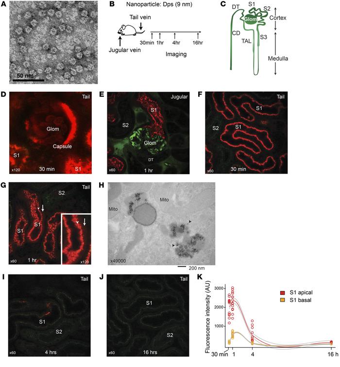
(A) Transmission electron microscopy image of S. solfataricus Dps. (B) Overview of intravital imaging protocol. (C) Schematic of renal tubule structure. Glom, glomerulus; S1, S2, and S3, proximal tubule subsegments; TAL, thick ascending loop of Henle; DT, distal tubule; CD, collecting duct. (D) Representative image of glomerulus and S1 proximal tubules 30 minutes after Dps i.v. (Texas red; n = 3 independent experiments). Mice were injected with Dps via tail vein, and the distribution of Dps was determined with intravital 2-photon microscopy. Note that male mice have extension of S1 segment into the Bowman’s capsule in the glomerulus. (E) Dps (red) and 70 kDa dextran (green) were injected via jugular vein. In this experiment, the mouse kidney was freshly dissected 60 minutes after Dps injection and imaged ex vivo in order to determine the distribution of Dps in the deep cortex that is beyond the reach of intravital 2-photon microscopy. (F and G) Intravital time-course imaging under indicated time points. Arrowheads point to the endosome/lysosome layer (S1 apical). Arrows point to cytoplasmic Dps signal beyond the endosome/lysosome layer (S1 basal). (H) Electron microscopy image of S1 proximal tubule 60 minutes after Dps i.v. Arrow points to Dps in the cytoplasm. (I and J) Intravital imaging of the kidney under indicated time points. (K) Quantification of Dps fluorescence intensity for the indicated time points and subcellular regions. Locally weighted regression curve fitting was applied for generating the trajectories and error lines (gray).
Follow JCI:
Copyright © 2021 American Society for Clinical Investigation
ISSN: 0021-9738 (print), 1558-8238 (online)

Sign up for email alerts