Go to JCI Insight
  • About
  • Editors
  • Consulting Editors
  • For authors
  • Publication ethics
  • Publication alerts by email
  • Advertising
  • Job board
  • Contact
  • Clinical Research and Public Health
  • Current issue
  • Past issues
  • By specialty
    • COVID-19
    • Cardiology
    • Gastroenterology
    • Immunology
    • Metabolism
    • Nephrology
    • Neuroscience
    • Oncology
    • Pulmonology
    • Vascular biology
    • All ...
  • Videos
    • Conversations with Giants in Medicine
    • Video Abstracts
  • Reviews
    • View all reviews ...
    • Complement Biology and Therapeutics (May 2025)
    • Evolving insights into MASLD and MASH pathogenesis and treatment (Apr 2025)
    • Microbiome in Health and Disease (Feb 2025)
    • Substance Use Disorders (Oct 2024)
    • Clonal Hematopoiesis (Oct 2024)
    • Sex Differences in Medicine (Sep 2024)
    • Vascular Malformations (Apr 2024)
    • View all review series ...
  • Viewpoint
  • Collections
    • In-Press Preview
    • Clinical Research and Public Health
    • Research Letters
    • Letters to the Editor
    • Editorials
    • Commentaries
    • Editor's notes
    • Reviews
    • Viewpoints
    • 100th anniversary
    • Top read articles

  • Current issue
  • Past issues
  • Specialties
  • Reviews
  • Review series
  • Conversations with Giants in Medicine
  • Video Abstracts
  • In-Press Preview
  • Clinical Research and Public Health
  • Research Letters
  • Letters to the Editor
  • Editorials
  • Commentaries
  • Editor's notes
  • Reviews
  • Viewpoints
  • 100th anniversary
  • Top read articles
  • About
  • Editors
  • Consulting Editors
  • For authors
  • Publication ethics
  • Publication alerts by email
  • Advertising
  • Job board
  • Contact
Soluble epoxide hydrolase promotes astrocyte survival in retinopathy of prematurity
Jiong Hu, … , Rüdiger Popp, Ingrid Fleming
Published September 3, 2019
Citation Information: J Clin Invest. 2019;129(12):5204-5218. https://doi.org/10.1172/JCI123835.
View: Text | PDF
Research Article Angiogenesis Ophthalmology

Soluble epoxide hydrolase promotes astrocyte survival in retinopathy of prematurity

  • Text
  • PDF
Abstract

Polyunsaturated fatty acids such as docosahexaenoic acid (DHA) positively affect the outcome of retinopathy of prematurity (ROP). Given that DHA metabolism by cytochrome P450 and soluble epoxide hydrolase (sEH) enzymes affects retinal angiogenesis and vascular stability, we investigated the role of sEH in a mouse model of ROP. In WT mice, hyperoxia elicited tyrosine nitration and inhibition of sEH and decreased generation of the DHA-derived diol 19,20-dihydroxydocosapentaenoic acid (19,20-DHDP). Correspondingly, in a murine model of ROP, sEH–/– mice developed a larger central avascular zone and peripheral pathological vascular tuft formation than did their WT littermates. Astrocytes were the cells most affected by sEH deletion, and hyperoxia increased astrocyte apoptosis. In rescue experiments, 19,20-DHDP prevented astrocyte loss by targeting the mitochondrial membrane to prevent the hyperoxia-induced dissociation of presenilin-1 and presenilin-1–associated protein to attenuate poly ADP-ribose polymerase activation and mitochondrial DNA damage. Therapeutic intravitreal administration of 19,20-DHDP not only suppressed astrocyte loss, but also reduced pathological vascular tuft formation in sEH–/– mice. Our data indicate that sEH activity is required for mitochondrial integrity and retinal astrocyte survival in ROP. Moreover, 19,20-DHDP may be more effective than DHA as a nutritional supplement for preventing retinopathy in preterm infants.

Authors

Jiong Hu, Sofia-Iris Bibli, Janina Wittig, Sven Zukunft, Jihong Lin, Hans-Peter Hammes, Rüdiger Popp, Ingrid Fleming

×

Figure 6

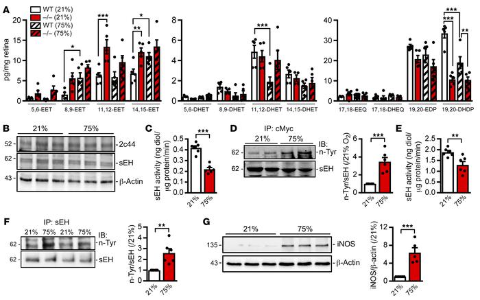
Consequences of hyperoxia on sEH activity.

Options: View larger image (or click on image) Download as PowerPoint
Consequences of hyperoxia on sEH activity.
(A) Fatty acid epoxide and di...
(A) Fatty acid epoxide and diol levels in retinas from WT and sEH–/– mice exposed to normoxia (21% O2) or hyperoxia (75% O2) for 24 hours starting on P7. n = 5 samples per group and each sample represents a pool of 6 retinas (2-way ANOVA with Tukey’s multiple comparisons test). 17,18-EEQ, 17,18-epoxy eicosatetraenoic acid; 17,18-DHEQ, 17,18-dihydroxy-eicosatetraenoic acid. (B) Immunoblot showing sEH and Cyp2c44 expression in retinas from WT mice after exposure to normoxia or hyperoxia from P7 for 24 hours. Comparable results were observed in 3 additional mice per group. (C) sEH activity in retinas from WT mice after exposure to normoxia or hyperoxia from P7 for 24 hours. n = 6 samples/group (Student’s t test). (D) Tyrosine nitration of sEH in sEH-cMyc expressing-HEK-293 cells exposed to 21% O2 or 75% O2 for 24 hours. n = 6 independent experiments (Student’s t test). (E) sEH activity in sEH expressing HEK-293 cells after exposure to 21% O2 or 75% O2 for 24 hours. n = 6 independent experiments (Student’s t test). (F) Tyrosine nitration of sEH immunoprecipitated from retinas of WT mice after exposure to normoxia or hyperoxia for 24 hours. n = 6 animals/group (Student’s t test). (G) iNOS expression in retinas from WT mice after exposure to normoxia or hyperoxia for 24 hours. n = 6 animals/group (Student’s t test). *P < 0.05, **P < 0.01, and ***P < 0.001. IB, immunoblot.

Copyright © 2025 American Society for Clinical Investigation
ISSN: 0021-9738 (print), 1558-8238 (online)

Sign up for email alerts