Technical Advance
Citation Information: J Clin Invest. 2011. https://doi.org/10.1172/JCI57235.
Abstract
Mycobacterium tuberculosis causes widespread, persistent infection, often residing in macrophages that neither sterilize the bacilli nor allow them to cause disease. How macrophages restrict growth of pathogens is one of many aspects of human phagocyte biology whose study relies largely on macrophages differentiated from monocytes in vitro. However, such cells fail to recapitulate the phenotype of tissue macrophages in key respects, including that they support early, extensive replication of M. tuberculosis and die in several days. Here we found that human macrophages could survive infection, kill Mycobacterium bovis BCG, and severely limit the replication of M. tuberculosis for several weeks if differentiated in 40% human plasma under 5%–10% (physiologic) oxygen in the presence of GM-CSF and/or TNF-α followed by IFN-γ. Control was lost with fetal bovine serum, 20% oxygen, M-CSF, higher concentrations of cytokines, or premature exposure to IFN-γ. We believe that the new culture method will enable inquiries into the antimicrobial mechanisms of human macrophages.
Authors
Guillaume Vogt, Carl Nathan
Citation Information: J Clin Invest. 2011. https://doi.org/10.1172/JCI58447.
A genetically engineered human pancreatic β cell line exhibiting glucose-inducible insulin secretion
Abstract
Despite intense efforts over the past 30 years, human pancreatic β cell lines have not been available. Here, we describe a robust technology for producing a functional human β cell line using targeted oncogenesis in human fetal tissue. Human fetal pancreatic buds were transduced with a lentiviral vector that expressed SV40LT under the control of the insulin promoter. The transduced buds were then grafted into SCID mice so that they could develop into mature pancreatic tissue. Upon differentiation, the newly formed SV40LT-expressing β cells proliferated and formed insulinomas. The resulting β cells were then transduced with human telomerase reverse transcriptase (hTERT), grafted into other SCID mice, and finally expanded in vitro to generate cell lines. One of these cell lines, EndoC-βH1, expressed many β cell–specific markers without any substantial expression of markers of other pancreatic cell types. The cells secreted insulin when stimulated by glucose or other insulin secretagogues, and cell transplantation reversed chemically induced diabetes in mice. These cells represent a unique tool for large-scale drug discovery and provide a preclinical model for cell replacement therapy in diabetes. This technology could be generalized to generate other human cell lines when the cell type–specific promoter is available.
Authors
Philippe Ravassard, Yasmine Hazhouz, Séverine Pechberty, Emilie Bricout-Neveu, Mathieu Armanet, Paul Czernichow, Raphael Scharfmann
Citation Information: J Clin Invest. 2011. https://doi.org/10.1172/JCI45557.
Abstract
The drug development process for CNS indications is hampered by a paucity of preclinical tests that accurately predict drug efficacy in humans. Here, we show that a wide variety of CNS-active drugs induce characteristic alterations in visual stimulus–induced and/or spontaneous eye movements in mice. Active compounds included sedatives and antipsychotic, antidepressant, and antiseizure drugs as well as drugs of abuse, such as cocaine, morphine, and phencyclidine. The use of quantitative eye-movement analysis was demonstrated by comparing it with the commonly used rotarod test of motor coordination and by using eye movements to monitor pharmacokinetics, blood-brain barrier penetration, drug-receptor interactions, heavy metal toxicity, pharmacologic treatment in a model of schizophrenia, and degenerative CNS disease. We conclude that eye-movement analysis could complement existing animal tests to improve preclinical drug development.
Authors
Hugh Cahill, Amir Rattner, Jeremy Nathans
Citation Information: J Clin Invest. 2011. https://doi.org/10.1172/JCI45600.
Abstract
Nanoparticle-based materials, such as drug delivery vehicles and diagnostic probes, currently under evaluation in oncology clinical trials are largely not tumor selective. To be clinically successful, the next generation of nanoparticle agents should be tumor selective, nontoxic, and exhibit favorable targeting and clearance profiles. Developing probes meeting these criteria is challenging, requiring comprehensive in vivo evaluations. Here, we describe our full characterization of an approximately 7-nm diameter multimodal silica nanoparticle, exhibiting what we believe to be a unique combination of structural, optical, and biological properties. This ultrasmall cancer-selective silica particle was recently approved for a first-in-human clinical trial. Optimized for efficient renal clearance, it concurrently achieved specific tumor targeting. Dye-encapsulating particles, surface functionalized with cyclic arginine–glycine–aspartic acid peptide ligands and radioiodine, exhibited high-affinity/avidity binding, favorable tumor-to-blood residence time ratios, and enhanced tumor-selective accumulation in αvβ3 integrin–expressing melanoma xenografts in mice. Further, the sensitive, real-time detection and imaging of lymphatic drainage patterns, particle clearance rates, nodal metastases, and differential tumor burden in a large-animal model of melanoma highlighted the distinct potential advantage of this multimodal platform for staging metastatic disease in the clinical setting.
Authors
Miriam Benezra, Oula Penate-Medina, Pat B. Zanzonico, David Schaer, Hooisweng Ow, Andrew Burns, Elisa DeStanchina, Valerie Longo, Erik Herz, Srikant Iyer, Jedd Wolchok, Steven M. Larson, Ulrich Wiesner, Michelle S. Bradbury
Citation Information: J Clin Invest. 2011. https://doi.org/10.1172/JCI57377.
Abstract
Leber congenital amaurosis (LCA) is a rare degenerative eye disease, linked to mutations in at least 14 genes. A recent gene therapy trial in patients with LCA2, who have mutations in RPE65, demonstrated that subretinal injection of an adeno-associated virus (AAV) carrying the normal cDNA of that gene (AAV2-hRPE65v2) could markedly improve vision. However, it remains unclear how the visual cortex responds to recovery of retinal function after prolonged sensory deprivation. Here, 3 of the gene therapy trial subjects, treated at ages 8, 9, and 35 years, underwent functional MRI within 2 years of unilateral injection of AAV2-hRPE65v2. All subjects showed increased cortical activation in response to high- and medium-contrast stimuli after exposure to the treated compared with the untreated eye. Furthermore, we observed a correlation between the visual field maps and the distribution of cortical activations for the treated eyes. These data suggest that despite severe and long-term visual impairment, treated LCA2 patients have intact and responsive visual pathways. In addition, these data suggest that gene therapy resulted in not only sustained and improved visual ability, but also enhanced contrast sensitivity.
Authors
Manzar Ashtari, Laura L. Cyckowski, Justin F. Monroe, Kathleen A. Marshall, Daniel C. Chung, Alberto Auricchio, Francesca Simonelli, Bart P. Leroy, Albert M. Maguire, Kenneth S. Shindler, Jean Bennett
Citation Information: J Clin Invest. 2011. https://doi.org/10.1172/JCI45081.
Generating mouse models of degenerative diseases using Cre/lox-mediated in vivo mosaic cell ablation
Abstract
Most degenerative diseases begin with a gradual loss of specific cell types before reaching a threshold for symptomatic onset. However, the endogenous regenerative capacities of different tissues are difficult to study, because of the limitations of models for early stages of cell loss. Therefore, we generated a transgenic mouse line (Mos-iCsp3) in which a lox-mismatched Cre/lox cassette can be activated to produce a drug-regulated dimerizable caspase-3. Tissue-restricted Cre expression yielded stochastic Casp3 expression, randomly ablating a subset of specific cell types in a defined domain. The limited and mosaic cell loss led to distinct responses in 3 different tissues targeted using respective Cre mice: reversible, impaired glucose tolerance with normoglycemia in pancreatic β cells; wound healing and irreversible hair loss in the skin; and permanent moderate deafness due to the loss of auditory hair cells in the inner ear. These mice will be important for assessing the repair capacities of tissues and the potential effectiveness of new regenerative therapies.
Authors
Masato Fujioka, Hisashi Tokano, Keiko Shiina Fujioka, Hideyuki Okano, Albert S.B. Edge
Citation Information: J Clin Invest. 2011. https://doi.org/10.1172/JCI45284.
Abstract
Acute promyelocytic leukemia (APL) is a subtype of acute myeloid leukemia (AML). It is characterized by the t(15;17)(q22;q11.2) chromosomal translocation that creates the promyelocytic leukemia–retinoic acid receptor α (PML-RARA) fusion oncogene. Although this fusion oncogene is known to initiate APL in mice, other cooperating mutations, as yet ill defined, are important for disease pathogenesis. To identify these, we used a mouse model of APL, whereby PML-RARA expressed in myeloid cells leads to a myeloproliferative disease that ultimately evolves into APL. Sequencing of a mouse APL genome revealed 3 somatic, nonsynonymous mutations relevant to APL pathogenesis, of which 1 (Jak1 V657F) was found to be recurrent in other affected mice. This mutation was identical to the JAK1 V658F mutation previously found in human APL and acute lymphoblastic leukemia samples. Further analysis showed that JAK1 V658F cooperated in vivo with PML-RARA, causing a rapidly fatal leukemia in mice. We also discovered a somatic 150-kb deletion involving the lysine (K)-specific demethylase 6A (Kdm6a, also known as Utx) gene, in the mouse APL genome. Similar deletions were observed in 3 out of 14 additional mouse APL samples and 1 out of 150 human AML samples. In conclusion, whole genome sequencing of mouse cancer genomes can provide an unbiased and comprehensive approach for discovering functionally relevant mutations that are also present in human leukemias.
Authors
Lukas D. Wartman, David E. Larson, Zhifu Xiang, Li Ding, Ken Chen, Ling Lin, Patrick Cahan, Jeffery M. Klco, John S. Welch, Cheng Li, Jacqueline E. Payton, Geoffrey L. Uy, Nobish Varghese, Rhonda E. Ries, Mieke Hoock, Daniel C. Koboldt, Michael D. McLellan, Heather Schmidt, Robert S. Fulton, Rachel M. Abbott, Lisa Cook, Sean D. McGrath, Xian Fan, Adam F. Dukes, Tammi Vickery, Joelle Kalicki, Tamara L. Lamprecht, Timothy A. Graubert, Michael H. Tomasson, Elaine R. Mardis, Richard K. Wilson, Timothy J. Ley
Citation Information: J Clin Invest. 2011. https://doi.org/10.1172/JCI44909.
Abstract
Tuberous sclerosis complex (TSC) is an autosomal dominant disorder characterized by mutations in Tsc1 or Tsc2 that lead to mammalian target of rapamycin (mTOR) hyperactivity. Patients with TSC suffer from intractable seizures resulting from cortical malformations known as tubers, but research into how these tubers form has been limited because of the lack of an animal model. To address this limitation, we used in utero electroporation to knock out Tsc1 in selected neuronal populations in mice heterozygous for a mutant Tsc1 allele that eliminates the Tsc1 gene product at a precise developmental time point. Knockout of Tsc1 in single cells led to increased mTOR activity and soma size in the affected neurons. The mice exhibited white matter heterotopic nodules and discrete cortical tuber-like lesions containing cytomegalic and multinucleated neurons with abnormal dendritic trees resembling giant cells. Cortical tubers in the mutant mice did not exhibit signs of gliosis. Furthermore, phospho-S6 immunoreactivity was not upregulated in Tsc1-null astrocytes despite a lower seizure threshold. Collectively, these data suggest that a double-hit strategy to eliminate Tsc1 in discrete neuronal populations generates TSC-associated cortical lesions, providing a model to uncover the mechanisms of lesion formation and cortical hyperexcitability. In addition, the absence of glial reactivity argues against a contribution of astrocytes to lesion-associated hyperexcitability.
Authors
David M. Feliciano, Tiffany Su, Jean Lopez, Jean-Claude Platel, Angélique Bordey
Citation Information: J Clin Invest. 2011. https://doi.org/10.1172/JCI43457.
Abstract
Malaria caused by Plasmodium falciparum results in approximately 1 million annual deaths worldwide, with young children and pregnant mothers at highest risk. Disease severity might be related to parasite virulence factors, but expression profiling studies of parasites to test this hypothesis have been hindered by extensive sequence variation in putative virulence genes and a preponderance of host RNA in clinical samples. We report here the application of RNA sequencing to clinical isolates of P. falciparum, using not-so-random (NSR) primers to successfully exclude human ribosomal RNA and globin transcripts and enrich for parasite transcripts. Using NSR-seq, we confirmed earlier microarray studies showing upregulation of a distinct subset of genes in parasites infecting pregnant women, including that encoding the well-established pregnancy malaria vaccine candidate var2csa. We also describe a subset of parasite transcripts that distinguished parasites infecting children from those infecting pregnant women and confirmed this observation using quantitative real-time PCR and mass spectrometry proteomic analyses. Based on their putative functional properties, we propose that these proteins could have a role in childhood malaria pathogenesis. Our study provides proof of principle that NSR-seq represents an approach that can be used to study clinical isolates of parasites causing severe malaria syndromes as well other blood-borne pathogens and blood-related diseases.
Authors
Marissa Vignali, Christopher D. Armour, Jingyang Chen, Robert Morrison, John C. Castle, Matthew C. Biery, Heather Bouzek, Wonjong Moon, Tomas Babak, Michal Fried, Christopher K. Raymond, Patrick E. Duffy
Citation Information: J Clin Invest. 2011. https://doi.org/10.1172/JCI44605.
Abstract
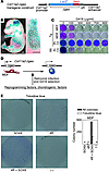
Repair of cartilage injury with hyaline cartilage continues to be a challenging clinical problem. Because of the limited number of chondrocytes in vivo, coupled with in vitro de-differentiation of chondrocytes into fibrochondrocytes, which secrete type I collagen and have an altered matrix architecture and mechanical function, there is a need for a novel cell source that produces hyaline cartilage. The generation of induced pluripotent stem (iPS) cells has provided a tool for reprogramming dermal fibroblasts to an undifferentiated state by ectopic expression of reprogramming factors. Here, we show that retroviral expression of two reprogramming factors (c-Myc and Klf4) and one chondrogenic factor (SOX9) induces polygonal chondrogenic cells directly from adult dermal fibroblast cultures. Induced cells expressed marker genes for chondrocytes but not fibroblasts, i.e., the promoters of type I collagen genes were extensively methylated. Although some induced cell lines formed tumors when subcutaneously injected into nude mice, other induced cell lines generated stable homogenous hyaline cartilage–like tissue. Further, the doxycycline-inducible induction system demonstrated that induced cells are able to respond to chondrogenic medium by expressing endogenous Sox9 and maintain chondrogenic potential after substantial reduction of transgene expression. Thus, this approach could lead to the preparation of hyaline cartilage directly from skin, without generating iPS cells.
Authors
Kunihiko Hiramatsu, Satoru Sasagawa, Hidetatsu Outani, Kanako Nakagawa, Hideki Yoshikawa, Noriyuki Tsumaki


Copyright © 2025 American Society for Clinical Investigation
ISSN: 0021-9738 (print), 1558-8238 (online)


