Go to JCI Insight
  • About
  • Editors
  • Consulting Editors
  • For authors
  • Publication ethics
  • Publication alerts by email
  • Advertising
  • Job board
  • Contact
  • Clinical Research and Public Health
  • Current issue
  • Past issues
  • By specialty
    • COVID-19
    • Cardiology
    • Gastroenterology
    • Immunology
    • Metabolism
    • Nephrology
    • Neuroscience
    • Oncology
    • Pulmonology
    • Vascular biology
    • All ...
  • Videos
    • Conversations with Giants in Medicine
    • Video Abstracts
  • Reviews
    • View all reviews ...
    • Clinical innovation and scientific progress in GLP-1 medicine (Nov 2025)
    • Pancreatic Cancer (Jul 2025)
    • Complement Biology and Therapeutics (May 2025)
    • Evolving insights into MASLD and MASH pathogenesis and treatment (Apr 2025)
    • Microbiome in Health and Disease (Feb 2025)
    • Substance Use Disorders (Oct 2024)
    • Clonal Hematopoiesis (Oct 2024)
    • View all review series ...
  • Viewpoint
  • Collections
    • In-Press Preview
    • Clinical Research and Public Health
    • Research Letters
    • Letters to the Editor
    • Editorials
    • Commentaries
    • Editor's notes
    • Reviews
    • Viewpoints
    • 100th anniversary
    • Top read articles

  • Current issue
  • Past issues
  • Specialties
  • Reviews
  • Review series
  • Conversations with Giants in Medicine
  • Video Abstracts
  • In-Press Preview
  • Clinical Research and Public Health
  • Research Letters
  • Letters to the Editor
  • Editorials
  • Commentaries
  • Editor's notes
  • Reviews
  • Viewpoints
  • 100th anniversary
  • Top read articles
  • About
  • Editors
  • Consulting Editors
  • For authors
  • Publication ethics
  • Publication alerts by email
  • Advertising
  • Job board
  • Contact

Microbiology

  • 57 Articles
  • 0 Posts
  • ← Previous
  • 1
  • 2
  • 3
  • 4
  • 5
  • 6
  • Next →
Endocarditis and biofilm-associated pili of Enterococcus faecalis
Sreedhar R. Nallapareddy, Kavindra V. Singh, Jouko Sillanpää, Danielle A. Garsin, Magnus Höök, Stanley L. Erlandsen, Barbara E. Murray
View: Text | PDF

Endocarditis and biofilm-associated pili of Enterococcus faecalis

  • Text
  • PDF
Abstract

Increasing multidrug resistance in Enterococcus faecalis, a nosocomial opportunist and common cause of bacterial endocarditis, emphasizes the need for alternative therapeutic approaches such as immunotherapy or immunoprophylaxis. In an earlier study, we demonstrated the presence of antibodies in E. faecalis endocarditis patient sera to recombinant forms of 9 E. faecalis cell wall–anchored proteins; of these, we have now characterized an in vivo–expressed locus of 3 genes and an associated sortase gene (encoding sortase C; SrtC). Here, using mutation analyses and complementation, we demonstrated that both the ebp (encoding endocarditis and biofilm-associated pili) operon and srtC are important for biofilm production of E. faecalis strain OG1RF. In addition, immunogold electron microscopy using antisera against EbpA–EbpC proteins as well as patient serum demonstrated that E. faecalis produces pleomorphic surface pili. Assembly of pili and their cell wall attachment appeared to occur via a mechanism of cross-linking of the Ebp proteins by the designated SrtC. Importantly, a nonpiliated, allelic replacement mutant was significantly attenuated in an endocarditis model. These biologically important surface pili, which are antigenic in humans during endocarditis and encoded by a ubiquitous E. faecalis operon, may be a useful immunotarget for studies aimed at prevention and/or treatment of this pathogen.

Authors

Sreedhar R. Nallapareddy, Kavindra V. Singh, Jouko Sillanpää, Danielle A. Garsin, Magnus Höök, Stanley L. Erlandsen, Barbara E. Murray

×

Bacterial neuraminidase facilitates mucosal infection by participating in biofilm production
Grace Soong, Amanda Muir, Marisa I. Gomez, Jonathan Waks, Bharat Reddy, Paul Planet, Pradeep K. Singh, Yukihiro Kanetko, Matthew C. Wolfgang, Yu-Shan Hsiao, Liang Tong, Alice Prince
View: Text | PDF | Corrigendum

Bacterial neuraminidase facilitates mucosal infection by participating in biofilm production

  • Text
  • PDF
Abstract

Many respiratory pathogens, including Hemophilus influenzae, Streptococcus pneumoniae, and Pseudomonas aeruginosa, express neuraminidases that can cleave α2,3-linked sialic acids from glycoconjugates. As mucosal surfaces are heavily sialylated, neuraminidases have been thought to modify epithelial cells by exposing potential bacterial receptors. However, in contrast to neuraminidase produced by the influenza virus, a role for bacterial neuraminidase in pathogenesis has not yet been clearly established. We constructed a mutant of P. aeruginosa PAO1 by deleting the PA2794 neuraminidase locus (Δ2794) and tested its virulence and immunostimulatory capabilities in a mouse model of infection. Although fully virulent when introduced i.p., the Δ2794 mutant was unable to establish respiratory infection by i.n. inoculation. The inability to colonize the respiratory tract correlated with diminished production of biofilm, as assessed by scanning electron microscopy and in vitro assays. The importance of neuraminidase in biofilm production was further demonstrated by showing that viral neuraminidase inhibitors in clinical use blocked P. aeruginosa biofilm production in vitro as well. The P. aeruginosa neuraminidase has a key role in the initial stages of pulmonary infection by targeting bacterial glycoconjugates and contributing to the formation of biofilm. Inhibiting bacterial neuraminidases could provide a novel mechanism to prevent bacterial pneumonia.

Authors

Grace Soong, Amanda Muir, Marisa I. Gomez, Jonathan Waks, Bharat Reddy, Paul Planet, Pradeep K. Singh, Yukihiro Kanetko, Matthew C. Wolfgang, Yu-Shan Hsiao, Liang Tong, Alice Prince

×

Immune sensing of Candida albicans requires cooperative recognition of mannans and glucans by lectin and Toll-like receptors
Mihai G. Netea, Neil A.R. Gow, Carol A. Munro, Steven Bates, Claire Collins, Gerben Ferwerda, Richard P. Hobson, Gwyneth Bertram, H. Bleddyn Hughes, Trees Jansen, Liesbeth Jacobs, Ed T. Buurman, Karlijn Gijzen, David L. Williams, Ruurd Torensma, Alistair McKinnon, Donna M. MacCallum, Frank C. Odds, Jos W.M. Van der Meer, Alistair J.P. Brown, Bart Jan Kullberg
View: Text | PDF

Immune sensing of Candida albicans requires cooperative recognition of mannans and glucans by lectin and Toll-like receptors

  • Text
  • PDF
Abstract

The fungal pathogen Candida albicans has a multilayered cell wall composed of an outer layer of proteins glycosylated with N- or O-linked mannosyl residues and an inner skeletal layer of β-glucans and chitin. We demonstrate that cytokine production by human mononuclear cells or murine macrophages was markedly reduced when stimulated by C. albicans mutants defective in mannosylation. Recognition of mannosyl residues was mediated by mannose receptor binding to N-linked mannosyl residues and by TLR4 binding to O-linked mannosyl residues. Residual cytokine production was mediated by recognition of β-glucan by the dectin-1/TLR2 receptor complex. C. albicans mutants with a cell wall defective in mannosyl residues were less virulent in experimental disseminated candidiasis and elicited reduced cytokine production in vivo. We concluded that recognition of C. albicans by monocytes/macrophages is mediated by 3 recognition systems of differing importance, each of which senses specific layers of the C. albicans cell wall.

Authors

Mihai G. Netea, Neil A.R. Gow, Carol A. Munro, Steven Bates, Claire Collins, Gerben Ferwerda, Richard P. Hobson, Gwyneth Bertram, H. Bleddyn Hughes, Trees Jansen, Liesbeth Jacobs, Ed T. Buurman, Karlijn Gijzen, David L. Williams, Ruurd Torensma, Alistair McKinnon, Donna M. MacCallum, Frank C. Odds, Jos W.M. Van der Meer, Alistair J.P. Brown, Bart Jan Kullberg

×

Trans -cyclopropanation of mycolic acids on trehalose dimycolate suppresses Mycobacterium tuberculosis –induced inflammation and virulence
Vivek Rao, Feng Gao, Bing Chen, William R. Jacobs, Michael S. Glickman
View: Text | PDF

Trans -cyclopropanation of mycolic acids on trehalose dimycolate suppresses Mycobacterium tuberculosis –induced inflammation and virulence

  • Text
  • PDF
Abstract

Recent studies have shown that fine structural modifications of Mycobacterium tuberculosis cell envelope lipids mediate host cell immune activation during infection. One such alteration in lipid structure is cis-cyclopropane modification of the mycolic acids on trehalose dimycolate (TDM) mediated by proximal cyclopropane synthase of α mycolates (pcaA), a proinflammatory lipid modification during early infection. Here we examine the pathogenetic role and immunomodulatory function of mycolic acid cyclopropane stereochemistry by characterizing an M. tuberculosis cyclopropane–mycolic acid synthase 2 (cmaA2) null mutant (ΔcmaA2) that lacks trans-cyclopropanation of mycolic acids. Although titers of WT and ΔcmaA2 organisms were identical during mouse infection, ΔcmaA2 bacteria were hypervirulent while inducing larger granulomas than WT M. tuberculosis. The hypervirulence of the ΔcmaA2 strain depended on host TNF-α and IFN-γ. Loss of trans-cyclopropanation enhanced M. tuberculosis–induced macrophage inflammatory responses, a phenotype that was transferable with petroleum ether extractable lipids. Finally, purified TDM lacking trans-cyclopropane rings was 5-fold more potent in stimulating macrophages. These results establish cmaA2-dependent trans-cyclopropanation of TDM as a suppressor of M. tuberculosis–induced inflammation and virulence. In addition, cyclopropane stereochemistries on mycolic acids interact directly with host cells to both positively and negatively influence host innate immune activation.

Authors

Vivek Rao, Feng Gao, Bing Chen, William R. Jacobs, Michael S. Glickman

×

Glucosylceramide synthase is an essential regulator of pathogenicity of Cryptococcus neoformans
Philipp C. Rittershaus, Talar B. Kechichian, Jeremy C. Allegood, Alfred H. Merrill, Mirko Hennig, Chiara Luberto, Maurizio Del Poeta
View: Text | PDF | Corrigendum

Glucosylceramide synthase is an essential regulator of pathogenicity of Cryptococcus neoformans

  • Text
  • PDF
Abstract

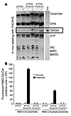
The pathogenic fungus Cryptococcus neoformans infects humans upon inhalation and causes the most common fungal meningoencephalitis in immunocompromised subjects worldwide. In the host, C. neoformans is found both intracellularly and extracellularly, but how these two components contribute to the development of the disease is largely unknown. Here we show that the glycosphingolipid glucosylceramide (GlcCer), which is present in C. neoformans, was essential for fungal growth in host extracellular environments, such as in alveolar spaces and in the bloodstream, which are characterized by a neutral/alkaline pH, but not in the host intracellular environment, such as in the phagolysosome of macrophages, which is characteristically acidic. Indeed, a C. neoformans mutant strain lacking GlcCer did not grow in vitro at a neutral/alkaline pH, yet it had no growth defect at an acidic pH. The mechanism by which GlcCer regulates alkali tolerance was by allowing the transition of C. neoformans through the cell cycle. This study establishes C. neoformans GlcCer as a key virulence factor of cryptococcal pathogenicity, with important implications for future development of new antifungal strategies.

Authors

Philipp C. Rittershaus, Talar B. Kechichian, Jeremy C. Allegood, Alfred H. Merrill, Mirko Hennig, Chiara Luberto, Maurizio Del Poeta

×

Staphylococcus aureus Panton-Valentine leukocidin directly targets mitochondria and induces Bax-independent apoptosis of human neutrophils
Anne-Laure Genestier, Marie-Cécile Michallet, Gilles Prévost, Gregory Bellot, Lara Chalabreysse, Simone Peyrol, Françoise Thivolet, Jerome Etienne, Gérard Lina, François M. Vallette, François Vandenesch, Laurent Genestier
View: Text | PDF

Staphylococcus aureus Panton-Valentine leukocidin directly targets mitochondria and induces Bax-independent apoptosis of human neutrophils

  • Text
  • PDF
Abstract

Panton-Valentine leukocidin (PVL) is a pore-forming toxin secreted by Staphylococcus aureus that has recently been associated with necrotizing pneumonia. In the present study, we report that in vitro, PVL induces polymorphonuclear cell death by necrosis or by apoptosis, depending on the PVL concentration. PVL-induced apoptosis was associated with a rapid disruption of mitochondrial homeostasis and activation of caspase-9 and caspase-3, suggesting that PVL-induced apoptosis is preferentially mediated by the mitochondrial pathway. Polymorphonuclear cell exposure to PVL leads to mitochondrial localization of the toxin, whereas Bax, 1 of the 2 essential proapoptotic members of the Bcl-2 family, was still localized in the cytosol. Addition of PVL to isolated mitochondria induced the release of the apoptogenic proteins cytochrome c and Smac/DIABLO. Therefore, we suggest that PVL, which belongs to the pore-forming toxin family, could act at the mitochondrion level by creating pores in the mitochondrial outer membrane. Furthermore, LukS-PV, 1 of the 2 components of PVL, was detected in lung sections of patients with necrotizing pneumonia together with DNA fragmentation, suggesting that PVL induces apoptosis in vivo and thereby is directly involved in the pathophysiology of necrotizing pneumonia.

Authors

Anne-Laure Genestier, Marie-Cécile Michallet, Gilles Prévost, Gregory Bellot, Lara Chalabreysse, Simone Peyrol, Françoise Thivolet, Jerome Etienne, Gérard Lina, François M. Vallette, François Vandenesch, Laurent Genestier

×

The crucial role of Campylobacter jejuni genes in anti-ganglioside antibody induction in Guillain-Barré syndrome
Peggy C.R. Godschalk, Astrid P. Heikema, Michel Gilbert, Tomoko Komagamine, C. Wim Ang, Jobine Glerum, Denis Brochu, Jianjun Li, Nobuhiro Yuki, Bart C. Jacobs, Alex van Belkum, Hubert P. Endtz
View: Text | PDF

The crucial role of Campylobacter jejuni genes in anti-ganglioside antibody induction in Guillain-Barré syndrome

  • Text
  • PDF
Abstract

Molecular mimicry of Campylobacter jejuni lipo-oligosaccharides (LOS) with gangliosides in nervous tissue is considered to induce cross-reactive antibodies that lead to Guillain-Barré syndrome (GBS), an acute polyneuropathy. To determine whether specific bacterial genes are crucial for the biosynthesis of ganglioside-like structures and the induction of anti-ganglioside antibodies, we characterized the C. jejuni LOS biosynthesis gene locus in GBS-associated and control strains. We demonstrated that specific types of the LOS biosynthesis gene locus are associated with GBS and with the expression of ganglioside-mimicking structures. Campylobacter knockout mutants of 2 potential GBS marker genes, both involved in LOS sialylation, expressed truncated LOS structures without sialic acid, showed reduced reactivity with GBS patient serum, and failed to induce an anti-ganglioside antibody response in mice. We demonstrate, for the first time, to our knowledge, that specific bacterial genes are crucial for the induction of anti-ganglioside antibodies.

Authors

Peggy C.R. Godschalk, Astrid P. Heikema, Michel Gilbert, Tomoko Komagamine, C. Wim Ang, Jobine Glerum, Denis Brochu, Jianjun Li, Nobuhiro Yuki, Bart C. Jacobs, Alex van Belkum, Hubert P. Endtz

×
  • ← Previous
  • 1
  • 2
  • 3
  • 4
  • 5
  • 6
  • Next →

No posts were found with this tag.

Advertisement

Copyright © 2025 American Society for Clinical Investigation
ISSN: 0021-9738 (print), 1558-8238 (online)

Sign up for email alerts