Go to JCI Insight
  • About
  • Editors
  • Consulting Editors
  • For authors
  • Publication ethics
  • Publication alerts by email
  • Advertising
  • Job board
  • Contact
  • Clinical Research and Public Health
  • Current issue
  • Past issues
  • By specialty
    • COVID-19
    • Cardiology
    • Gastroenterology
    • Immunology
    • Metabolism
    • Nephrology
    • Neuroscience
    • Oncology
    • Pulmonology
    • Vascular biology
    • All ...
  • Videos
    • Conversations with Giants in Medicine
    • Video Abstracts
  • Reviews
    • View all reviews ...
    • Clinical innovation and scientific progress in GLP-1 medicine (Nov 2025)
    • Pancreatic Cancer (Jul 2025)
    • Complement Biology and Therapeutics (May 2025)
    • Evolving insights into MASLD and MASH pathogenesis and treatment (Apr 2025)
    • Microbiome in Health and Disease (Feb 2025)
    • Substance Use Disorders (Oct 2024)
    • Clonal Hematopoiesis (Oct 2024)
    • View all review series ...
  • Viewpoint
  • Collections
    • In-Press Preview
    • Clinical Research and Public Health
    • Research Letters
    • Letters to the Editor
    • Editorials
    • Commentaries
    • Editor's notes
    • Reviews
    • Viewpoints
    • 100th anniversary
    • Top read articles

  • Current issue
  • Past issues
  • Specialties
  • Reviews
  • Review series
  • Conversations with Giants in Medicine
  • Video Abstracts
  • In-Press Preview
  • Clinical Research and Public Health
  • Research Letters
  • Letters to the Editor
  • Editorials
  • Commentaries
  • Editor's notes
  • Reviews
  • Viewpoints
  • 100th anniversary
  • Top read articles
  • About
  • Editors
  • Consulting Editors
  • For authors
  • Publication ethics
  • Publication alerts by email
  • Advertising
  • Job board
  • Contact

An unconventional EMT

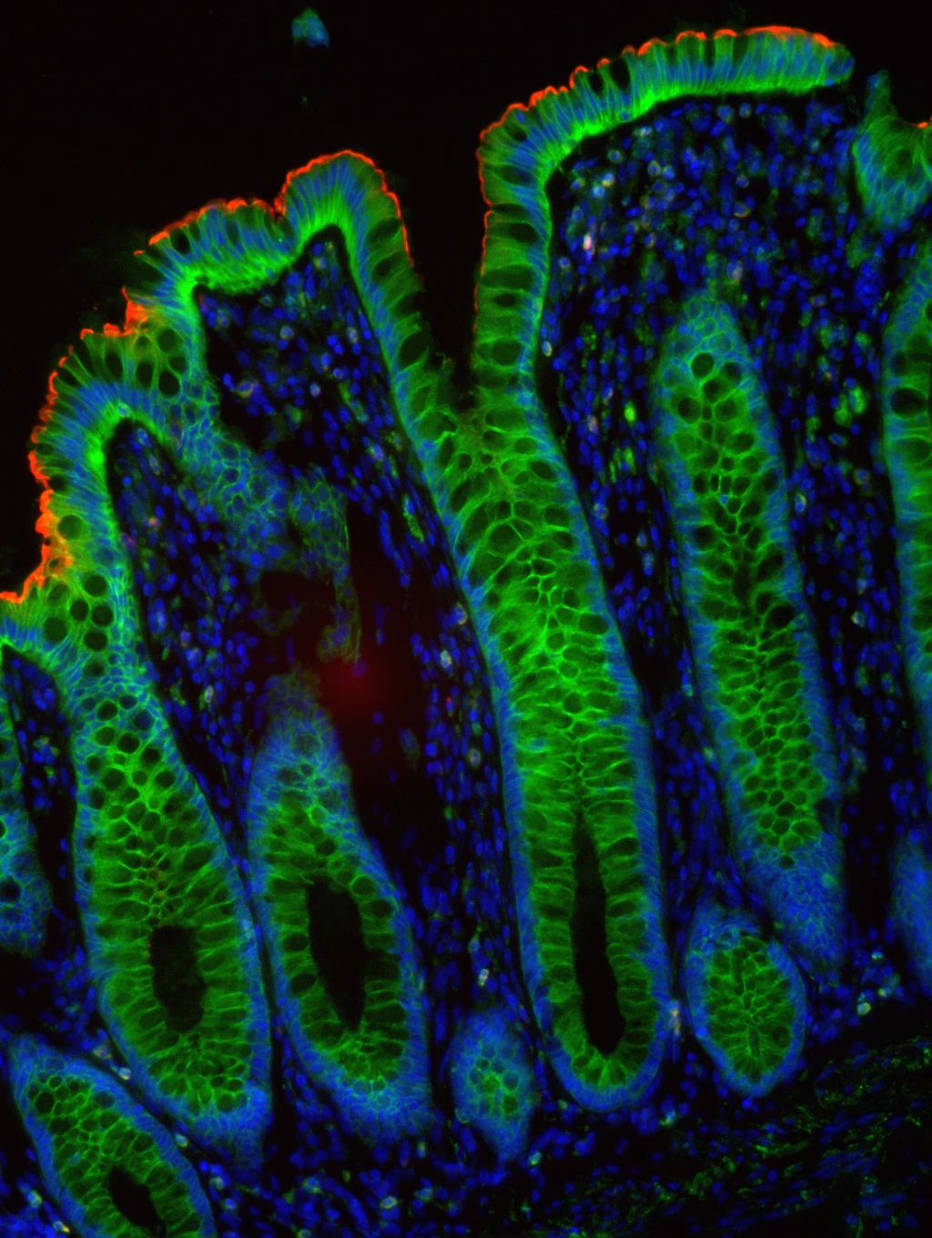
Inappropriate induction of endothelial to mesenchymal transition (EMT) confers migratory and stem-like properties to epithelial cells and enables cancer cells to metastasize and invade other sites. Enhanced expression of placenta-specific 8 (PLAC8) has been implicated in colorectal cancer (CRC); however, it is not clear how PLAC8 promotes CRC cell growth. Using zebrafish models and human tissue, Cunxi Li, Haiting Ma, and colleagues at Vanderbilt University and Washington University, respectively, determined that endogenous PLAC8 localizes to the apical membrane of the intestinal epithelium in healthy tissue, and that PLAC8 is cytosolic in CRC. CRC cells with elevated PLAC8 exhibited enhanced invasiveness, motility, growth, and mesenchylmal gene expression. PLAC8-induced EMT was linked to ERK2 phosphorylation, which was the result of PLAC8 binding to and inhibiting the ERK2 phosphatase DUSP6. Furthermore, multiplex imaging of the leading edge of a human colorectal tumor revealed cytosolic PLAC8 and expression of EMT markers, supporting a role for PLAC8 dysfunction in CRC invasion. The accompanying immunofluorescence image reveals that endogenous PLAC8 (red) in normal human colon localizes to the apical domain of the differentiated colonic epithelium (green) at the top of crypts. 

Published April 1, 2014, by Corinne Williams

Scientific Show Stopper

Related articles

Excess PLAC8 promotes an unconventional ERK2-dependent EMT in colon cancer
Cunxi Li, Haiting Ma, Yang Wang, Zheng Cao, Ramona Graves-Deal, Anne E. Powell, Alina Starchenko, Gregory D. Ayers, Mary Kay Washington, Vidya Kamath, Keyur Desai, Michael J. Gerdes, Lila Solnica-Krezel, Robert J. Coffey
View: Text | PDF
Research Article Oncology

Excess PLAC8 promotes an unconventional ERK2-dependent EMT in colon cancer

  • Text
  • PDF
Abstract

The epithelial-to-mesenchymal transition (EMT) transcriptional program is characterized by repression of E-cadherin (CDH1) and induction of N-cadherin (CDH2), and mesenchymal genes like vimentin (VIM). Placenta-specific 8 (PLAC8) has been implicated in colon cancer; however, how PLAC8 contributes to disease is unknown, and endogenous PLAC8 protein has not been studied. We analyzed zebrafish and human tissues and found that endogenous PLAC8 localizes to the apical domain of differentiated intestinal epithelium. Colon cancer cells with elevated PLAC8 levels exhibited EMT features, including increased expression of VIM and zinc finger E-box binding homeobox 1 (ZEB1), aberrant cell motility, and increased invasiveness. In contrast to classical EMT, PLAC8 overexpression reduced cell surface CDH1 and upregulated P-cadherin (CDH3) without affecting CDH2 expression. PLAC8-induced EMT was linked to increased phosphorylated ERK2 (p-ERK2), and ERK2 knockdown restored cell surface CDH1 and suppressed CDH3, VIM, and ZEB1 upregulation. In vitro, PLAC8 directly bound and inactivated the ERK2 phosphatase DUSP6, thereby increasing p-ERK2. In a murine xenograft model, knockdown of endogenous PLAC8 in colon cancer cells resulted in smaller tumors, reduced local invasion, and decreased p-ERK2. Using MultiOmyx, a multiplex immunofluorescence-based methodology, we observed coexpression of cytosolic PLAC8, CDH3, and VIM at the leading edge of a human colorectal tumor, supporting a role for PLAC8 in cancer invasion in vivo.

Authors

Cunxi Li, Haiting Ma, Yang Wang, Zheng Cao, Ramona Graves-Deal, Anne E. Powell, Alina Starchenko, Gregory D. Ayers, Mary Kay Washington, Vidya Kamath, Keyur Desai, Michael J. Gerdes, Lila Solnica-Krezel, Robert J. Coffey

×
Advertisement

Copyright © 2025 American Society for Clinical Investigation
ISSN: 0021-9738 (print), 1558-8238 (online)

Sign up for email alerts