
Advertisement
Review
Open Access |  10.1172/JCI208001
Hemiplegic migraine: genetics and pathophysiology
Daniela Pietrobon
Department of Biomedical Sciences and Padova Neuroscience Center, University of Padova, Padova, Italy.
Address correspondence to: Daniela Pietrobon, Dept. of Biomedical Sciences and Padova Neuroscience Center, University of Padova, Via Ugo Bassi 58, 35131, Padova, Italy. Email: daniela.pietrobon@unipd.it.
Find articles by Pietrobon, D. in: PubMed | Google Scholar
Published September 15, 2026 - More info
J Clin Invest. 2026;136(18):e208001. https://doi.org/10.1172/JCI208001.
© 2026 Pietrobon This work is licensed under the Creative Commons Attribution 4.0 International License. To view a copy of this license, visit http://creativecommons.org/licenses/by/4.0/.

-
Introduction
Migraine is a common disabling neurological disorder characterized by recurrent attacks of typically throbbing and unilateral, often severe, headache with associated features such as nausea and/or photophobia and phonophobia. In approximately one-third of patients, these symptoms are preceded by transient visual, sensory, or other CNS symptoms (migraine aura). When the aura includes motor weakness, the disorder is classified as hemiplegic migraine (HM) (1) (Table 1). Migraine is characterized by a global dysfunction in multisensory processing, which points to alterations in multiple brain networks in addition to those related to pain (2–4).
The mechanisms underlying migraine headache and aura are reasonably well understood and have been reviewed in detail elsewhere (2–9). Briefly, the headache depends on activation and sensitization of the trigeminovascular system, with a pivotal role for calcitonin gene related peptide (CGRP) release. The neurophysiological correlate of migraine aura is a slowly propagating (2–5 mm/min), self-sustaining wave of nearly complete depolarization of brain cells, named cortical spreading depression or spreading depolarization (CSD). Accumulating evidence indicates that CSD can initiate the headache mechanisms, as it can activate and sensitize trigeminal nociceptors and the trigeminovascular pain pathway, produce dural and cortical inflammation, and induce trigeminal pain behavior in rodents that responds to migraine therapies (6, 9–11). However, although most migraine attacks originate in the brain, the neurobiological mechanisms underlying their onset and the susceptibility to CSD remain largely unknown.
Migraine is a complex multifactorial genetic disorder, brought about by a combination of multiple genetic variants, each with a small effect size, and internal and external environmental factors (12, 13). Rare monogenic subtypes of migraine with aura (MA), which include familial HM (FHM), are caused by single exonic mutations (12, 13). Studies of the functional consequences of FHM mutations in cellular and animal models of the disease have enabled insights into the causative mechanisms at the molecular, cellular, and circuit level. Here, I review current knowledge of the genetics and pathophysiology of HM and discuss the translational implications.

-
HM genetics
A subset of patients with HM carry single pathogenic mutations in the CACNA1A (FHM1/SHM1), ATP1A2 (FHM2/SHM2), or SCN1A (FHM3/SHM3) genes (Table 1) (16–18). SHM can be caused by de novo mutations in genes causing FHM or by inheritance of a mutation from an asymptomatic parent with FHM (19, 20). In sporadic cases, early-onset and presence of associated neurological symptoms are associated with increased likelihood of an FHM mutation (14, 19, 21). Although HM attacks are similar regardless of the gene involved, the causative gene may influence the spectrum of associated manifestations. For example, both febrile comas and cerebellar ataxia are more frequent in FHM1 than FHM2 and are not described in FHM3; in contrast to FHM1 and FHM2, HM attacks and seizures do not overlap in FHM3, in which elicited repetitive daily blindness (ERDB) is a type-specific comorbidity (14, 15, 22–24). Distinct mutations in the same gene can result in pure FHM/SHM with typical attacks or severe FHM/SHM with atypical attacks and chronic symptoms (25). However, family members with the same mutation can show wide variability in clinical presentation, suggesting that unknown genetic or environmental factors can influence phenotype (26).
Population-based studies of FHM/SHM and next-generation sequencing approaches in large cohorts of patients suspected of having HM show that only a minority of families/patients have pathogenic variants in CACNA1A, ATP1A2, or SCNA1A (27–31) (Table 2). In contrast, in a cohort of mainly Dutch families, causative mutations in the three FHM genes were present in 63% of symptomatic individuals (21). Patients without such mutation had a milder phenotype, more similar to nonhemiplegic MA, and were older at disease onset than patients carrying a causative mutation (21). In the Finnish population, the clinical symptoms of patients with HM (most of whom did not carry mutations in CACNA1A, ATP1A2, or SCNA1A) could not clearly distinguish them from patients with MA but rather reflected an extreme phenotype in the MA continuum (29).
Based on the relatively high frequency of likely pathogenic PRRT2 gene variants in a large cohort of HM probands, Riant et al. (31) concluded that PRRT2 should be regarded as the fourth autosomal dominant gene for FHM/SHM (22). Loss-of-function mutations in PRRT2 are the leading cause for a wide spectrum of paroxysmal diseases, including benign familial infantile epilepsy (BFIE), infantile convulsions and choreoathetosis (ICCA), and paroxysmal kinesigenic dyskinesia (PKD) (32). The most common PRRT2 variant in patients with HM is the truncating c.649dupC frameshift variation, which is the main causative mutation in PKD and BFIE and can also cause ICCA (31, 32). The vast majority of individuals with this loss-of-function mutation do not have HM (32). Thus, a PRRT2 mutation alone does not seem sufficient to cause HM (21, 33), but since the frequency of HM in PRRT2 mutation carriers appears too high to be incidental (32, 34), PRRT2 might act as a main disease modifier gene for HM within a complex polygenic mechanism (21, 33). Indeed, an increased burden of common and/or rare variants in several genes contributes to familial aggregation or disease risk in large cohorts of families and patients with FHM referred for HM diagnosis without a disease-causing mutation in CACNA1A, ATP1A2, or SCNA1A (35–38). In a small number of patients with HM, mutations in other genes (mainly ion or solute carriers, including SLC1A3 and SLC4A4) have been reported (13, 30, 39–42), but additional data are needed before they can be considered causal (12, 43). Recently, a heterozygous missense mutation in SCN2A was found to cosegregate with FHM in a four-generation pedigree, and additional SCN2A variants were found in a second family and a sporadic case (44). Overall, the findings suggest that FHM is a dominantly inherited monogenic disease in some patients but may be polygenic or oligogenic in others (13, 21, 29, 35–37).

-
HM genes and proteins
FHM1/SHM1. CACNA1A encodes the pore-forming subunit of the voltage-gated calcium channel CaV2.1 (or P/Q-type). This channel is widely expressed in the nervous system, where it plays a prominent role in initiating neurotransmitter release, particularly at central synapses (ref. 45 and references therein). Expression is particularly high in the cerebellum, which may explain the cerebellar symptoms in several patients with FHM1 (45).
More than 40 missense mutations associated with FHM1/SHM1, affecting conserved amino acids in CaV2.1, have been identified (46). FHM1/SHM1-associated variants are gain-of-function mutations, resulting in increased open probability and activation at lower voltages of human recombinant CaV2.1 channels (45, 47, 48). Reduced G protein–mediated inhibition of mutant channels might lead to a further increase in Ca2+ influx during neuromodulation (49–51). The gain-of-function effect may depend on the specific CaV2.1 splice variant and/or auxiliary subunit (52–54), and thus may be neuron-type specific. Indeed, in Cacna1a-knockin mice carrying FHM1 mutations, the CaV2.1 current was enhanced in specific neurons, including cortical pyramidal cells and distinct trigeminal ganglion (TG) neurons (55–60), but was unaltered in others, including cortical inhibitory interneurons (58, 61) (Figure 1).
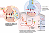 Figure 1
Functional alterations in the cerebral cortex in FHM1 knockin mouse models. In FHM1 mice, the CaV2.1 current is increased in pyramidal cells, but unaltered in inhibitory interneurons. Excitatory synaptic transmission is increased due to enhanced probability of glutamate release, while inhibitory synaptic transmission is unaltered. Both recurrent excitation and disynaptic feedback and feedforward inhibition are enhanced, and the excitation/inhibition (E/I) balance may be shifted toward inhibition in certain conditions. The heightened CaV2.1-dependent glutamate release may explain the enhanced susceptibility to experimentally induced CSD. Glut-R, glutamate receptor; NMDA-R, NMDA receptor; PC, pyramidal cell; IN, interneuron.
FHM2/SHM2. ATP1A2 encodes the α2 Na+/K+ ATPase (NKA). This NKA isoform is expressed primarily in neurons during embryonic development and at the time of birth and almost exclusively in astrocytes in the adult brain, where it plays a key role in K+ and glutamate clearance during neuronal activity (ref. 62 and references therein). The fundamental role of α2 NKA in clearance of synaptically released glutamate relies on its colocalization and physical coupling with the glutamate transporters GLT-1 and GLAST at the astrocytic processes surrounding cortical excitatory synapses and its relatively low affinity for [Na+]in (62–65). α2 NKA is also important for intracellular Na+ and Ca2+ homeostasis/signaling, pH regulation, and neuron-astrocyte neurometabolic coupling (66, 67).
More than 90 missense mutations in ATP1A2 have been associated with FHM2/SHM2 (46), and these cause complete or partial loss-of-function of human recombinant α2 NKA (68–71) (Figure 2). The brain expression of α2 NKA in heterozygous knockin mice carrying FHM2 mutations was either 50% reduced or unaltered, depending on whether the mutations impaired membrane targeting and caused complete loss of function of recombinant pumps (W887R, G301R) or caused partial loss of function (T345) (72–77).
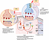 Figure 2
Functional alterations in the cerebral cortex in FHM2 and FHM3 knockin mouse models. In FHM2 mice, the 50% reduction of α2 NKA expression results in a similarly reduced density of glutamate transporters at perisynaptic astrocytic processes and in a reduced rate of clearance of both K+ and glutamate during neuronal activity. Spontaneous focal glutamate transients (glutamate plumes) occur in layer 1, as markers of inefficient glutamate clearance. The activation of extrasynaptic GluN1-N2B NMDARs in pyramidal cells elicited by stimulation of layer 1 afferents is enhanced and the generation of dendritic NMDA spikes in pyramidal cells is facilitated, leading to enhanced somatic burst firing of the pyramidal cells. The reduced rate of glutamate clearance and the increased activation of extrasynaptic GluN1-N2B NMDARs may explain the enhanced susceptibility to experimentally induced CSD. In FHM3 mice, inactivation of the NaV1.1 current is slowed, and the persistent Na+ current as well as the firing rate are increased in fast-spiking inhibitory interneurons. Glut-R, glutamate receptor; NMDA-R, NMDA receptor; PC, pyramidal cell; IN, interneuron.
FHM3/SHM3. SCNA1A encodes the pore-forming subunit of the voltage-gated sodium channel NaV1.1. This neuronal channel is predominantly expressed in inhibitory interneurons in several brain areas; it predominantly localizes at the axon initial segment where it plays a key role in interneuron excitability, particularly in sustaining high-frequency firing (78–81). Of potential relevance for migraine pain, NaV1.1 is also expressed in (and regulates excitability of) sensory mechanoreceptive A-δ fibers and a subset of TG neurons (82).
More than 10 missense mutations in SCNA1A have been associated with FHM3/SHM3 (46, 83). The initial functional studies of FHM3 mutations on human recombinant NaV1.1 channels generated conflicting results (reviewed in ref. 24), but it is now understood that FHM3 mutations produce an overall gain-of-function effect, mainly due to destabilization of the NaV1.1-inactivated state (24, 84–87). Accordingly, knockin of a FHM3 mutation in mice resulted in increased persistent Na+ current and slower Na+ current inactivation in GABAergic neurons (88) (Figure 2). Stabilization of the NaV1.1-inactivated state by the late Na+ current inhibitor GS967 rescued the gain of function of recombinant human NaV1.1 channels (86, 87), and GS967 inhibited the persistent Na+ current in fast-spiking interneurons of FHM3 mice (88).
-
Genetic mouse models of FHM
Eight FHM mouse models have been generated by introducing human mutations in the orthologous genes (Table 3) (55, 56, 72, 73, 77, 88–90). Most carry mutations causing pure HM, and heterozygous mice of these lines did not show overt behavioral phenotypes. Homozygous Cacna1aR192Q mice also did not display an overt phenotype (55), but homozygous Scna1aL1649Q and Atp1a2T345A mice had reduced lifespan (77, 88), and homozygous Atp1a2W887R mice (with barely observable brain expression of α2 NKA) died at birth (72). Heterozygous Scna1aL263V mice (90), which carry a mutation identified in two families in which most carriers had pure HM and one family branch showed independent attacks of epilepsy (91), died prematurely due to spontaneous brainstem depolarization-associated apnea, which was prevented by administration of GS967 (87, 90). Notably, GS967-sensitive life-threatening apneic events were reported in an infant homozygous for SCNA1AL263V, whereas the heterozygous family members had pure FHM (87).
Cacna1aS218L and Atp1a2G301R (56, 73) mice carry mutations associated with severe HM attacks, which include prolonged coma/torpor, confusional state, cerebral edema (often triggered by minor head trauma in S218L carriers), fever, seizures, and transient or permanent cerebellar signs, including progressive ataxia (76, 92–94). Homozygous Cacna1aS218L mice model the main features of the severe human clinical syndrome, including spontaneous attacks of hemiparesis and sometimes fatal seizures, brain edema after mild head impact, and ataxia (56, 95). The severe clinical/behavioral phenotype correlates with the particularly low activation threshold of human S218L CaV2.1 channels (48). Heterozygous Atp1a2G301R mice show several behavioral alterations, including increased startle response to aversive acoustic stimuli, decreased sociability, stress-induced depression-like phenotypes, and increased compulsive behavior specifically in females (73). Atp1a2G301R mice also exhibit dysfunction of the glymphatic system, possibly linked to excessive neurovascular coupling (96, 97).
Increased susceptibility to CSD. A key migraine-relevant phenotype shared by all FHM mouse models is increased susceptibility to CSD induced by focal stimulation (although only as a trend in heterozygous Atp1a2T345A and Atp1a2E700K mice) (Table 3) (55–57, 64, 72, 77, 88–90, 98–102). The rate of CSD propagation was increased in all FHM1 and FHM2 models. In Cacna1aR192Q and Cacna1aS218L mice, a single CSD caused contralateral hemiplegia with leaning and circling, and CSD readily propagated into the striatum, suggesting facilitation of corticostriatal CSD propagation as a possible explanation for the motor deficits (99, 103, 104). The typical reduction of cerebral blood flow after CSD was more prolonged and the metabolic oxygen consumption during CSD was larger and resulted in a larger decrease of tissue oxygenation in Cacna1aR192Q mice compared with WT mice (105). In both FHM1 models, CSD facilitation was larger in female than male mice (101), and both stress hormone administration and relief from stress (but not acute or chronic stress) facilitated CSD (106, 107). These mice also showed increased susceptibility to CSDs after traumatic brain injury (108).
Correlating with the clinical phenotypes, facilitation of CSD initiation and propagation as well as severity of the post-CSD neurological motor deficits and propensity of CSD to propagate into subcortical structures were greater in Cacna1aS218L mice than in Cacna1aR192Q mice (56, 101, 103, 104, 108). Moreover, multiple CSDs after a single CSD-inducing stimulus were frequently observed in Cacna1aS218L (but not Cacna1aR192Q) mice, and such events were more frequent in homozygotes than in heterozygotes (56, 99). Homozygous Cacna1aS218L and heterozygous Atp1a2G301R mice also developed generalized seizures after CSDs (98, 101). These unique CSD features might contribute to the severe clinical phenotype in humans carrying S218L or G301R mutations.
Long-term recordings with implanted electrodes revealed only rare spontaneous CSDs in homozygous Ccacna1aR192Q and both heterozygous Cacna1aS218L and Atp1a2T345A mice, whereas frequent spontaneous CSDs, typically originating from the hippocampus and inhibited by GS967, were revealed in homozygous Atp1a2T345A mice (77, 99). Frequent spontaneous CSDs were also recorded in heterozygous Scna1aL263V mice (90).
The mechanisms underlying the enhanced susceptibility to CSDs in the FHM1 and FHM2 models involve alterations in glutamate signaling. In the FHM1 mice, excitatory synaptic transmission at different intracortical and thalamocortical synapses was enhanced, due to increased probability of glutamate release (57, 109–111). In contrast, GABAergic transmission at different cortical inhibitory synapses, although initiated by CaV2.1 channels, was unaltered (57, 61, 109, 111) (Figure 1). A causative link between gain of function of glutamate release at cortical synapses and facilitation of CSD was established in Cacna1aR192Q mice (57). In addition to a larger evoked glutamate release in Cacna1aS218L mice compared with Cacna1aR192Q mice, a larger metabolic burden on cortical neurons likely contributes to the larger facilitation of CSD and the unique CSD features in Cacna1aS218L mice (109). Indeed, in contrast with R192Q, the S218L mutation leads to CaV2.1 channel opening at resting potentials and to very slow channel inactivation during prolonged depolarizations (48, 59, 109, 112). Opening of S218L CaV2.1 channels at resting potentials also contributes to the irregular spontaneous Purkinje cell firing and the ataxia phenotype in Cacna1aS218L mice (113).
The importance of excessive glutamatergic transmission in CSD susceptibility is underscored and supported by the functional alterations in the cerebral cortex of Atp1a2W887R mice. In this model, the 50% reduction of α2 NKA expression results in a similarly reduced density of GLT-1 glutamate transporters at cortical perisynaptic astrocytic processes and in a reduction of the rate of clearance of both K+ and glutamate at cortical synapses during neuronal activity (64, 102, 114, 115). As a consequence, awake Atp1a2W887R mice showed spontaneous high-amplitude, focal glutamate transients (“glutamate plumes”) not present in WT mice (102). The activation of NMDA receptors (NMDARs) in layer 2/3 pyramidal cells elicited by stimulation of layer 1 afferents was increased and prolonged in Atp1a2W887R mice, owing to activation of extrasynaptic diheteromeric GluN1-N2B receptors (115). In addition, the generation of long-lasting NMDA spikes in the tuft dendrites of L5 pyramidal cells was facilitated, leading to enhanced output somatic burst firing (114) (Figure 2).
The reduced rate of glutamate clearance at cortical synapses can account for most of the facilitation of CSD initiation and a large fraction of the facilitation of CSD propagation in Atp1a2W887R mice (64). The remainder is likely related to the reduced rate of K+ clearance in this model. Extracellular glutamate imaging at the site of CSD initiation in awake head-fixed mice revealed that an increase in basal glutamate and in frequency of glutamate plumes precedes and may predict CSD initiation (102). The critical threshold level of glutamate at which CSD ignited was similar in Atp1a2W887R and WT mice but was reached with a lower stimulation intensity and more rapidly in Atp1a2W887R mice, accounting for their facilitated CSD initiation (102).
Recent studies showed that a critical threshold level of CaV2.1-dependent NMDAR activation is necessary for CSD ignition; this critical level is similar in Cacna1aR192Q and WT mice but is reached with a lower stimulation intensity and more rapidly in Cacna1aR192Q mice (116, 117). These findings point to excessive activation of NMDARs by low-intensity stimulation, as the key mechanism underlying facilitation of CSD initiation in FHM1 and FHM2. Indeed, both the increased and prolonged activation of NMDARs and the facilitation of CSD initiation in Atp1a2W887R mice were rescued by specific inhibition of GluN1-N2B NMDARs (115).
Overall, the findings suggest a model of CSD initiation in which a threshold depolarizing stimulus leads to CaV-dependent glutamate release that overwhelms the reuptake capacity of the astrocytic glutamate transporters and activates synaptic and extrasynaptic NMDARs above the level necessary to generate a net self-sustaining inward current. This initiates the positive feedback cycle that ignites CSD by making the neuronal depolarization and the K+/glutamate elevations self-regenerative (5, 100, 118). This amplificatory cycle of glutamate/K+ release is potentiated in the FHM1 and FHM2 models as a consequence of either increased glutamate release or reduced rate of glutamate clearance. In both cases, the CSD-threshold levels of glutamate and NMDAR activation are reached with stimuli of lower intensity and more rapidly, resulting in increased susceptibility to CSD (Figure 3). Notably, increased glutamate release can also account for CSD facilitation in a genetic mouse model of nonhemiplegic MA (119).
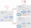 Figure 3
Excessive glutamatergic neurotransmission and excessive activation of NMDA receptors underlie the facilitation of CSD in FHM1 and FHM2 mouse models. The threshold depolarizing stimulus that initiates CSD in FHM1 and FHM2 mice does not induce CSD in WT mice. In the mutants, this stimulus increases glutamate and activates NMDARs above the critical levels necessary to generate a net self-sustaining inward current and initiate the CSD positive feedback cycle. The amplificatory cycle of glutamate/K+ release is potentiated as a consequence of either increased glutamate release (FHM1 models) or reduced glutamate clearance (FHM2 models), resulting in increased susceptibility to CSD. Adapted with permission from Journal of Headache and Pain (100).
A different mechanism may underlie facilitation of CSD in FHM3 mice. The facilitation of CSD in heterozygous Scna1aL1649Q mice was rescued by preferential inhibition of the persistent Na+ current with GS967 (88). It has been suggested that the larger accumulation of extracellular K+ at each action potential, due to increased persistent NaV1.1 current in interneurons, and the consequent build-up of [K+]e and increased firing of excitatory neurons may underly the enhanced susceptibility to CSD in FHM3 mice (86, 88, 100, 120). Given the reciprocal relationship between K+ and glutamate, whereby an increased [K+]e leads to increased glutamate release and decreased efficiency of glutamate reuptake, the proposed explanation for facilitation of CSD in FHM3 mice might be consistent with the model of CSD initiation and facilitation described above (100). In addition, it was proposed that an increased persistent NaV1.1 current in cortical pyramidal cells (121) might contribute to CSD facilitation in FHM3 (100).
Although these studies in FHM mouse models give insights into the key players likely involved in initiation of spontaneous CSDs in the brain of patients with HM (and possibly migraine), the mechanisms whereby specific triggering factors lead to CSD eruption in the brain of migraineurs remain largely unknown. Given the differential effect of FHM1 mutations on cortical excitatory and inhibitory neurotransmission and on synaptic plasticity at different synapses (57, 61, 109–111), it was proposed that the key pathogenic mechanism in HM (and possibly migraine) may be dysfunctional regulation of the excitatory-inhibitory (E/I) balance in specific neuronal circuits (57, 122). Considering that CSD is most easily induced in cortical upper dendritic layers (123–125), to favor CSD ignition, such dysfunction should probably lead to hyperactive excitatory synapses in upper layers and disinhibition of apical dendrites (perhaps with some metabolic compromise) (100). Whether this occurs, in which conditions and which neural circuits are involved, remain unknown.
Cacna1aR192Q mice show evidence of dysfunctional regulation of the E/I balance in specific cortical microcircuits, which are essential to prevent overexcitation (110, 111). Unexpectedly, the E/I balance was not skewed toward excitation, indeed during repetitive thalamic stimulation it was skewed toward inhibition. This is due to enhanced excitatory transmission at the synapses onto inhibitory interneurons and consequent enhancement of disynaptic feedforward and feedback inhibition, which may or may not counterbalance the increased recurrent excitation, depending on conditions (110, 111) (Figure 1). The responses to visual stimulation in awake Cacna1aR192Q mice were also consistent with a shift of the E/I balance toward inhibition (126). While the circuit alterations revealed by these and other studies in FHM1 mice (99, 127) may lead to alterations in sensory processing, it is difficult to envision how they may favor initiation of spontaneous CSDs (100). At any rate, considering the FHM1 cortex as hyperexcitable might be an oversimplification.
Migraine-relevant pain behavior. Some of the FHM mouse models exhibit migraine-relevant pain behavior. When subjected to novelty or restraint stress, homozygous Cacna1aR192Q mice showed behavioral changes suggestive of unilateral head pain, which were more frequent in females and normalized after administration of the antimigraine drug rizatriptan (128, 129). Some of these behaviors were more frequent and severe in Cacna1aS218L mice (129). Optogenetic induction of CSDs in freely behaving mice evoked a pain mimic that was longer lasting in heterozygous Cacna1aS218L mice than in WT mice (130). Atp1a2W887R mice showed enhanced cranial pain responses to systemic administration of the nitric oxide donor nitroglycerin (NTG) (114), which is known to induce a delayed migraine-like headache in migraineurs (131–133). In mice, NTG produces thermal and mechanical allodynia, responsive to the antimigraine drug sumatriptan (134). In Atp1a2W887R mice, orofacial mechanical hypersensitivity was elicited by low doses of NTG that were ineffective in WT mice (114).
Given the key role of CGRP in the trigeminovascular pain pathway, CGRP release has been analyzed in Cacna1aR192Q mice. Neither basal nor K+-evoked CGRP release from dura mater were altered in adult homozygous Cacna1aR192Q mice (58, 135). This is likely consistent with the unaltered CaV2.1 current in small capsaicin-sensitive Cacna1aR192Q TG neurons, which constitute the majority of small dural afferents (58), and with the lower fraction of CGRP-expressing neurons in TG of Cacna1aR192Q mice (136). Dural artery vasodilation induced in vivo by either endogenous (evoked by capsaicin) or exogenous CGRP was decreased in Cacna1aR192Q mice (135), suggesting unaltered CGRP release from trigeminal afferents together with downregulation and/or desensitization of blood vessel CGRP receptors. Thus, it is unlikely that enhanced CGRP-dependent meningeal neurogenic inflammation (8) contributes to the migraine-relevant pain phenotypes in FHM1 mice (Figure 4). K+-evoked (but not basal) CGRP release from intact trigeminal ganglia was increased in adult Cacna1aR192Q mice, consistent with the enhanced CaV2.1 current and prolonged duration of action potentials in a subpopulation of TG neurons not innervating the dura (58). Basal CGRP release was increased in cultured TG neurons from Cacna1aR192Q pups (137), and multiple studies on these cultures (reviewed in refs. 138, 139) led to the suggestion that, in the ganglion, CGRP may function in a paracrine manner to stimulate satellite glial cells and neurons, which, by releasing signals like NO and cytokines, may further stimulate CGRP release. This would promote a positive feedback neuron-glia inflammatory cycle that may mediate sensitization of TG neurons, and this inflammatory cycle may be amplified in Cacna1aR192Q mice (Figure 4). Further work is necessary to establish whether enhanced release of CGRP (and other algogenic mediators) in the adult FHM1 TG leads to enhanced and prolonged peripheral trigeminal sensitization and contributes to migraine-relevant pain phenotypes.
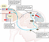 Figure 4
Functional alterations in the trigeminovascular pain pathway in FHM1 knockin mouse models. In FHM1 mice, a number of alterations in the trigeminovascular system might contribute to migraine-relevant pain behavior, including enhanced and longer lasting cortical and subcortical inflammatory responses induced by CSD; enhanced CGRP release from trigeminal ganglia and, possibly, enhanced neuron-glia inflammatory cycle at the ganglion level; enhanced c-fos expression in centromedial/posterior thalamic nuclei elicited by dural stimulation and enhanced synaptic transmission from thalamocortical neurons of the ventrobasal nucleus to layer 4 neurons in the primary somatosensory cortex. In contrast, migraine-relevant pain behavior is likely unrelated to CGRP-dependent meningeal neurogenic inflammation.
Synaptic transmission from thalamocortical neurons of the ventrobasal nucleus (which comprises the primary thalamus sensory relay nucleus of trigeminovascular activation, ref. 4) to layer 4 neurons in the primary somatosensory cortex was enhanced in Cacna1aR192Q mice (110), as was c-fos expression elicited by dural electrical stimulation in centromedian/posterior thalamic nuclei (140) (Figure 4). It remains to be established whether these alterations contribute to migraine-relevant pain behavior in FHM1 mice.
CSD induced an enhanced and longer lasting cortical and subcortical inflammatory response in heterozygous Cacna1aS218L mice compared with that in WT mice, which was reduced by local block of NMDARs (130, 141) (Figure 4). This might contribute to the longer lasting CSD-triggered pain phenotype in FHM1 mice (130).
The cingulate cortex is activated during both spontaneous migraine attacks and during the premonitory phase of the migraine-like headache induced by NTG (132, 142). Some of the downstream regions to which the cingulate cortex is connected are involved in descending modulatory pathways implicated in central sensitization of trigeminovascular neurons in the trigeminal nucleus caudalis (TNC) (2, 3, 143). Sensitization of TNC neurons receiving convergent input from the meningeal nociceptors and facial skin underlies migraine cephalic allodynia (2). In Atp1a2W887R mice, restoring α2 NKA expression in the cingulate cortex (via local viral delivery of the Atp1a2 gene) not only reversed the defective K+ and glutamate clearance and the increased output firing in layer 5 pyramidal cells but also strongly reduced NTG-induced orofacial mechanical hypersensitivity (114). This shows that altered neural activity in the cingulate cortex contributes to the enhanced cranial pain responses to NTG treatment in Atp1a2W887R mice.


Copyright © 2026 American Society for Clinical Investigation
ISSN: 0021-9738 (print), 1558-8238 (online)