
Advertisement
Review
Open Access |  10.1172/JCI208000
Sterol biosynthesis, brain development, and disease
Eric S. Peeples,1,2,3 Zeljka Korade,1,2 and Karoly Mirnics1,2,4
1Department of Pediatrics, University of Nebraska Medical Center, Omaha, Nebraska, USA.
2Child Health Research Institute, Omaha, Nebraska, USA.
3Division of Neonatology, Children’s Nebraska, Omaha, Nebraska, USA.
4Munroe-Meyer Institute for Genetics and Rehabilitation, University of Nebraska Medical Center, Omaha, Nebraska, USA.
Address correspondence to: Karoly Mirnics, Munroe-Meyer Institute for Genetics and Rehabilitation, 985450 University of Nebraska Medical Center, Omaha, Nebraska 68198-5450, USA. Phone: 402.559.5702; Email: karoly.mirnics@unmc.edu.
Find articles by Peeples, E. in: PubMed | Google Scholar
1Department of Pediatrics, University of Nebraska Medical Center, Omaha, Nebraska, USA.
2Child Health Research Institute, Omaha, Nebraska, USA.
3Division of Neonatology, Children’s Nebraska, Omaha, Nebraska, USA.
4Munroe-Meyer Institute for Genetics and Rehabilitation, University of Nebraska Medical Center, Omaha, Nebraska, USA.
Address correspondence to: Karoly Mirnics, Munroe-Meyer Institute for Genetics and Rehabilitation, 985450 University of Nebraska Medical Center, Omaha, Nebraska 68198-5450, USA. Phone: 402.559.5702; Email: karoly.mirnics@unmc.edu.
Find articles by Korade, Z. in: PubMed | Google Scholar
1Department of Pediatrics, University of Nebraska Medical Center, Omaha, Nebraska, USA.
2Child Health Research Institute, Omaha, Nebraska, USA.
3Division of Neonatology, Children’s Nebraska, Omaha, Nebraska, USA.
4Munroe-Meyer Institute for Genetics and Rehabilitation, University of Nebraska Medical Center, Omaha, Nebraska, USA.
Address correspondence to: Karoly Mirnics, Munroe-Meyer Institute for Genetics and Rehabilitation, 985450 University of Nebraska Medical Center, Omaha, Nebraska 68198-5450, USA. Phone: 402.559.5702; Email: karoly.mirnics@unmc.edu.
Find articles by Mirnics, K. in: PubMed | Google Scholar
Published July 15, 2026 - More info
J Clin Invest. 2026;136(14):e208000. https://doi.org/10.1172/JCI208000.
© 2026 Peeples et al. This work is licensed under the Creative Commons Attribution 4.0 International License. To view a copy of this license, visit http://creativecommons.org/licenses/by/4.0/.

-
Cholesterol biosynthesis
At the molecular level, developmental production of cholesterol involves complex, coordinated expression of enzymes across the two interconnected sterol biosynthesis pathways (Kandutsch-Russell and Bloch branches) (6, 7) (Figure 1). The condensation of acetyl-CoA units into mevalonate, catalyzed by HMG-CoA reductase, is the rate-limiting step and a major point of control. Subsequent reactions yield lanosterol, which then undergoes multiple postsqualene modifications mediated by lanosterol synthase (LSS), 24-dehydrocholesterol reductase (DHCR24), 7-dehydrocholesterol reductase (DHCR7), and other sterol isomerases and reductases. During perinatal brain development, expression of DHCR24 and DHCR7 is particularly elevated, consistent with the high demand for cholesterol in axonal growth and synaptogenesis (24). Transcriptional regulation of these enzymes is further modulated by multiple SREBPs, liver X receptors, and other lipid-sensitive transcription factors (25–29).
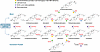 Figure 1
Enzymes and intermediates of the postlanosterol biosynthetic pathway. Enzymes are denoted in red font; sterol intermediates are in black. Bloch branch intermediates are in the top row; Kandutsch-Russell branch intermediates are in the bottom row. Colored shapes denote genetic inhibition associated with four inborn errors of postlanosterol biosynthesis (also see Table 1).
Sterol biosynthesis can be perturbed through multiple mechanistic pathways. Transcriptional downregulation of sterol biosynthesis genes — via suppression of SREBP or other regulatory transcription factors — can reduce pathway capacity (9, 25, 30, 31). Direct enzymatic inhibition of postsqualene reductases and isomerases can further disrupt both the Kandutsch-Russell and Bloch branches (32). Perturbations of NADPH availability can impair essential reductive steps, while mitochondrial dysfunction or oxidative stress may exert secondary inhibition across the pathway (33–35). Decreased flux through the mevalonate pathway, as well as competition for cytochrome P450 enzymes that modify sterols and oxysterols, can additionally shift metabolite pools. All of these mechanisms can destabilize sterol homeostasis and alter downstream sterol accumulation and/or oxysterol signaling.
-
Sterol biosynthesis gene variants result in neurodevelopmental disorders
Inborn errors of cholesterol metabolism encompass a group of rare but severe genetic disorders resulting from loss-of-function pathogenic variants (mutations) that impair specific enzymatic steps in the sterol biosynthetic pathway (22). The spectrum of clinical severity correlates with residual enzymatic activity and the degree of sterol intermediate accumulation. Notable examples of inborn errors of sterol biosynthesis include Smith-Lemli-Opitz syndrome (SLOS; DHCR7 deficiency) (56), desmosterolosis (DHCR24 deficiency) (57), lathosterolosis (SC5D pathogenic variants) (58), squalene synthase deficiency (59), LSS deficiency (60), X-linked chondrodysplasia punctata 2 (61–63), and 17β-hydroxysteroid dehydrogenase type 7 deficiency (64–66). These diseases are characterized by distinct biochemical profiles and overlapping neurological and dysmorphological phenotypes.
Most of these disorders are rare, and only variants sparing some enzymatic activity are compatible with life. Clinically affected individuals typically present with a combination of craniofacial anomalies, limb malformations, cataracts, ichthyosis, skeletal dysplasias, growth restriction, and variable other organ defects (Table 1). Neurologic manifestations range from hypotonia and seizures to structural brain anomalies and intellectual disability. Biochemical diagnosis relies on sterol profiling by gas chromatography–mass spectrometry or liquid chromatography–tandem mass spectrometry, which identify characteristic precursor accumulation patterns. Genetic diagnosis is established by DNA sequencing. Treatment options remain limited and largely supportive; however, cholesterol supplementation might benefit some patients, and emerging strategies aim to reduce toxic sterol intermediates or enhance residual enzymatic activity (67, 68).
Desmosterol is the immediate precursor to cholesterol in the Bloch pathway, and it is used as a marker of endogenous cholesterol synthesis. It also serves as a ligand for liver X receptors. Desmosterol is converted to cholesterol by DHCR24 (Figure 1). Desmosterolosis is a rare autosomal recessive disorder caused by biallelic pathogenic variants in DHCR24 (69–71). Pathogenic variants lead to markedly reduced enzyme activity and consequent accumulation of desmosterol in plasma and tissues. Clinically, affected individuals present with a constellation of multiple congenital anomalies, including corpus callosum agenesis or hypoplasia, white matter loss, microcephaly or macrocephaly, arthrogryposis, spasticity, cleft palate, facial dysmorphism, ocular abnormalities, and seizures (72). Intellectual disability, delayed motor development, and failure to thrive are reported across all cases. Due to its rarity, phenotypic overlap with inborn errors of postlanosterol biosynthesis, and limited availability of testing, many cases might never be diagnosed.
SLOS is the most common and best characterized inborn error of sterol biosynthesis, caused by homozygous loss-of-function pathogenic variants in DHCR7, which encodes the enzyme responsible for reducing 7-DHC to cholesterol in the final biosynthetic step (56). SLOS manifests with microcephaly, intellectual disability, dysmorphic features, and behavioral disturbances. SLOS can be considered a sonic hedgehog (SHH) pathway disease because the SHH protein needs cholesterol to become active, and without sufficient cholesterol, the SHH signaling pathway is disrupted, leading to many of the developmental abnormalities associated with the disorder (73). In mouse models of SLOS and lathosterolosis, reduced cellular cholesterol correlates with diminished Hh responsiveness (55). Sever et al. (74) identified B-ring 7-DHC–derived oxysterols that inhibit Hh signaling by blocking the Hh receptor Smoothened, providing a mechanistic link between sterol biosynthesis defects and impaired morphogen signaling.
Unique to SLOS is the accumulation of 7-DHC oxysterols that have biological activity and likely contribute to the phenotype seen in this disorder (75, 76). Notably, many SLOS patients exhibit a behavioral phenotype consistent with autism spectrum disorder (ASD) (77). Furthermore, in humans with SLOS, cerebrospinal fluid levels of serotonin metabolite 5-HIAA and dopamine metabolite homovanillic acid are reduced relative to age-matched controls (78), suggesting a sterol-related defect in synaptic vesicle formation and neurotransmitter release.
Beyond SLOS and other inborn errors of sterol biosynthesis, additional neurodevelopmental and autism-related disorders have also been linked to disruptions in cholesterol metabolism. In a small study of children with ASD, 19% (19/100) had serum cholesterol levels below the fifth percentile for age (79). A landmark study from the Kohane lab in 2020 found that 6.5% of individuals with ASD had abnormal lipid levels — nearly twice the number of those without autism (80). In addition, when the fathers in the families studied had lipid abnormalities, the risk of having a child with autism was 13% higher. This comprehensive study provided strong evidence for a dyslipidemia-associated ASD subtype (80). Individuals with Rett syndrome (MECP2 variants) also exhibit elevated circulating cholesterol (81, 82), whereas patients with Fragile X syndrome show reduced total cholesterol and HDL levels (83). Taken together, these studies highlight the potential that dysregulation of cholesterol homeostasis is a shared underlying mechanism in neurodevelopmental disorders.

-
Conclusions and future directions
The work described here firmly establishes the critical requirement for cholesterol biosynthesis during brain development and indicates that exposure to SBIMs during pregnancy may have neurodevelopmental effects. The concern that medication used during pregnancy could predispose to adverse outcomes for the baby is not new (112), and medication package inserts developed by pharmaceutical companies clearly state the risks of medications if taken during pregnancy. Unfortunately, although the package inserts are easily accessible for physicians and patients, the relevant data are often buried in the large amount of information in them. Notably, in 2015, the FDA retired the A, B, C, D, and X risk pregnancy categories, which had been used since 1979 (113). The categories were replaced with narrative sections and subsections to allow for more nuance and data transparency. However, these narrative sections may cause more confusion, especially for patients with low health literacy and for physicians attempting to balance the time burden of their clinical duties with interpreting the often complex and sometimes conflicting study results listed in the inserts.
Importantly, the data from our recent study (23) suggest only one of likely several important associations with ASD and do not conflict with the extensively documented, strong genetic component of ASD (114–116). Not all pregnancies prescribed SBIMs resulted in a child with ASD, and it is not yet clear which factors may exacerbate the sterol-inhibiting effects (or side effects) of these medications. The variables that could interact with SBIM use are nearly endless, including the sterol biosynthesis gene variants of the mother and developing baby, comorbidities, polypharmacy, environmental influences, socioeconomical circumstances, and nutritional factors. Furthermore, the dose, duration, and timing of SBIM administration are likely to also be critical determinants of ASD risk.
Notably, one potential risk factor is widely overlooked. There is extremely limited data on the effects of paternal SBIM use in the context of reproductive health. Cholesterol is essential for male fertility as a precursor to testosterone and a structural component of sperm membranes (117). Sperm divide extremely fast, and animal model studies show a negative effect of aripiprazole on male fertility, sperm motility, and sperm quality (118, 119). Similarly, atorvastatin treatment has been shown to alter semen parameters in healthy males, including the total number of spermatozoa, vitality, total motility, and kinetics (120). The effect of paternal SBIM use on fetal development is thus far untested. However, given that paternal alcohol consumption before conception can disrupt offspring’s brain, skull, and face development (121), investigating the impact of environmental factors including paternal SBIM use on neurodevelopmental outcomes should become a priority (122).
It is also important to acknowledge that in addition to its neurodevelopmental roles, cholesterol is also critical for adult brain function. As in the developing brain, cholesterol is critical in the adult brain for structural integrity and to enable efficient synaptic transmission. Dysregulated cholesterol homeostasis may contribute to the development of neurodegenerative diseases, including Alzheimer’s disease (123, 124). How interventions including SBIMs may interact with this biology is another area of intense interest in the field (125).
Finally, the combined findings above raise a critical question: Are we only seeing the proverbial tip of the iceberg? Can prenatal SBIM exposure lead to other developmental challenges, such as dysmorphologies, obsessive-compulsive disorder, learning difficulties, or anxiety later in childhood or as an adult? Does fetal sterol biosynthesis inhibition permanently rewire the developing brain, with functional/behavioral outcomes that may only reveal themselves years after the initial insult? We propose that the strongest inhibition of sterol biosynthesis might result in earlier and more clinically apparent changes (e.g., dysmorphologies), while more moderate inhibition may have primarily microanatomical and behavioral consequences (Figure 2). This complex framework deserves further investigation.
 Figure 2
Proposed neurodevelopmental pathophysiology and consequences of developmental sterol inhibition. SBIMs utilized during pregnancy can interact with the normal developmental trajectory of the fetal brain and body. The magnitude of the sterol inhibition depends on many factors highlighted in the circle. We propose that the strongest sterol inhibition would result in SLOS-like dysmorphologies, while a more moderate inhibition would result in functional consequences with a delayed manifestation of functional connectivity changes that could persist through a lifetime. ADHD, attention-deficit/hyperactivity disorder; ASD, autism spectrum disorder; OCD, obsessive-compulsive disorder.
In summary, developmental cholesterol biosynthesis is fundamental for CNS maturation. The spatiotemporal regulation of sterol synthesis, enzyme expression, and intercellular lipid transport ensures adequate cholesterol supply for membrane biogenesis and signaling throughout neurodevelopment. Prenatal disruption of this finely tuned system — whether by medications or genetic, metabolic, or environmental factors — can have lasting consequences for brain structure and function. Prescription of SBIMs to pregnant women increased considerably over the period of our study, from 4.6% in 2014 to 16.8% in 2023 (23). Given the potential risks of the sterol biosynthesis–inhibiting effects, the increased utilization of SBIMs, likely reflecting increased provider comfort with prescribing these medications during pregnancy, is a cause for concern (126, 127).

Article tools
- Download citation information
- Send a comment
- Terms of use
- Standard abbreviations
- Need help? Email the journal
Metrics
Go to
- Top
- Abstract
- Introduction
- Cholesterol biosynthesis
- Cholesterol and the developing brain
- Sterol biosynthesis gene variants result in neurodevelopmental disorders
- Sterol biosynthesis–inhibiting side effects of currently approved medications
- Human biomaterial and epidemiological studies
- Sterol biosynthesis inhibition in fetal fentanyl syndrome
- SBIM prescription during pregnancy and risk of ASD
- Interaction between sterol inhibition and genetic vulnerability
- Conclusions and future directions
- Conflict of interest
- Funding support
- Footnotes
- References
- Version history


Copyright © 2026 American Society for Clinical Investigation
ISSN: 0021-9738 (print), 1558-8238 (online)