
Go to JCI Insight
  • About
  • Editors
  • Consulting Editors
  • For authors
  • Publication ethics
  • Publication alerts by email
  • Advertising
  • Job board
  • Contact
  • Clinical Research and Public Health
  • Current issue
  • Past issues
  • By specialty
    • COVID-19
    • Cardiology
    • Gastroenterology
    • Immunology
    • Metabolism
    • Nephrology
    • Neuroscience
    • Oncology
    • Pulmonology
    • Vascular biology
    • All ...
  • Videos
    • ASCI Milestone Awards
    • Video Abstracts
    • Conversations with Giants in Medicine
  • Reviews
    • View all reviews ...
    • The cGAS-STING pathway: DNA sensing in health and disease (Jun 2026)
    • Neurodegeneration (Mar 2026)
    • Clinical innovation and scientific progress in GLP-1 medicine (Nov 2025)
    • Pancreatic Cancer (Jul 2025)
    • Complement Biology and Therapeutics (May 2025)
    • Evolving insights into MASLD and MASH pathogenesis and treatment (Apr 2025)
    • Microbiome in Health and Disease (Feb 2025)
    • View all review series ...
  • Viewpoint
  • Collections
    • In-Press Preview
    • Clinical Research and Public Health
    • Research Letters
    • Letters to the Editor
    • Editorials
    • Commentaries
    • Editor's notes
    • Reviews
    • Viewpoints
    • 100th anniversary
    • Top read articles

  • Current issue
  • Past issues
  • Specialties
  • Reviews
  • Review series
  • ASCI Milestone Awards
  • Video Abstracts
  • Conversations with Giants in Medicine
  • In-Press Preview
  • Clinical Research and Public Health
  • Research Letters
  • Letters to the Editor
  • Editorials
  • Commentaries
  • Editor's notes
  • Reviews
  • Viewpoints
  • 100th anniversary
  • Top read articles
  • About
  • Editors
  • Consulting Editors
  • For authors
  • Publication ethics
  • Publication alerts by email
  • Advertising
  • Job board
  • Contact
Top
  • View PDF
  • Download citation information
  • Send a comment
  • Terms of use
  • Standard abbreviations
  • Need help? Email the journal
  • Top
  • Abstract
  • Introduction
  • Mechanisms of transcript structure diversity
  • Regulation of transcript isoform diversity
  • Pathological splicing alterations and disease phenotypes
  • Technologies characterizing alternative splicing and isoforms
  • Therapeutic strategies targeting alternative splicing and isoforms
  • Conclusion and future directions
  • Conflict of interest
  • Funding support
  • Footnotes
  • References
  • Version history
  • Article usage
  • Citations to this article

Advertisement

Review Open Access | 10.1172/JCI207476

The splice of life: an isoform-centric view of disease, technology, and therapeutics

Timothy Pan,1,2,3 Lina Lu,1,2 and Ruli Gao1,2,3

1Department of Biochemistry and Molecular Genetics;

2Center for Cancer Genomics, Robert H. Lurie Cancer Center; and

3The Driskill Graduate Program, Northwestern University Feinberg School of Medicine, Chicago, Illinois, USA.

Address correspondence to: Ruli Gao, 303 E Superior St, SQ7-527, Chicago, Illinois, USA 60611. Email: ruli.gao@northwestern.edu.

Find articles by Pan, T. in: PubMed | Google Scholar

1Department of Biochemistry and Molecular Genetics;

2Center for Cancer Genomics, Robert H. Lurie Cancer Center; and

3The Driskill Graduate Program, Northwestern University Feinberg School of Medicine, Chicago, Illinois, USA.

Address correspondence to: Ruli Gao, 303 E Superior St, SQ7-527, Chicago, Illinois, USA 60611. Email: ruli.gao@northwestern.edu.

Find articles by Lu, L. in: PubMed | Google Scholar

1Department of Biochemistry and Molecular Genetics;

2Center for Cancer Genomics, Robert H. Lurie Cancer Center; and

3The Driskill Graduate Program, Northwestern University Feinberg School of Medicine, Chicago, Illinois, USA.

Address correspondence to: Ruli Gao, 303 E Superior St, SQ7-527, Chicago, Illinois, USA 60611. Email: ruli.gao@northwestern.edu.

Find articles by Gao, R. in: PubMed | Google Scholar

Published July 15, 2026 - More info

Published in Volume 136, Issue 14 on July 15, 2026
J Clin Invest. 2026;136(14):e207476. https://doi.org/10.1172/JCI207476.
© 2026 Pan et al. This work is licensed under the Creative Commons Attribution 4.0 International License. To view a copy of this license, visit http://creativecommons.org/licenses/by/4.0/.
Published July 15, 2026 - Version history
View PDF
Abstract

Alternative splicing is a pervasive mechanism that expands the coding potential and functional complexity of the human genome. Dysregulated isoform usage alters gene functions and contributes broadly to human disease across developmental, neurodegenerative, and cancer settings. Technologies for characterizing splicing and isoforms have advanced rapidly, evolving from Sanger sequencing of individual cDNA clones to high-throughput next-generation sequencing of splice junctions, and more recently to long-read sequencing that resolves full-length transcripts at bulk, single-cell, and spatial resolutions. With the growing recognition of their critical roles in human disease, multiple therapeutic modalities have been developed to precisely target splicing and isoform regulation at the DNA, RNA, and protein levels. Clinical-grade small molecules and antisense oligonucleotides that modulate aberrant RNA splicing and isoform switching have become available, offering new hope for previously incurable diseases. Here, we review this crucial yet underexplored layer of transcriptomic regulation in human disease, encompassing regulatory mechanisms, technological advances, therapeutic strategies, and future directions.

Introduction

The human genome catalogs more than 20,000 protein-coding genes, more than 95% of which are multiexonic, with an average of approximately 9 exons per gene (1). Through selective inclusion or exclusion of exons, alternative splicing generates transcript isoforms that directly impact protein sequences and modulate functional elements, including transcript stability, localization, translational efficiency, and the retention or loss of regulatory elements. These processes are tightly coordinated to support physiological demands of distinct tissues, defining and reinforcing cell identity, plasticity, and intercellular interaction. While these diverse attributes grant functional complexity and adaptability at the cellular and organismal level, splicing dysregulation is known to compromise normal cell physiology, carrying serious implications for disease progression. Indeed, altered splicing machinery and mis-spliced isoforms have been implicated in a broad range of human diseases, including neurological and muscular disorders, cardiovascular diseases, autoimmune diseases, fibrotic and metabolic diseases, and cancer (2–5). Moreover, altered peptide sequences may serve as neoantigen substrates, rewiring immune microenvironments, particularly in cancer (6, 7).

Splicing abnormalities were first characterized via gel electrophoresis and Sanger sequencing of cDNA clones. The launch of next-generation sequencers led to an era of high-throughput profiling of splicing events at large scale. More recently, long-read sequencing has emerged as a transformative approach for resolving full-length transcripts across bulk, single-cell, and spatial contexts. With the growing recognition of splicing-related pathogenesis, therapeutic strategies that modulate RNA splicing and isoform switching have become a new class of interventions for previously incurable diseases.

Although the origins, mechanistic basis, regulation, and pathophysiological significance of alternative splicing have been extensively reviewed (8–13), here, we focus on the functional impact of transcript isoforms in human disease, particularly the insights gained from genomics-driven efforts. We examine how technological advances in RNA sequencing have transformed the identification and interpretation of transcript isoforms. Last, we provide an overview of the current state of splicing-based therapeutics.

Mechanisms of transcript structure diversity

While alternative splicing serves as the predominant contributor of transcript diversity, nonsplicing sources of diversity, such as alternative promoters, transcription start sites (TSSs), transcription termination sites (TTSs), and polyadenylation, represent an emerging focus in understanding transcriptome complexity (Figure 1). Moreover, recent data highlight the roles of transcriptional rate, chromatin state, and epigenetic modifications in splicing outcomes, suggesting a higher degree of coordination between RNA processing and gene expression than previously thought. The mechanistic and regulatory basis of these processes lays the foundation for the extensive repertoire of transcript isoforms, supporting both broad and specialized functions at the cellular, tissue, and organismal levels.
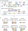
A dense regulatory network dictates isoform diversity.Figure 1

A dense regulatory network dictates isoform diversity. (A) Cis-regulatory factors encompass the 5′ and 3′ UTRs, exonic and intronic splicing enhancer and silencers (ESEs, ESSs, ISEs, ISSs), the 5′ and 3′ splice site consensus sequences, and RNA secondary structures. Sources of trans-splicing regulation include spliceosomal machinery, RNA-binding proteins (RBPs), splicing factors, long noncoding RNAs (lncRNAs), RNA editases, and alternative splicing with nonsense-mediated decay (AS-NMD) coupling. The coordination between cis- and trans-regulation determines alternative splicing event decisions. Epigenetic, chromatin, and transcriptional regulation encompass alternative promoters, transcription start sites (TSSs), histone modifications, transcriptional elongation rate, alternative transcription termination sites (TTSs), and alternative polyadenylation sites (APAs). (B) Splicing events can be categorized as exon skipping, alternative 5′ and 3′ splice sites, intron retention, mutually exclusive exons, alternative promoters, alternative TSSs, and APAs. snRNPs, small nuclear ribonucleoproteins; SRSFs, serine and arginine rich splicing factors; hnRNPs, heterogeneous nuclear ribonucleoproteins; Pol, polymerase.

Alternative splicing–derived transcript diversity. Alternative splicing generates transcript isoforms that vary in exon and intron composition from the same gene. The majority of introns (i.e., U2-type introns) are bound by 5′ and 3′ splice sites, which feature the core dinucleotides GU and AG, respectively. These splice sites, together with a core branch point adenosine and polypyrimidine tract located upstream of the 3′ splice site, constitute the essential elements of splicing by the major spliceosome. The spliceosome, composed of the core snRNPs U1, U2, U4, U5, and U6, along with associated splicing factors, assembles onto splice site motifs on pre-mRNA to catalyze two sequential transesterification reactions, producing the lariat intermediate and completing exon ligation (8, 14, 15). Minor introns (i.e., U12-type), accounting for <0.4%–1% of introns in human genes, feature less efficient AU-AC splice site motifs with a distinct branch point sequence, which mediate excision by the minor spliceosome (16, 17). The precise patterns of intron removal and exon ligation largely determine the identity of the mature transcript isoform and can be classified into several distinct alternative splicing events (18).

A common event generating alternative isoforms is cassette exon splicing, or exon skipping, in which an exon is selectively removed from the pre-mRNA. While constitutive exons are essential and almost always included in mature mRNA, cassette exons predominantly function as modular elements that encode tissue-specific protein domains, functional motifs, and flexible regions, providing a major source of protein diversity (19, 20). Cassette exons are often characterized by weaker splice sites, shorter length, and lower GC content compared with constitutive exons (21). A widely studied cassette exon is exon 7 of survival motor neuron 2 (SMN2), which produces a nonfunctional protein isoform when skipped (22). As reviewed in detail below, therapeutic interventions in spinal muscular atrophy (SMA) aim to promote SMN2 exon 7 inclusion to rescue the function of its paralog, SMN1, due to homozygous loss.

The spliceosome may also mediate cleavage proximal to canonical splice sites, known as alternative splice sites. Splicing at these alternative sites may shift the 3′ and 5′ boundaries of the upstream and downstream exon, respectively, producing transcript isoforms with altered protein-coding sequences, open reading frames (ORFs), or regulatory motifs (23, 24). The exact cleavage site is determined by regulatory elements embedded in the pre-mRNA and their interaction with splicing factors. Alternative splice sites can also naturally occur within transcript bodies, and their selective recognition by splicing factors contributes to shifts in isoform usage. A representative example is the alternative 5′ splice site selection of exon 2 of the BCLX (aka, BCL2L1) gene, which produces two splice variants that exert opposing effects on apoptosis in cancer (25).

Intron retention events, also widely observed, occur when an intron that is normally excised during splicing is retained in the mature transcript. These introns often disrupt the frame of the transcript and render it nonproductive. Accordingly, the mechanism is a common mode of regulation that controls transcript abundance in a tissue- and cell type–specific manner, as well as in response to developmental cues, stress, and disease (26–28). Retained introns may also serve as a signal for nuclear retention, affecting turnover and regulating splicing completion (29–31). In some cases, transcripts harboring retained introns maintain an ORF and are translated to produce functional protein isoforms. These unique introns usually encode amino acids within disordered regions and are termed “exitrons” (exonic introns) (32, 33).

A small subset of alternative splicing events involves mutually exclusive exons (MXEs), in which only one exon from a set of two or more is retained in the mature mRNA, while the others are excised. MXEs often encode alternative versions of a protein domain and were previously thought to originate from the same ancestral exon. Large-scale RNA-seq studies characterizing more than 1,300 MXEs have since shown little sequence homology across MXEs, many of which are embedded within secondary structural elements (34). A classic example of MXEs is found in the fibronectin gene FN1, which contains 2 MXEs encoding either extra domain A or extra domain B, conferring distinct functions in extracellular matrix remodeling, tissue repair, and cell differentiation (35).

Non-splicing-derived transcript structure diversity. A large portion of transcript variability is introduced outside of spliceosome-mediated cleavage and ligation. These differences emerge predominantly at the transcriptional level, encompassing alternative promoters, alternative TSSs, and alternative TTSs. In many cases, alternative promoters produce transcript isoforms that differ in the first exon, resulting in the presence or absence of distinct N-terminal protein domains that contribute specialized functional activities (36–38). In comparison, alternative TSSs represent a finer scale of transcript variation, distinguished by the initiation of transcription at different positions within a common promoter. This mechanism can generate transcript variation that influences posttranscriptional regulation in the form of increased 5′ UTR length, the inclusion of repressive elements, and translationally repressive upstream ORFs (39). Alternative TTSs, also referred to as alternative cleavage and polyadenylation sites, contribute to the production of distinct isoforms by defining heterogeneity at the 3′ UTR, a critical component that regulates RNA subcellular localization and turnover (40, 41). Additionally, variation of the terminal exon may produce key protein isoform derivations, as exemplified by the secreted and membrane-bound forms of the IgM heavy chain in B cells (42). More than half of human genes have been reported to encode two or more canonical polyadenylation signals, with differential usage associated with cell and tissue types, extracellular cues, developmental stage, and disease (43, 44).
Regulation of transcript isoform diversity

The mechanisms generating transcript diversity are regulated through a coordinated interplay among cis-acting elements, the trans-acting factors that bind or influence them, and more recently appreciated cotranscriptional, epigenetic, and biophysical dynamics. Further regulatory mechanisms include RNA editing and NMD, as well as splicing regulation mediated by lncRNAs.

Cis-regulatory elements. By convention, cis-acting elements are motifs embedded within the pre-mRNA, with the consensus sequences of 5′ and 3′ splice sites, branch points, and polypyrimidine tracts representing the archetypal features that regulate splicing activity. These motifs vary in conservation, strongly influencing direct recognition and binding by core spliceosome machinery (45). Less evolutionarily constrained cis-acting elements are exonic and intronic splicing enhancers and silencers (ESEs, ESSs, ISEs, ISSs), which are often short motifs that recruit a diverse array of RBPs and splicing factors (13, 46, 47). As mentioned, pre-mRNA may also harbor multiple polyadenylation signals, canonically AAUAAA hexamers flanked by upstream or downstream auxiliary elements, that mediate cleavage and polyadenylation efficiency capable of exerting strong influence on the 3′ UTR (40, 48). While not strictly encoded, RNA secondary structures are a unique class of cis-acting elements increasingly recognized as splicing regulators, functioning by sterically masking linear cis-acting motifs. These structures (e.g., hairpins, stem-loops, and G-quadruplexes) have also been shown to serve as binding sites for RBPs and play an active role impeding transcription, allowing for dynamic regulation of splicing events (49, 50). The significance of pre-mRNA secondary structures is demonstrated by silent mutations that destabilize a stem-loop proximal to the 5′ splice site of Tau exon 10, leading to increased exon inclusion and pathogenesis of frontotemporal dementia (51).

Trans-acting regulatory factors. Trans-acting factors traditionally encompass the spliceosome components, splicing factors, and RBPs, which collectively regulate splicing activity by recognizing cis-acting motifs and modulating spliceosome activity. The regulatory roles of splicing factors and RBPs largely depend on their expression patterns, which are tissue and cell type dependent (52). Individual splicing factors can recognize a broad array of RNA motifs, further amplifying their complex roles in regulation (53). These properties are largely dictated by RNA-binding motifs, enabling broad classification of splicing factors into families such as serine-arginine–rich (SR) proteins and hnRNPs (54, 55).

Given the programmed expression and broad specificity of splicing factors, recent attention has shifted toward transcriptional dynamics and the chromatin landscape to explain how specific interactions between cis- and trans-acting factors are achieved in regulating splicing outcomes. In addition, increasing data support the kinetic coupling of alternative splicing and RNA Pol II elongation leading to preference of distinct splicing events, with the inclusion of exon 33 of fibronectin transcripts among the most well-characterized cases (56). The nonrandom enrichment of nucleosomes at exon-intron boundaries also suggests chromatin structure as a regulator of splice site decisions, modulating elongation kinetics and indirectly recruiting splicing factors through epigenetic modifications (57, 58). Emerging models describe the impact and coordination of RNA Pol II elongation kinetics, chromatin structure, and histone modifications on isoform diversity (59, 60).

RNA editases (e.g., ADARs and APOBECs) directly modify RNA sequences, affecting over 60% of human transcripts via RNA-editing events, typically A-to-I or C-to-U conversions on single-stranded or double-stranded RNA (61). In addition to their role in antiviral immunity, these conversions are known to tune cis-regulatory elements and, consequently, splice site decisions (62, 63). Furthermore, the expression of many genes encoding RNA editases is tissue restricted, allowing for context-dependent editing tailored to specific tissue and cell conditions (64).

NMD, a posttranscriptional surveillance mechanism, serves as a key pathway that modulates isoform abundance (65, 66). In this process, premature termination codons (PTCs) within exons encountered during translation are recognized by degradation factors (e.g., UPF1), which recruit decay factors that promote decapping and exonucleolytic degradation. Controlled inclusion of PTCs couples alternative splicing with NMD (AS-NMD), generating unproductive isoforms selectively targeted for degradation, thereby autoregulating gene and protein expression under various cellular conditions (67). Indeed, large-scale analyses have predicted that over one-third of alternatively spliced isoforms in humans are prone to NMD (68), with a more recent study establishing a link between aberrant splicing and high production of isoforms that bear a PTC, further underscoring the widespread regulatory role of AS-NMD (69).

The lncRNA class represents another potent regulator of alternative splicing dynamics. Direct interactions with splicing factors modulate their phosphorylation state and nuclear localization, as exemplified by the NEAT1 and MALAT1 lncRNAs (70, 71). Alternatively, many lncRNAs form DNA-RNA heteroduplexes (i.e., R-loops) that remodel chromatin, affecting histone modifications and RNA Pol II kinetics, both of which tune splicing decisions (59, 72). Directly influencing splice site selection, lncRNAs may duplex with pre-mRNA, as observed between SAF lncRNA and FAS pre-mRNA. That interaction promotes exon 6 skipping, producing an isoform that decreases sensitivity toward FasL-induced apoptosis in tumor cells (73).

Pathological splicing alterations and disease phenotypes

While alternative splicing provides the genome with immense transcriptional versatility, it also introduces vulnerabilities to dysregulation and pathogenic errors. Here, we illustrate well-characterized disorders arising from the mechanisms described above, highlighting the etiological implications of splicing pathogenesis in human diseases.

Localized cis-acting mutations. A large portion of disease-associated mutations exert their primary functional impact through the disruption of RNA splicing rather than direct alterations to protein-coding sequences (74). These variants primarily occur within cis-regulatory elements, particularly through the destruction of the 5′ or 3′ splice boundaries, leading to exon skipping or intron retention. A more complex class of mutations involves the activation of cryptic splice sites within either introns or the affected exons, creating de novo splice site consensus sequences that disrupt normal splicing regulation. When they occur in introns, these aberrant sites lead to the recognition of noncoding sequences as pseudo-exons. Conversely, when activated within exons, they trigger the use of an alternative, internal boundary, leading to the partial deletion of exonic sequences. Both outcomes typically introduce premature stop codons or internal deletions that compromise protein integrity. In addition, mutations may occur within auxiliary regulatory elements (e.g., ESEs, ISEs, ESSs, ISSs) that bind SR proteins/hnRNPs to regulate pre-mRNA splicing, promoting or inhibiting exon inclusion.

Such splicing defects have been documented across a wide range of human diseases (Table 1). Many mutations linked to Duchenne muscular dystrophy (DMD) target the canonical splice junctions of the dystrophin (DMD) gene, leading to exon skipping and disruption of the ORF. This frameshift introduces a premature stop codon, resulting in the loss of functional dystrophin and progressive muscle wasting characteristic of the DMD phenotype (75–77). Another classic example is β-thalassemia, an inherited disease driven by cis-acting splicing mutations in the HBB gene, which activate cryptic splice sites and eventually cause the loss or reduction of β-globin production (78, 79). The two most common variants involved in β-thalassemia are the IVS1-110G>A and IVS2-654C>T mutations. The IVS1-110G>A variant, prevalent in the Middle East, the Mediterranean, and Cyprus, generates a novel splice acceptor within the first intron of HBB, leading to the inclusion of a 19-nucleotide intronic fragment that triggers a premature stop codon (80, 81). The IVS2-654C>T variant, common in East Asian populations, activates a de novo 5′ splice site deep within the second intron, resulting in the recognition of a 73-nucleotide pseudo-exon (82, 83). Similarly, in Hutchinson-Gilford progeria syndrome (HGPS), the common causal factor is a de novo point mutation in exon 11 of the LMNA gene (c.1824C>T). Although this synonymous substitution does not alter the encoded amino acid, it activates a cryptic 5′ splice donor site within the exon, leading to a 150-nucleotide internal deletion in the mature mRNA. The resulting progerin protein exhibits a truncated C-terminus devoid of a critical endoproteolytic cleavage site required for proper lamin A maturation, causing the accelerated aging phenotype characteristic of HGPS (84, 85).

Table 1

Examples of pathological splicing alterations

Disruption of auxiliary splicing regulatory elements is also frequently implicated in human disease. As mentioned above, homozygous mutation or deletion of SMN1 cannot be fully compensated for by its paralog, SMN2, due to a single nucleotide substitution (c.840C>T) in exon 7. Although this mutation is synonymous, it disrupts an ESE and creates an ESS. This shift reduces SRSF1 binding while recruiting hnRNP A1/A2, ultimately promoting exon 7 skipping. Consequently, the protein isoform lacks an essential C-terminal site necessary for SMN protein stability and self-oligomerization, leading to the loss of crucial motor functions and rapid degradation (22, 86, 87). In frontotemporal dementia with parkinsonism linked to chromosome 17 (FTDP-17), mutations within the MAPT (Tau) gene disturb the delicate balance of alternative splicing. Normal brain function depends on a precise 1:1 ratio of Tau isoforms containing either three (3R) or four (4R) microtubule-binding repeats, a balance maintained by the regulated inclusion of exon 10. In the diseased condition, pathogenic mutations in MAPT strengthen an ESE or weaken an ESS, leading to increased inclusion of exon 10 and skewing the equilibrium toward the 4R-Tau isoform. Excessive 4R-Tau triggers toxic neurofibrillary tangles and progressive neuronal death, driving severe neurodegenerative phenotypes (51, 88, 89).

In addition, cis-acting mutations are widely observed in human cancers (90–94). In many malignancies, mutations create de novo splice sites that alter the function of tumor suppressors or oncogenes. A prominent example is the tumor-suppressive gene TP53, in which a variety of point mutations target nucleotides adjacent to splice sites that cause exon skipping, intron retention, or activation of cryptic splice sites, leading to the production of p53 isoforms that lack transcriptional activity (95–98). Dysregulation of auxiliary splicing elements can also drive oncogenic isoform switching, as exemplified by an alteration in fibroblast growth factor receptor 2 (FGFR2), in which disruption of intronic regulatory elements promotes the usage shift from isoforms associated with the epithelial (IIIb) to the mesenchymal (IIIc) phenotype, thereby enhancing epithelial-mesenchymal transition, a critical step in tumor metastasis and increased invasiveness (99).

Systemic trans-acting splicing dysregulations. In contrast with cis-acting mutations, which typically affect individual genes, trans-acting dysregulations exert broader, systemic effects through alterations in the proteins that orchestrate spliceosome assembly and splice site selection. These factors include the core snRNP components of the spliceosome and auxiliary splicing factors like SR proteins and hnRNPs. Disruptions in their expression, activity, or sequence can lead to widespread and coordinated splicing alterations across numerous transcripts, driving complex disease phenotypes.

A notable class of trans-acting disorders, termed spliceosomopathies, results from mutations in the ubiquitous components of the spliceosome (100). Despite the essentiality of these proteins, their abnormalities often demonstrate tissue-specific outcomes. A prototypical example is retinitis pigmentosa (RP), which is frequently driven by mutations in genes encoding core components of the U4/U6.U5 tri-snRNP complex, including PRPF8, PRPF31, PRPF3, and SNRNP200 (101, 102). The mis-splicing events arising from the abnormality of these components is associated with the apoptosis of retinal photoreceptor cells, a key pathological feature in RP. In addition to core spliceosomal defects, aberrant expression or mutation of splicing regulatory factors may underlie tumorigenesis of many human cancers. For instance, recurrent somatic mutations in SF3B1, U2AF1, and SRSF2 are known to drive a large portion of myelodysplastic syndromes (MDS) and are associated with myeloid leukemias (103, 104). The most prevalent MDS mutation occurs in SF3B1, which causes aberrant recognition of alternative 3′ splice sites, typically ~20 nucleotides upstream of the canonical site, leading to the widespread inclusion of cryptic sequences and resulting in widespread mis-splicing of genes involved in hematopoiesis, mitochondrial function, and DNA repair. Similarly, mutations in U2AF1 and SRSF2 disrupt sequence recognition at the 3′ splice site and exon definition, leading to transcriptome-wide changes in exon inclusion and splice site choice.

Beyond genomic mutations, dysregulated expression of splicing regulatory genes is also frequently implicated in human malignancy. For example, overexpression of SRSF1 has been reported in multiple cancers, promoting splicing dysregulation of genes such as BIN1, MNK2, and S6K1. These aberrant isoforms collectively enhance cell proliferation, inhibit apoptosis, and promote malignant transformation (105, 106). Conversely, altered expression of hnRNP family members can repress exon inclusion and contribute to malignant phenotypes through coordinated changes in alternative splicing networks. For instance, high levels of hnRNPA1 and hnRNPA2 bind to the pyruvate kinase (PKM) pre-mRNA to suppress exon 9 inclusion in favor of exon 10. This orchestrated switching to the PKM2 isoform is recognized as a hallmark of the Warburg effect, facilitating the aerobic glycolysis required for rapid tumor growth (107). Together, these cases demonstrate that even in the absence of primary sequence mutations, disruption of splicing factors can collapse healthy alternative splicing networks and drive complex disease phenotypes.

Technologies characterizing alternative splicing and isoforms

Pre-NGS era. Before the launch of next-generation sequencing (NGS) platforms, the characterization of alternative splicing and transcript isoforms relied primarily on conventional low-throughput molecular techniques (Figure 2). One widely used approach was a cDNA amplification and cloning method that distinguished splicing isoforms based on differences in product sizes (108). Following the establishment of the EST platform (109) by Adams et al., in 1991, advances in full-length cDNA-cloning methods, such as the CAP trapper strategy (110, 111), further drove isoform studies beyond mere gene identification. These developments enabled the systematic discovery of alternative splicing events through EST-to-genome alignment analysis in the late 1990s and early 2000s (112–116). In parallel, pioneering computational frameworks, such as splice graphs by Xing et al. (117), were developed to support genome-wide isoform assembly. Later, microarray hybridization-based exon and splicing arrays provided higher-throughput methodologies for measuring exon-level expression across the genome (118–122). These early approaches established fundamental principles of isoform characterization, yet they were largely limited by low-throughput, incomplete transcript coverage and dependence on prior annotations.
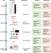
Technological advancements driving isoform-level investigations.Figure 2

Technological advancements driving isoform-level investigations. Pre-NGS era approaches to study transcript isoforms included cDNA cloning, expressed sequence tag (EST) sequencing, and microarray. These methods offered valuable insights but were generally limited by throughput and high labor demands. NGS technologies introduced high-throughput, transcriptome-wide studies of isoforms but were constrained by read fragmentation, which created ambiguity in full-length transcript abundances. While early platforms exhibited high error rates, accuracy has improved. Tissue and cell resolution can now be achieved when integrated with single-cell and spatial technologies. TGS, third-generation sequencing.

NGS-based splicing profiling. The advent of NGS marked a major shift in splicing analysis, enabling large-scale systematic interrogation of transcriptomes. Launched in 2005, the 454 platform was the first successfully commercialized NGS system, which was soon overtaken by Illumina platforms that utilized bridge amplification to generate millions to billions of clonal clusters on a glass flow cell. Due to the inherent constraints of bridge amplification and sequencing-by-synthesis chemistry, Illumina sequencing is limited to short reads, typically ~50–300 bp lengths. Nevertheless, its high yield, accuracy, and throughput enabled deep coverage and precise quantification of splicing events. The deep surveys of alternative splicing in the human transcriptome were reported in 2008 by Pan et al. (123), Sultan et al. (124), and Wang et al. (125), which revealed that approximately 95% of multiexon genes undergo alternative splicing across major human tissues.

As sequencing depth and data scale increased, quantitative characterization of splicing features from large-scale RNA-seq data became challenging. The percent spliced in (PSI or Ψ) metric was first introduced by Wang et al. in 2008 to quantify the ratio of reads supporting the inclusion of an alternative exon to the total number of reads (inclusion + exclusion) (125). As a statistical model, mixture-of-isoforms was applied to estimate the relative isoform dosages and refine PSI values (126). DEXSeq was developed to quantify differential exon usages by testing the changes in exon-level read counts (127). Replicate multivariate analysis of transcript splicing (rMATS), developed by Shen et al. in 2014, introduced replicate-aware models for detecting differential splicing across conditions (128). Along with the development of robust computational methods, large-scale initiatives such as GENCODE (129) and GTEx (130–132) were established to systematically catalog splicing events across human tissues, providing foundational references for modern transcriptome analysis.

Entering the long-read sequencing era. While NGS led to advancements in the understanding of alternative splicing and its roles in human disease, studies were limited to analyzing individual splicing sites without informing complete isoform structures. Only a small portion of isoforms could be confidently reconstructed despite advancements in computational methodologies (133–135). These limitations drove the development of long-read RNA-sequencing (LR-RNA-seq) technologies, primarily pioneered by Pacific Biosciences (PacBio) and the Oxford Nanopore Technologies (ONT), which enable direct, isoform-resolved transcriptome profiling.

Introduced in 2011, the first commercialized long-read platform (PacBio RS) employed a single-molecule real-time (SMRT) sequencing technique to enable real-time observation of DNA Pol activity. A key milestone was the development of high-fidelity reads, which drastically improved sequencing accuracy (136). Newer PacBio platforms (Revio and Sequel IIe) have achieved up to 100 gigabase yields per SMRT Cell. Through the Multiplexed Arrays Sequencing (MAS-Seq) workflow, the throughput of transcriptomic isoform sequencing (ISO-Seq) is increased by concatenating individual amplicons of cDNAs into long molecules for sequencing.

In a fundamentally different approach, the ONT nanopore-based sequencing technology determines nucleotide bases by measuring characteristic changes in the ionic current as individual DNA or RNA molecules pass through a protein nanopore under an applied voltage. The first commercial ONT sequencer, MinION, supported ultralong reads exceeding 100 kilobases and the direct sequencing of native RNA molecules (137). The technology has since advanced toward higher throughput and improved accuracy for large-scale sequencing applications. PromethION platforms now achieve ultrahigh yields, ranging from hundreds of gigabases to multi-terabase scales, supporting large-scale full-length transcriptome studies.

Full-length transcript characterization via advanced computational tools, such as IsoQuant (138), Bambu (139), and others (140–150), has enabled the profiling and discovery of thousands of transcript isoforms, both known and novel. To date, a tremendous number of LR-RNA-seq datasets (>300 studies in humans) have been deposited in the NCBI GEO, encompassing full-length isoform assemblies across diverse tissues and conditions (147, 151, 152). However, the vast majority represent bulk approaches that average signals across heterogeneous cell populations, obscuring cell type– and cell state–specific isoform patterns.

Across single-cell and spatial dimensions. Emerging long-read single-cell RNA-sequencing (LR-scRNA-seq) technologies barcode full-length cDNAs of single cells and sequence them on high-yield long-read sequencing platforms. To address high error rates in barcode sequences, early LR-scRNA-seq approaches often relied on matched short-read data to facilitate accurate barcode detection and data demultiplexing, as leveraged by ScISOr-Seq in 2018 and ScNaUmi-seq in 2020 (153, 154). In 2023, Shiau et al. presented scNanoRNAseq and an accompanying computational tool, scNanoGPS, which fully removed the dependence on matched short-read data (155), representing a transition to a long-read-only, single-cell workflow. Applying this approach, Pan et al. reported a comprehensive cell-level isoform atlas of the adult human heart and heart failure, revealing hundreds of cell type–specific and disease-associated isoform usage-shifting events (156). Concurrently, Al’Khafaji et al. introduced MAS-ISO-seq, combining PacBio MAS-seq with single-cell barcoding to enable isoform-level quantification in individual cells (157). In parallel with technical advances, analytical approaches have shifted from categorizing splicing events toward more accurate quantification of isoform usage and diversity in cells. Computational frameworks, such as DEXSeq and Hypatia (127, 158), have been adapted or developed to enable comparative analysis of isoform complexity across cell populations. Similarly, long-read spatial transcriptomics have advanced by combining cDNA-based spatial transcriptomic platforms such as Visium (10x Genomics) with long-read sequencing to reveal the spatial mapping of full-length isoforms and splicing regulation (150, 159, 160). Despite these advances, current methods remain limited by dropout, although targeted enrichment strategies partially mitigate data sparsity.

New insights enabled by emerging technologies. Recent long-read sequencing studies have led to the identification of previously unappreciated classes of splicing defects, such as multiexon skipping, exonic intron creation, intronic polyadenylation, combinatorial splicing defects, and thousands of novel isoforms that are missed by short reads (161–163). Notably, direct linkages between genetic variants and splice isoforms established from LR-RNA-seq data enabled the diagnosis of multiple rare diseases and revealed new RNA-processing events, such as cryptic intronic polyadenylation activation and haplotype-specific splicing variants (164).

Moreover, emerging single-cell and spatial LR-RNA-seq technologies have further advanced the discovery of isoform usage shifts across cell types, cell states, and spatial niches, providing a foundational framework for understanding previously unrecognized disease mechanisms. In cancer, the identification of hundreds of tumor cell–enriched isoforms (155, 158) offers a rich source of candidate targets for selective tumor cell killing, with the potential to minimize effects on normal tissues. In heart failure, the discovery of cardiomyocyte-specific isoform-switching events (156) may inform more translationally relevant studies and therapeutic strategies. In addition, neoantigens derived from tumor cell–specific, protein-coding isoforms present new opportunities for oncoimmunotherapies. Furthermore, the resolution of cell type– and cell state–specific isoforms with full-length characteristics may enhance the fidelity of in vitro disease models, enabling more accurate recapitulation of cellular phenotypes and fate trajectories.
Therapeutic strategies targeting alternative splicing and isoforms

Growing recognition of the critical roles of alternative splicing and isoforms in human diseases has driven the development of multiple therapeutic strategies (Figure 3). The most widely studied approaches are small-molecule inhibitors and antisense oligonucleotides (ASOs). CRISPR-based approaches have recently expanded rapidly. Splicing-antigen– based immunotherapies and PROTAC-mediated targeted degradation of aberrant isoforms have emerged as novel therapeutics. Clinical-grade modulators of RNA splicing have become available, providing new hope for isoform-driven diseases that were previously considered incurable. Here we focus on the unique isoform-targeting paradigms that have demonstrated therapeutic benefits, highlighting approaches that directly target splicing regulation and isoform usage.
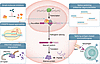
Therapeutic modalities targeting alternative splicing and isoforms in humanFigure 3

Therapeutic modalities targeting alternative splicing and isoforms in human diseases. Small-molecule inhibitors bind spliceosome and splicing factors, preventing generation of aberrant isoforms. Antisense oligonucleotides (ASOs) base-pair to pre-mRNA, blocking aberrant splicing sites. CRISPR-based approaches target pre-mRNA or DNA to correct/disrupt aberrant splice sites. Splicing-antigen–based immunotherapies involve engineering immune cells to recognize isoform-derived neoantigens. PROTAC-mediated degradation approaches recruit an E3 ligase for ubiquitination and proteasomal degradation of aberrant proteins. PROTAC, proteolysis-targeting chimera.

Small-molecule modifiers. Early splicing-targeting strategies focused on identifying natural products that inhibit functional components of the spliceosome. In 2008, O’Brien et al. reported the discovery of isoginkgetin, a natural biflavonoid isolated from Ginkgo biloba leaves that is membrane permeable and inhibits pre-mRNA splicing by disrupting recruitment of the U4/U5/U6 tri-snRNP to the pre-spliceosome (165). The antitumor drug E7107 is another natural product that can block spliceosome assembly (166). As research continued, more compounds targeting splicing factors and spliceosomes were identified through in vitro splicing assays (165, 167), including H3B-8800, an FDA-approved orally available small-molecule splicing modulator that targets mutated SF3B1 to selectively kill cancer cells (168).

Modulation of pre-mRNA structure offers an additional strategy to selectively target mRNAs by binding to secondary or tertiary structural elements, thereby promoting or inhibiting the inclusion of specific exons in pathogenic genes. In 2020, risdiplam (Evrysdi) became the first FDA-approved, orally administered small-molecule splicing modulator for treating SMA. It functions by targeting a weak secondary structure at the 5′ splice site of the pre-mRNA of SMN2 to stabilize its interaction with the spliceosome and overcome an inhibitory stem-loop structure. This precise structural stabilization ensures the inclusion of exon 7, allowing for the production of the full-length functional SMN protein essential for motor neuron survival (169).

ASOs. Splice-switching antisense oligonucleotides (SSOs) are a widely studied and clinically proven therapeutic strategy for selective targeting of pathogenic splicing and isoforms. SSOs are a distinct class of ASOs (typically 15–30 nucleotides) that complement specific regions of pre-mRNA to interfere with splice site recognition, modulating splicing and preventing the production of pathogenic isoforms. The first generation of naked (unmodified) ASOs were highly unstable in vivo as they were subject to various endo- and exonuclease degradation (170, 171). A foundational optimization was achieved through the introduction of phosphorothioate (PS) modification, which greatly enhanced ASO resistance to RNase H cleavage and in vivo half-life (172, 173). However, the PS modification was frequently associated with off-target interactions and toxicities (174, 175). To ameliorate these effects, the PS backbone modification was combined with 2′ sugar modifications and locked nucleic acid (LNA) chemistry (176). These approaches resulted in higher binding affinity and specificity (177, 178) and are widely adopted in modern SSO drugs.

A successful example of SSOs is nusinersen (Spinraza), approved for SMA, an 18-mer 2′-MOE-PS oligonucleotide that binds to an intronic splicing silencer (ISS-N1) in SMN2, forcing the inclusion of exon 7 to produce stable, full-length protein. In cancer, SSOs are applied to rewire BCL2L1 isoform usage, as the isoform BCL-xL drives antiapoptotic signaling and therapeutic resistance, whereas the isoform BCL-xS promotes apoptosis and treatment sensitivity. SSO-mediated redirection of splicing from BCL-xL to BCL-xS demonstrated robust antitumor effects in multiple studies (179). Further examples include SSO targeting of exon 15 of HER2 to reduce the full-length transcripts in breast cancer (180). Similarly, LNA-based oligonucleotides targeting androgen receptor expression exhibit antitumor activity in prostate cancer cells (181). However, although the number of genes successfully targeted with PS backbone SSOs is growing, their cellular toxicity and retention remain incompletely resolved.

A safer alternative, charge-neutral phosphorodiamidate morpholinos (PMOs), exhibit reduced nonspecific protein binding and lower risk of hepatotoxicity and immune activation (177). Due to their weak interactions with cell membranes, PMO-based strategies often require high doses to mitigate inefficient cellular uptake and limited tissue penetration, especially in nonmuscle tissues. In contrast with the wide usage of PS-based SSO agents across tissue types, the effective and approved PMOs predominantly target neuromuscular disorders. Eteplirsen (Exondys 51) is approved for DMD, which employs a PMO-based strategy to induce the skipping of mutated exon 51 in the dystrophin gene (DMD) and restore the ORF. Casimersen (Amondys 45), another PMO, is approved for treating DMD via exon 45 skipping of DMD. Moreover, both golodirsen (Vyondys 53) and viltolarsen (Viltepso) are approved PMOs for exon 53 skipping of DMD.

CRISPR/Cas-based genome editing. CRISPR/Cas-based technology has recently evolved into a precise tool for transcriptomic engineering by enabling the genomic editing of cis-regulatory elements in pre-mRNAs. Unlike SSOs or small-molecule drugs that act transiently at the RNA level, CRISPR/Cas-based strategies allow for durable and potentially permanent correction of aberrant splicing programs in the genome. Although still in active development, they have shown great potential for precise editing of transcript isoforms. Yue et al. successfully rewired the expression of the long- or short-splicing isoforms of the mouse Xist gene by modifying the 5′ splice site in intron 7 using the CRISPR/Cas9 system (182). Du et al. reported an engineering system of CRISPR Artificial Splicing Factors (CASFx) that enhanced inclusion of exon 7 of SMN2 in SMA patient fibroblasts (183). Yuan et al. reported a versatile genetic platform for modulating RNA splicing by using CRISPR-guided cytidine deaminase to convert guanines to adenines at 5′ or 3′ splice sites, thereby correcting the aberrant splicing in disease (184). These early studies reveal considerable promise of applying CRISPR/Cas-based systems for precise isoform corrections yet raise safety concerns because of the off-target effects and permanent genomic alterations.

Recent advances in RNA-targeting and -editing technologies have expanded the capability of the CRISPR-based therapeutic toolkit. CRISPR-Csm (Type III) systems are programmable, RNA-guided, RNA-targeting tools for precise knockdown of nuclear and cytoplasmic transcripts without high off-target cleavage or permanent genome editing (185). Combinatorial RNA Engineering via Scaffold Tagged gRNA (CREST) further extends this approach by enabling simultaneous alternative splicing modulation and RNA base editing through multifunctional transcriptome engineering, while reducing off-target editing by nearly 99% (186). In parallel, CasRx/dCasRx-based platforms have emerged to regulate alternative splicing by recruiting splicing effectors or sterically modulating splice site recognition, allowing for precise, reversible exon-level isoform modulation while avoiding direct genome editing (187). Collectively, these RNA-targeting platforms offer powerful and programmable tools for precisely regulating pathogenic isoforms and address off-target concerns associated with earlier genome-editing methods.

Targeted protein degradation. PROTACs offer a transformative strategy to selectively eliminate aberrant splicing factors and protein isoforms, particularly those previously considered undruggable (188). Isoform-specific targeting is possible with ligand-driven PROTACs, in which ligands uniquely bind to the targeted protein isoforms to recruit E3 ligases that confer degradation. Qiu et al. demonstrated that the PROTAC-based degrader SIAIS361034 can selectively degrade the antiapoptotic protein BCL-xL and inhibit tumor growth while having low side platelet toxicity compared with conventional inhibitors (189). Interestingly, Ghidini et al. introduced a new concept of RNA-PROTACs for targeting RBPs by employing small RNA mimics as targeting groups that dock the RNA-binding site of the RBP while conjugating with E3-recruiting peptides for proteasomal degradation (190). Although currently under development and evaluation, PROTAC-based approaches address a key limitation of conventional small-molecule inhibitors by overcoming the challenge of high sequence homology between protein isoforms, enabling improved selectivity.
Conclusion and future directions

Alternative splicing and isoform switching represent critical mechanisms of gene regulation and protein diversity in humans. Integrating isoform biology with disease mechanisms opens new avenues for precision medicine. Advances in long-read single-cell and spatial transcriptomics are transforming our ability to resolve full-length isoforms across diverse cell populations and spatial contexts, enabling a more comprehensive understanding of transcript diversity. Further integration with genomic and proteomic alterations and functional annotation will enhance the identification of disease-relevant isoforms and therapeutic targets. Translating isoform-level insights into clinically actionable therapeutic strategies remains a key objective. While new classes of therapeutics have rapidly emerged, the development of precise targeting strategies via integrative genomic and proteomic approaches holds promise for more durable treatments of splicing-associated diseases.

Conflict of interest

The authors have declared that no conflict of interest exists.

Funding support

This work is the result of NIH funding, in whole or in part, and is subject to the NIH Public Access Policy. Through acceptance of this federal funding, the NIH has been given a right to make the work publicly available in PubMed Central.

  • Support provided to RG by the National Institute of General Medical Sciences (NIH R35GM142539) and the National Heart, Lung, and Blood Institute (NIH 1R01HL160552-01).
Footnotes

Copyright: © 2026, Pan et al. This is an open access article published under the terms of the Creative Commons Attribution 4.0 International License.

Reference information: J Clin Invest. 2026;136(14):e207476. https://doi.org/10.1172/JCI207476.

References
  1. Mudge JM, et al. GENCODE 2025: reference gene annotation for human and mouse. Nucleic Acids Res. 2025;53(d1):D966–D975.
    View this article via: CrossRef PubMed Google Scholar
  2. Nikom D, Zheng S. Alternative splicing in neurodegenerative disease and the promise of RNA therapies. Nat Rev Neurosci. 2023;24(8):457–473.
    View this article via: CrossRef PubMed Google Scholar
  3. Xu X, et al. ASF/SF2-regulated CaMKIIdelta alternative splicing temporally reprograms excitation-contraction coupling in cardiac muscle. Cell. 2005;120(1):59–72.
    View this article via: CrossRef PubMed Google Scholar
  4. Tsokos GC. Systemic lupus erythematosus. N Engl J Med. 2011;365(22):2110–2121.
    View this article via: CrossRef PubMed Google Scholar
  5. Kahraman A, et al. Pathogenic impact of transcript isoform switching in 1,209 cancer samples covering 27 cancer types using an isoform-specific interaction network. Sci Rep. 2020;10(1):14453.
    View this article via: CrossRef PubMed Google Scholar
  6. Kwok DW, et al. Tumour-wide RNA splicing aberrations generate actionable public neoantigens. Nature. 2025;639(8054):463–473.
    View this article via: CrossRef PubMed Google Scholar
  7. Rosenberg-Mogilevsky A, et al. Generation of tumor neoantigens by RNA splicing perturbation. Trends Cancer. 2025;11(1):12–24.
    View this article via: CrossRef PubMed Google Scholar
  8. Lee Y, Rio DC. Mechanisms and regulation of alternative pre-mRNA splicing. Annu Rev Biochem. 2015;84:291–323.
    View this article via: CrossRef PubMed Google Scholar
  9. Marasco LE, Kornblihtt AR. The physiology of alternative splicing. Nat Rev Mol Cell Biol. 2023;24(4):242–254.
    View this article via: CrossRef PubMed Google Scholar
  10. Tazi J, et al. Alternative splicing and disease. Biochim Biophys Acta. 2009;1792(1):14–26.
    View this article via: CrossRef PubMed Google Scholar
  11. Chen M, Manley JL. Mechanisms of alternative splicing regulation: insights from molecular and genomics approaches. Nat Rev Mol Cell Biol. 2009;10(11):741–754.
    View this article via: CrossRef PubMed Google Scholar
  12. Ule J, Blencowe BJ. Alternative splicing regulatory networks: functions, mechanisms, and evolution. Mol Cell. 2019;76(2):329–345.
    View this article via: CrossRef PubMed Google Scholar
  13. Wang Z, Burge CB. Splicing regulation: from a parts list of regulatory elements to an integrated splicing code. RNA. 2008;14(5):802–813.
    View this article via: CrossRef PubMed Google Scholar
  14. Wilkinson ME, et al. RNA splicing by the spliceosome. Annu Rev Biochem. 2020;89:359–388.
    View this article via: CrossRef PubMed Google Scholar
  15. Will CL, Lührmann R. Spliceosome structure and function. Cold Spring Harb Perspect Biol. 2011;3(7):a003707.
    View this article via: CrossRef PubMed Google Scholar
  16. Turunen JJ, et al. The significant other: splicing by the minor spliceosome. Wiley Interdiscip Rev RNA. 2013;4(1):61–76.
    View this article via: CrossRef PubMed Google Scholar
  17. Sharp PA, Burge CB. Classification of introns: U2-type or U12-type. Cell. 1997;91(7):875–879.
    View this article via: CrossRef PubMed Google Scholar
  18. Black DL. Mechanisms of alternative pre-messenger RNA splicing. Annu Rev Biochem. 2003;72:291–336.
    View this article via: CrossRef PubMed Google Scholar
  19. Buljan M, et al. Tissue-specific splicing of disordered segments that embed binding motifs rewires protein interaction networks. Mol Cell. 2012;46(6):871–883.
    View this article via: CrossRef PubMed Google Scholar
  20. Ellis JD, et al. Tissue-specific alternative splicing remodels protein-protein interaction networks. Mol Cell. 2012;46(6):884–892.
    View this article via: CrossRef PubMed Google Scholar
  21. Cui Y, et al. Comparative analysis and classification of cassette exons and constitutive exons. Biomed Res Int. 2017;2017:7323508.
    View this article via: PubMed Google Scholar
  22. Cartegni L, et al. Determinants of exon 7 splicing in the spinal muscular atrophy genes, SMN1 and SMN2. Am J Hum Genet. 2006;78(1):63–77.
    View this article via: CrossRef PubMed Google Scholar
  23. Dou Y, et al. Genomic splice-site analysis reveals frequent alternative splicing close to the dominant splice site. RNA. 2006;12(12):2047–2056.
    View this article via: CrossRef PubMed Google Scholar
  24. Roca X, et al. Pick one, but be quick: 5’ splice sites and the problems of too many choices. Genes Dev. 2013;27(2):129–144.
    View this article via: CrossRef PubMed Google Scholar
  25. Stevens M, Oltean S. Modulation of the apoptosis gene Bcl-x function through alternative splicing. Front Genet. 2019;10:804.
    View this article via: CrossRef PubMed Google Scholar
  26. Jacob AG, Smith CWJ. Intron retention as a component of regulated gene expression programs. Hum Genet. 2017;136(9):1043–1057.
    View this article via: CrossRef PubMed Google Scholar
  27. Girardini KN, et al. Introns: the “dark matter” of the eukaryotic genome. Front Genet. 2023;14:1150212.
    View this article via: CrossRef PubMed Google Scholar
  28. Braunschweig U, et al. Widespread intron retention in mammals functionally tunes transcriptomes. Genome Res. 2014;24(11):1774–1786.
    View this article via: CrossRef PubMed Google Scholar
  29. Ni T, et al. Global intron retention mediated gene regulation during CD4+ T cell activation. Nucleic Acids Res. 2016;44(14):6817–6829.
    View this article via: CrossRef PubMed Google Scholar
  30. Dumbović G, et al. Nuclear compartmentalization of TERT mRNA and TUG1 lncRNA is driven by intron retention. Nat Commun. 2021;12(1):3308.
    View this article via: CrossRef PubMed Google Scholar
  31. Mazille M, et al. Stimulus-specific remodeling of the neuronal transcriptome through nuclear intron-retaining transcripts. EMBO J. 2022;41(21):e110192.
    View this article via: CrossRef PubMed Google Scholar
  32. Marquez Y, et al. Unmasking alternative splicing inside protein-coding exons defines exitrons and their role in proteome plasticity. Genome Res. 2015;25(7):995–1007.
    View this article via: CrossRef PubMed Google Scholar
  33. Staiger D, Simpson GG. Enter exitrons. Genome Biol. 2015;16(1):136.
    View this article via: CrossRef PubMed Google Scholar
  34. Hatje K, et al. The landscape of human mutually exclusive splicing. Mol Syst Biol. 2017;13(12):959.
    View this article via: CrossRef PubMed Google Scholar
  35. White ES, et al. New insights into form and function of fibronectin splice variants. J Pathol. 2008;216(1):1–14.
    View this article via: CrossRef PubMed Google Scholar
  36. Arce L, et al. Diversity of LEF/TCF action in development and disease. Oncogene. 2006;25(57):7492–7504.
    View this article via: CrossRef PubMed Google Scholar
  37. Goossens S, et al. Truncated isoform of mouse alphaT-catenin is testis-restricted in expression and function. FASEB J. 2007;21(3):647–655.
    View this article via: CrossRef PubMed Google Scholar
  38. Killick R, et al. p73: a multifunctional protein in neurobiology. Mol Neurobiol. 2011;43(2):139–146.
    View this article via: CrossRef PubMed Google Scholar
  39. Alfonso-Gonzalez C, Hilgers V. (Alternative) transcription start sites as regulators of RNA processing. Trends Cell Biol. 2024;34(12):1018–1028.
    View this article via: CrossRef PubMed Google Scholar
  40. Di Giammartino DC, et al. Mechanisms and consequences of alternative polyadenylation. Mol Cell. 2011;43(6):853–866.
    View this article via: CrossRef PubMed Google Scholar
  41. Arora A, et al. The role of alternative polyadenylation in the regulation of subcellular RNA localization. Front Genet. 2022;12:818668.
    View this article via: CrossRef PubMed Google Scholar
  42. Takagaki Y, et al. The polyadenylation factor CstF-64 regulates alternative processing of IgM heavy chain pre-mRNA during B cell differentiation. Cell. 1996;87(5):941–952.
    View this article via: CrossRef PubMed Google Scholar
  43. Tian B, et al. A large-scale analysis of mRNA polyadenylation of human and mouse genes. Nucleic Acids Res. 2005;33(1):201–212.
    View this article via: CrossRef PubMed Google Scholar
  44. Tian B, Manley JL. Alternative cleavage and polyadenylation: the long and short of it. Trends Biochem Sci. 2013;38(6):312–320.
    View this article via: CrossRef PubMed Google Scholar
  45. Sheth N, et al. Comprehensive splice-site analysis using comparative genomics. Nucleic Acids Res. 2006;34(14):3955–3967.
    View this article via: CrossRef PubMed Google Scholar
  46. Blencowe BJ. Exonic splicing enhancers: mechanism of action, diversity and role in human genetic diseases. Trends Biochem Sci. 2000;25(3):106–110.
    View this article via: CrossRef PubMed Google Scholar
  47. Wang Y, et al. Intronic splicing enhancers, cognate splicing factors and context-dependent regulation rules. Nat Struct Mol Biol. 2012;19(10):1044–1052.
    View this article via: CrossRef PubMed Google Scholar
  48. Proudfoot NJ. Ending the message: poly(A) signals then and now. Genes Dev. 2011;25(17):1770–1782.
    View this article via: CrossRef PubMed Google Scholar
  49. Fay MM, et al. RNA G-quadruplexes in biology: principles and molecular mechanisms. J Mol Biol. 2017;429(14):2127–2147.
    View this article via: CrossRef PubMed Google Scholar
  50. Buratti E, Baralle FE. Influence of RNA secondary structure on the pre-mRNA splicing process. Mol Cell Biol. 2004;24(24):10505–10514.
    View this article via: CrossRef PubMed Google Scholar
  51. Donahue CP, et al. Stabilization of the tau exon 10 stem loop alters pre-mRNA splicing. J Biol Chem. 2006;281(33):23302–23306.
    View this article via: CrossRef PubMed Google Scholar
  52. Gerstberger S, et al. A census of human RNA-binding proteins. Nat Rev Genet. 2014;15(12):829–845.
    View this article via: CrossRef PubMed Google Scholar
  53. Van Nostrand EL, et al. A large-scale binding and functional map of human RNA-binding proteins. Nature. 2020;583(7818):711–719.
    View this article via: CrossRef PubMed Google Scholar
  54. Martinez-Contreras R, et al. hnRNP proteins and splicing control. Adv Exp Med Biol. 2007;623:123–147.
    View this article via: PubMed Google Scholar
  55. Jeong S. SR proteins: binders, regulators, and connectors of RNA. Mol Cells. 2017;40(1):1–9.
    View this article via: CrossRef PubMed Google Scholar
  56. de la Mata M, et al. A slow RNA polymerase II affects alternative splicing in vivo. Mol Cell. 2003;12(2):525–532.
    View this article via: CrossRef PubMed Google Scholar
  57. Andersson R, et al. Nucleosomes are well positioned in exons and carry characteristic histone modifications. Genome Res. 2009;19(10):1732–1741.
    View this article via: CrossRef PubMed Google Scholar
  58. Schwartz S, et al. Chromatin organization marks exon-intron structure. Nat Struct Mol Biol. 2009;16(9):990–995.
    View this article via: CrossRef PubMed Google Scholar
  59. Luco RF, et al. Epigenetics in alternative pre-mRNA splicing. Cell. 2011;144(1):16–26.
    View this article via: CrossRef PubMed Google Scholar
  60. Gehring NH, Roignant JY. Anything but ordinary - emerging splicing mechanisms in eukaryotic gene regulation. Trends Genet. 2021;37(4):355–372.
    View this article via: CrossRef PubMed Google Scholar
  61. Nishikura K. Functions and regulation of RNA editing by ADAR deaminases. Annu Rev Biochem. 2010;79:321–349.
    View this article via: CrossRef PubMed Google Scholar
  62. Rueter SM, et al. Regulation of alternative splicing by RNA editing. Nature. 1999;399(6731):75–80.
    View this article via: CrossRef PubMed Google Scholar
  63. Tang SJ, et al. Cis- and trans-regulations of pre-mRNA splicing by RNA editing enzymes influence cancer development. Nat Commun. 2020;11(1):799.
    View this article via: CrossRef PubMed Google Scholar
  64. Piazzi M, et al. Alternative splicing, RNA editing, and the current limits of next generation sequencing. Genes (Basel). 2023;14(7):1386.
    View this article via: CrossRef PubMed Google Scholar
  65. Lykke-Andersen S, Jensen TH. Nonsense-mediated mRNA decay: an intricate machinery that shapes transcriptomes. Nat Rev Mol Cell Biol. 2015;16(11):665–677.
    View this article via: CrossRef PubMed Google Scholar
  66. Hug N, et al. Mechanism and regulation of the nonsense-mediated decay pathway. Nucleic Acids Res. 2016;44(4):1483–1495.
    View this article via: CrossRef PubMed Google Scholar
  67. da Costa PJ, et al. The role of alternative splicing coupled to nonsense-mediated mRNA decay in human disease. Int J Biochem Cell Biol. 2017;91(pt b):168–175.
    View this article via: CrossRef PubMed Google Scholar
  68. Lewis BP, et al. Evidence for the widespread coupling of alternative splicing and nonsense-mediated mRNA decay in humans. Proc Natl Acad Sci U S A. 2003;100(1):189–192.
    View this article via: CrossRef PubMed Google Scholar
  69. Fair B, et al. Global impact of unproductive splicing on human gene expression. Nat Genet. 2024;56(9):1851–1861.
    View this article via: CrossRef PubMed Google Scholar
  70. Romero-Barrios N, et al. Splicing regulation by long noncoding RNAs. Nucleic Acids Res. 2018;46(5):2169–2184.
    View this article via: CrossRef PubMed Google Scholar
  71. Tripathi V, et al. The nuclear-retained noncoding RNA MALAT1 regulates alternative splicing by modulating SR splicing factor phosphorylation. Mol Cell. 2010;39(6):925–938.
    View this article via: CrossRef PubMed Google Scholar
  72. Pisignano G, Ladomery M. Epigenetic regulation of alternative splicing: how LncRNAs tailor the message. Noncoding RNA. 2021;7(1):21.
    View this article via: PubMed Google Scholar
  73. Villamizar O, et al. Long noncoding RNA Saf and splicing factor 45 increase soluble Fas and resistance to apoptosis. Oncotarget. 2016;7(12):13810–13826.
    View this article via: CrossRef PubMed Google Scholar
  74. Wang GS, Cooper TA. Splicing in disease: disruption of the splicing code and the decoding machinery. Nat Rev Genet. 2007;8(10):749–761.
    View this article via: CrossRef PubMed Google Scholar
  75. Hoffman EP, et al. Dystrophin: the protein product of the Duchenne muscular dystrophy locus. Cell. 1987;51(6):919–928.
    View this article via: CrossRef PubMed Google Scholar
  76. Ahn AH, Kunkel LM. The structural and functional diversity of dystrophin. Nat Genet. 1993;3(4):283–291.
    View this article via: CrossRef PubMed Google Scholar
  77. Tuffery-Giraud S, et al. Genotype-phenotype analysis in 2,405 patients with a dystrophinopathy using the UMD-DMD database: a model of nationwide knowledgebase. Hum Mutat. 2009;30(6):934–945.
    View this article via: CrossRef PubMed Google Scholar
  78. Xu S, et al. Editing aberrant splice sites efficiently restores β-globin expression in β-thalassemia. Blood. 2019;133(21):2255–2262.
    View this article via: CrossRef PubMed Google Scholar
  79. Busslinger M, et al. Beta + thalassemia: aberrant splicing results from a single point mutation in an intron. Cell. 1981;27(2 pt 1):289–298.
    View this article via: CrossRef PubMed Google Scholar
  80. Cao A, et al. Beta thalassaemia mutations in Mediterranean populations. Br J Haematol. 1989;71(3):309–312.
    View this article via: CrossRef PubMed Google Scholar
  81. Hardouin G, et al. Adenine base editor-mediated correction of the common and severe IVS1-110 (G&gt;A) β-thalassemia mutation. Blood. 2023;141(10):1169–1179.
    View this article via: CrossRef PubMed Google Scholar
  82. Chan V, et al. Distribution of beta-thalassemia mutations in south China and their association with haplotypes. Am J Hum Genet. 1987;41(4):678–685.
    View this article via: PubMed Google Scholar
  83. Zhang JZ, et al. Molecular basis of beta thalassemia in south China. Strategy for DNA analysis. Hum Genet. 1988;78(1):37–40.
    View this article via: CrossRef PubMed Google Scholar
  84. De Sandre-Giovannoli A, et al. Lamin a truncation in Hutchinson-Gilford progeria. Science. 2003;300(5628):2055.
    View this article via: CrossRef PubMed Google Scholar
  85. Eriksson M, et al. Recurrent de novo point mutations in lamin A cause Hutchinson-Gilford progeria syndrome. Nature. 2003;423(6937):293–298.
    View this article via: CrossRef PubMed Google Scholar
  86. Lorson CL, et al. A single nucleotide in the SMN gene regulates splicing and is responsible for spinal muscular atrophy. Proc Natl Acad Sci U S A. 1999;96(11):6307–6311.
    View this article via: CrossRef PubMed Google Scholar
  87. Kashima T, Manley JL. A negative element in SMN2 exon 7 inhibits splicing in spinal muscular atrophy. Nat Genet. 2003;34(4):460–463.
    View this article via: CrossRef PubMed Google Scholar
  88. Liu F, Gong CX. Tau exon 10 alternative splicing and tauopathies. Mol Neurodegener. 2008;3:8.
    View this article via: CrossRef PubMed Google Scholar
  89. Hutton M, et al. Association of missense and 5’-splice-site mutations in tau with the inherited dementia FTDP-17. Nature. 1998;393(6686):702–705.
    View this article via: CrossRef PubMed Google Scholar
  90. Wang E, Aifantis I. RNA splicing and cancer. Trends Cancer. 2020;6(8):631–644.
    View this article via: CrossRef PubMed Google Scholar
  91. Bradley RK, Anczukow O. RNA splicing dysregulation and the hallmarks of cancer. Nat Rev Cancer. 2023;23(3):135–155.
    View this article via: CrossRef PubMed Google Scholar
  92. Supek F, et al. Synonymous mutations frequently act as driver mutations in human cancers. Cell. 2014;156(6):1324–1335.
    View this article via: CrossRef PubMed Google Scholar
  93. Kahles A, et al. Comprehensive analysis of alternative splicing across tumors from 8,705 patients. Cancer Cell. 2018;34(2):211–224.
    View this article via: CrossRef PubMed Google Scholar
  94. Venables JP. Aberrant and alternative splicing in cancer. Cancer Res. 2004;64(21):7647–7654.
    View this article via: CrossRef PubMed Google Scholar
  95. Escobar-Hoyos LF, et al. Altered RNA splicing by mutant p53 activates oncogenic RAS signaling in pancreatic cancer. Cancer Cell. 2020;38(2):198–211.
    View this article via: CrossRef PubMed Google Scholar
  96. Bourdon JC. p53 and its isoforms in cancer. Br J Cancer. 2007;97(3):277–282.
    View this article via: CrossRef PubMed Google Scholar
  97. Solomon H, et al. Modulation of alternative splicing contributes to cancer development: focusing on p53 isoforms, p53β and p53γ. Cell Death Differ. 2014;21(9):1347–1349.
    View this article via: CrossRef PubMed Google Scholar
  98. Chen J, Weiss WA. Alternative splicing in cancer: implications for biology and therapy. Oncogene. 2015;34(1):1–14.
    View this article via: CrossRef PubMed Google Scholar
  99. Warzecha CC, et al. ESRP1 and ESRP2 are epithelial cell-type-specific regulators of FGFR2 splicing. Mol Cell. 2009;33(5):591–601.
    View this article via: CrossRef PubMed Google Scholar
  100. Griffin C, Saint-Jeannet JP. Spliceosomopathies: diseases and mechanisms. Dev Dyn. 2020;249(9):1038–1046.
    View this article via: CrossRef PubMed Google Scholar
  101. Zimmann F, et al. PRPF8-associated retinitis pigmentosa variant induces human neural retina-autonomous photoreceptor defects. Sci Rep. 2026;16(1):10264.
    View this article via: CrossRef PubMed Google Scholar
  102. Mordes D, et al. Pre-mRNA splicing and retinitis pigmentosa. Mol Vis. 2006;12:1259–1271.
    View this article via: PubMed Google Scholar
  103. Papaemmanuil E, et al. Clinical and biological implications of driver mutations in myelodysplastic syndromes. Blood. 2013;122(22):3616–27; quiz 3699.
    View this article via: CrossRef PubMed Google Scholar
  104. Yoshida K, et al. Frequent pathway mutations of splicing machinery in myelodysplasia. Nature. 2011;478(7367):64–69.
    View this article via: CrossRef PubMed Google Scholar
  105. Anczukow O, et al. The splicing factor SRSF1 regulates apoptosis and proliferation to promote mammary epithelial cell transformation. Nat Struct Mol Biol. 2012;19(2):220–228.
    View this article via: CrossRef PubMed Google Scholar
  106. Anczukow O, et al. SRSF1-regulated alternative splicing in breast cancer. Mol Cell. 2015;60(1):105–117.
    View this article via: CrossRef PubMed Google Scholar
  107. David CJ, et al. HnRNP proteins controlled by c-Myc deregulate pyruvate kinase mRNA splicing in cancer. Nature. 2010;463(7279):364–368.
    View this article via: CrossRef PubMed Google Scholar
  108. Frohman MA, et al. Rapid production of full-length cDNAs from rare transcripts: amplification using a single gene-specific oligonucleotide primer. Proc Natl Acad Sci U S A. 1988;85(23):8998–9002.
    View this article via: CrossRef PubMed Google Scholar
  109. Adams MD, et al. Complementary DNA sequencing: expressed sequence tags and human genome project. Science. 1991;252(5013):1651–1656.
    View this article via: CrossRef PubMed Google Scholar
  110. Carninci P, et al. High-efficiency full-length cDNA cloning by biotinylated CAP trapper. Genomics. 1996;37(3):327–336.
    View this article via: CrossRef PubMed Google Scholar
  111. Carninci P, Hayashizaki Y. High-efficiency full-length cDNA cloning. Methods Enzymol. 1999;303:19–44.
    View this article via: PubMed Google Scholar
  112. Mironov AA, et al. Frequent alternative splicing of human genes. Genome Res. 1999;9(12):1288–1293.
    View this article via: CrossRef PubMed Google Scholar
  113. Brett D, et al. EST comparison indicates 38% of human mRNAs contain possible alternative splice forms. FEBS Lett. 2000;474(1):83–86.
    View this article via: CrossRef PubMed Google Scholar
  114. Kan Z, et al. Gene structure prediction and alternative splicing analysis using genomically aligned ESTs. Genome Res. 2001;11(5):889–900.
    View this article via: CrossRef PubMed Google Scholar
  115. Modrek B, et al. Genome-wide detection of alternative splicing in expressed sequences of human genes. Nucleic Acids Res. 2001;29(13):2850–2859.
    View this article via: CrossRef PubMed Google Scholar
  116. Xu Q, et al. Genome-wide detection of tissue-specific alternative splicing in the human transcriptome. Nucleic Acids Res. 2002;30(17):3754–3766.
    View this article via: CrossRef PubMed Google Scholar
  117. Xing Y, et al. The multiassembly problem: reconstructing multiple transcript isoforms from EST fragment mixtures. Genome Res. 2004;14(3):426–441.
    View this article via: CrossRef PubMed Google Scholar
  118. Kapranov P, et al. Large-scale transcriptional activity in chromosomes 21 and 22. Science. 2002;296(5569):916–919.
    View this article via: CrossRef PubMed Google Scholar
  119. Johnson JM, et al. Genome-wide survey of human alternative pre-mRNA splicing with exon junction microarrays. Science. 2003;302(5653):2141–2144.
    View this article via: CrossRef PubMed Google Scholar
  120. Kampa D, et al. Novel RNAs identified from an in-depth analysis of the transcriptome of human chromosomes 21 and 22. Genome Res. 2004;14(3):331–342.
    View this article via: CrossRef PubMed Google Scholar
  121. Lee C, Roy M. Analysis of alternative splicing with microarrays: successes and challenges. Genome Biol. 2004;5(7):231.
    View this article via: CrossRef PubMed Google Scholar
  122. Kapur K, et al. Exon arrays provide accurate assessments of gene expression. Genome Biol. 2007;8(5):R82.
    View this article via: CrossRef PubMed Google Scholar
  123. Pan Q, et al. Deep surveying of alternative splicing complexity in the human transcriptome by high-throughput sequencing. Nat Genet. 2008;40(12):1413–1415.
    View this article via: CrossRef PubMed Google Scholar
  124. Sultan M, et al. A global view of gene activity and alternative splicing by deep sequencing of the human transcriptome. Science. 2008;321(5891):956–960.
    View this article via: CrossRef PubMed Google Scholar
  125. Wang ET, et al. Alternative isoform regulation in human tissue transcriptomes. Nature. 2008;456(7221):470–476.
    View this article via: CrossRef PubMed Google Scholar
  126. Katz Y, et al. Analysis and design of RNA sequencing experiments for identifying isoform regulation. Nat Methods. 2010;7(12):1009–1015.
    View this article via: CrossRef PubMed Google Scholar
  127. Anders S, et al. Detecting differential usage of exons from RNA-seq data. Genome Res. 2012;22(10):2008–2017.
    View this article via: CrossRef PubMed Google Scholar
  128. Shen S, et al. rMATS: robust and flexible detection of differential alternative splicing from replicate RNA-Seq data. Proc Natl Acad Sci U S A. 2014;111(51):5593–5601.
    View this article via: PubMed Google Scholar
  129. ENCODE Project Consortium. An integrated encyclopedia of DNA elements in the human genome. Nature. 2012;489(7414):57–74.
    View this article via: CrossRef PubMed Google Scholar
  130. GTEx Consortium. Human genomics. The Genotype-Tissue Expression (GTEx) pilot analysis: multitissue gene regulation in humans. Science. 2015;348(6235):648–660.
    View this article via: CrossRef PubMed Google Scholar
  131. GTEx Consortium, et al. Genetic effects on gene expression across human tissues. Nature. 2017;550(7675):204–213.
    View this article via: CrossRef PubMed Google Scholar
  132. GTEx Consortium. The GTEx Consortium atlas of genetic regulatory effects across human tissues. Science. 2020;369(6509):1318–1330.
    View this article via: CrossRef PubMed Google Scholar
  133. Sharon D, et al. A single-molecule long-read survey of the human transcriptome. Nat Biotechnol. 2013;31(11):1009–1014.
    View this article via: CrossRef PubMed Google Scholar
  134. Tilgner H, et al. Defining a personal, allele-specific, and single-molecule long-read transcriptome. Proc Natl Acad Sci U S A. 2014;111(27):9869–9874.
    View this article via: CrossRef PubMed Google Scholar
  135. Veiga DFT, et al. A comprehensive long-read isoform analysis platform and sequencing resource for breast cancer. Sci Adv. 2022;8(3):eabg6711.
    View this article via: CrossRef PubMed Google Scholar
  136. Hon T, et al. Highly accurate long-read HiFi sequencing data for five complex genomes. Sci Data. 2020;7(1):399.
    View this article via: CrossRef PubMed Google Scholar
  137. Wang Y, et al. Nanopore sequencing technology, bioinformatics and applications. Nat Biotechnol. 2021;39(11):1348–1365.
    View this article via: CrossRef PubMed Google Scholar
  138. Prjibelski AD, et al. Accurate isoform discovery with IsoQuant using long reads. Nat Biotechnol. 2023;41(7):915–918.
    View this article via: CrossRef PubMed Google Scholar
  139. Chen Y, et al. Context-aware transcript quantification from long-read RNA-seq data with Bambu. Nat Methods. 2023;20(8):1187–1195.
    View this article via: CrossRef PubMed Google Scholar
  140. Kovaka S, et al. Transcriptome assembly from long-read RNA-seq alignments with StringTie2. Genome Biol. 2019;20(1):278.
    View this article via: CrossRef PubMed Google Scholar
  141. Tang AD, et al. Full-length transcript characterization of SF3B1 mutation in chronic lymphocytic leukemia reveals downregulation of retained introns. Nat Commun. 2020;11(1):1438.
    View this article via: CrossRef PubMed Google Scholar
  142. Tian L, et al. Comprehensive characterization of single-cell full-length isoforms in human and mouse with long-read sequencing. Genome Biol. 2021;22(1):310.
    View this article via: CrossRef PubMed Google Scholar
  143. Orabi B, et al. Freddie: annotation-independent detection and discovery of transcriptomic alternative splicing isoforms using long-read sequencing. Nucleic Acids Res. 2023;51(2):e11.
    View this article via: CrossRef PubMed Google Scholar
  144. Wyman D, et al. A technology-agnostic long-read analysis pipeline for transcriptome discovery and quantification [preprint]. https://doi.org/10.1101/672931 Posted on bioRxiv March 24, 2020.
  145. Al Kadi M, et al. UNAGI: an automated pipeline for nanopore full-length cDNA sequencing uncovers novel transcripts and isoforms in yeast. Funct Integr Genomics. 2020;20(4):523–536.
    View this article via: CrossRef PubMed Google Scholar
  146. Kuo RI, et al. Illuminating the dark side of the human transcriptome with long read transcript sequencing. BMC Genomics. 2020;21(1):751.
    View this article via: CrossRef PubMed Google Scholar
  147. Gao Y, et al. ESPRESSO: Robust discovery and quantification of transcript isoforms from error-prone long-read RNA-seq data. Sci Adv. 2023;9(3):eabq5072.
    View this article via: CrossRef PubMed Google Scholar
  148. Pardo-Palacios FJ, et al. Systematic assessment of long-read RNA-seq methods for transcript identification and quantification. Nat Methods. 2024;21(7):1349–1363.
    View this article via: CrossRef PubMed Google Scholar
  149. Su Y, et al. Comprehensive assessment of mRNA isoform detection methods for long-read sequencing data. Nat Commun. 2024;15(1):3972.
    View this article via: CrossRef PubMed Google Scholar
  150. Fu Y, et al. Single cell and spatial alternative splicing analysis with Nanopore long read sequencing. Nat Commun. 2025;16(1):6654.
    View this article via: CrossRef PubMed Google Scholar
  151. Fu Y, et al. Single cell and spatial alternative splicing analysis with long read sequencing. Nat Commun. 2025;16:6654.
    View this article via: CrossRef PubMed Google Scholar
  152. Hardwick SA, et al. Single-nuclei isoform RNA sequencing unlocks barcoded exon connectivity in frozen brain tissue. Nat Biotechnol. 2022;40(7):1082–1092.
    View this article via: CrossRef PubMed Google Scholar
  153. Gupta I, et al. Single-cell isoform RNA sequencing characterizes isoforms in thousands of cerebellar cells. Nat Biotechnol. 2018;
    View this article via: PubMed Google Scholar
  154. Lebrigand K, et al. High throughput error corrected Nanopore single cell transcriptome sequencing. Nat Commun. 2020;11(1):4025.
    View this article via: CrossRef PubMed Google Scholar
  155. Shiau CK, et al. High throughput single cell long-read sequencing analyses of same-cell genotypes and phenotypes in human tumors. Nat Commun. 2023;14(1):4124.
    View this article via: CrossRef PubMed Google Scholar
  156. Pan T, et al. Single-cell splicing isoform atlas of the adult human heart and heart failure. Circulation. 2025;152(21):1501–1514.
    View this article via: CrossRef PubMed Google Scholar
  157. Al’Khafaji AM, et al. High-throughput RNA isoform sequencing using programmed cDNA concatenation. Nat Biotechnol. 2024;42(4):582–586.
    View this article via: CrossRef PubMed Google Scholar
  158. Pan T, et al. Hypatia: comparative isoform profiling across cell populations from long-read single-cell transcriptomes [preprint]. https://doi.org/10.64898/2026.01.13.699341 Posted on bioRxiv January 25, 2026.
  159. Belchikov N, et al. Understanding isoform expression by pairing long-read sequencing with single-cell and spatial transcriptomics. Genome Res. 2024;34(11):1735–1746.
    View this article via: CrossRef PubMed Google Scholar
  160. Foord C, et al. A spatial long-read approach at near-single-cell resolution reveals developmental regulation of splicing and polyadenylation sites in distinct cortical layers and cell types. Nat Commun. 2025;16(1):8093.
    View this article via: CrossRef PubMed Google Scholar
  161. Tilgner H, et al. Comprehensive transcriptome analysis using synthetic long-read sequencing reveals molecular co-association of distant splicing events. Nat Biotechnol. 2015;33(7):736–742.
    View this article via: CrossRef PubMed Google Scholar
  162. Workman RE, et al. Nanopore native RNA sequencing of a human poly(A) transcriptome. Nat Methods. 2019;16(12):1297–1305.
    View this article via: CrossRef PubMed Google Scholar
  163. Glinos DA, et al. Transcriptome variation in human tissues revealed by long-read sequencing. Nature. 2022;608(7922):353–359.
    View this article via: CrossRef PubMed Google Scholar
  164. Wang R, et al. Targeted long-read RNA sequencing for rare disease diagnosis and variant interpretation. Sci Adv. 2026;12(16):eady9895.
    View this article via: CrossRef PubMed Google Scholar
  165. O’Brien K, et al. The biflavonoid isoginkgetin is a general inhibitor of Pre-mRNA splicing. J Biol Chem. 2008;283(48):33147–33154.
    View this article via: CrossRef PubMed Google Scholar
  166. Folco EG, et al. The anti-tumor drug E7107 reveals an essential role for SF3b in remodeling U2 snRNP to expose the branch point-binding region. Genes Dev. 2011;25(5):440–444.
    View this article via: CrossRef PubMed Google Scholar
  167. Pawellek A, et al. Identification of small molecule inhibitors of pre-mRNA splicing. J Biol Chem. 2014;289(50):34683–34698.
    View this article via: CrossRef PubMed Google Scholar
  168. Seiler M, et al. H3B-8800, an orally available small-molecule splicing modulator, induces lethality in spliceosome-mutant cancers. Nat Med. 2018;24(4):497–504.
    View this article via: CrossRef PubMed Google Scholar
  169. Ratni H, et al. Risdiplam, the first approved small molecule splicing modifier drug as a blueprint for future transformative medicines. ACS Med Chem Lett. 2021;12(6):874–877.
    View this article via: CrossRef PubMed Google Scholar
  170. Fisher TL, et al. Intracellular disposition and metabolism of fluorescently-labeled unmodified and modified oligonucleotides microinjected into mammalian cells. Nucleic Acids Res. 1993;21(16):3857–3865.
    View this article via: CrossRef PubMed Google Scholar
  171. Ruchi R, et al. Evolution of antisense oligonucleotides: navigating nucleic acid chemistry and delivery challenges. Expert Opin Drug Discov. 2025;20(1):63–80.
    View this article via: CrossRef PubMed Google Scholar
  172. Stein CA, et al. Physicochemical properties of phosphorothioate oligodeoxynucleotides. Nucleic Acids Res. 1988;16(8):3209–3221.
    View this article via: CrossRef PubMed Google Scholar
  173. Eckstein F. Phosphorothioates, essential components of therapeutic oligonucleotides. Nucleic Acid Ther. 2014;24(6):374–387.
    View this article via: CrossRef PubMed Google Scholar
  174. Crooke ST, et al. Phosphorothioate modified oligonucleotide-protein interactions. Nucleic Acids Res. 2020;48(10):5235–5253.
    View this article via: CrossRef PubMed Google Scholar
  175. Shen W, et al. Acute hepatotoxicity of 2’ fluoro-modified 5-10-5 gapmer phosphorothioate oligonucleotides in mice correlates with intracellular protein binding and the loss of DBHS proteins. Nucleic Acids Res. 2018;46(5):2204–2217.
    View this article via: CrossRef PubMed Google Scholar
  176. Campbell MA, Wengel J. Locked vs. unlocked nucleic acids (LNA vs. UNA): contrasting structures work towards common therapeutic goals. Chem Soc Rev. 2011;40(12):5680–5689.
    View this article via: CrossRef PubMed Google Scholar
  177. Summerton J. Morpholino antisense oligomers: the case for an RNase H-independent structural type. Biochim Biophys Acta. 1999;1489(1):141–158.
    View this article via: CrossRef PubMed Google Scholar
  178. Rigo F, et al. Antisense oligonucleotide-based therapies for diseases caused by pre-mRNA processing defects. Adv Exp Med Biol. 2014;825:303–352.
    View this article via: PubMed Google Scholar
  179. Bauman JA, et al. Anti-tumor activity of splice-switching oligonucleotides. Nucleic Acids Res. 2010;38(22):8348–8356.
    View this article via: CrossRef PubMed Google Scholar
  180. Wan J. Antisense-mediated exon skipping to shift alternative splicing to treat cancer. Methods Mol Biol. 2012;867:201–208.
    View this article via: PubMed Google Scholar
  181. Castanotto D, et al. A multifunctional LNA oligonucleotide-based strategy blocks AR expression and transactivation activity in PCa cells. Mol Ther Nucleic Acids. 2021;23:63–75.
    View this article via: CrossRef PubMed Google Scholar
  182. Yue M, Ogawa Y. CRISPR/Cas9-mediated modulation of splicing efficiency reveals short splicing isoform of Xist RNA is sufficient to induce X-chromosome inactivation. Nucleic Acids Res. 2018;46(5):e26.
    View this article via: CrossRef PubMed Google Scholar
  183. Du M, et al. CRISPR artificial splicing factors. Nat Commun. 2020;11(1):2973.
    View this article via: CrossRef PubMed Google Scholar
  184. Yuan J, et al. Genetic modulation of RNA splicing with a CRISPR-guided cytidine deaminase. Mol Cell. 2018;72(2):380–394.
    View this article via: CrossRef PubMed Google Scholar
  185. Colognori D, et al. Precise transcript targeting by CRISPR-Csm complexes. Nat Biotechnol. 2023;41(9):1256–1264.
    View this article via: CrossRef PubMed Google Scholar
  186. Liu Z, et al. Simultaneous multifunctional transcriptome engineering by CRISPR RNA scaffold. Nucleic Acids Res. 2023;51(14):e77.
    View this article via: CrossRef PubMed Google Scholar
  187. Nunez-Alvarez Y, et al. A CRISPR-dCas13 RNA-editing tool to study alternative splicing. Nucleic Acids Res. 2024;52(19):11926–11939.
    View this article via: CrossRef PubMed Google Scholar
  188. Bekes M, et al. PROTAC targeted protein degraders: the past is prologue. Nat Rev Drug Discov. 2022;21(3):181–200.
    View this article via: CrossRef PubMed Google Scholar
  189. Qiu F, et al. The PROTAC selectively degrading BCL-XL inhibits the growth of tumors and significantly synergizes with Paclitaxel. Biochem Pharmacol. 2025;232:116731.
    View this article via: CrossRef PubMed Google Scholar
  190. Ghidini A, et al. RNA-PROTACs: degraders of RNA-binding proteins. Angew Chem Int Ed Engl. 2021;60(6):3163–3169.
    View this article via: CrossRef PubMed Google Scholar
Version history
  • Version 1 (July 15, 2026): Electronic publication

Article tools

  • View PDF
  • Download citation information
  • Send a comment
  • Terms of use
  • Standard abbreviations
  • Need help? Email the journal

Metrics

  • Article usage
  • Citations to this article

Go to

  • Top
  • Abstract
  • Introduction
  • Mechanisms of transcript structure diversity
  • Regulation of transcript isoform diversity
  • Pathological splicing alterations and disease phenotypes
  • Technologies characterizing alternative splicing and isoforms
  • Therapeutic strategies targeting alternative splicing and isoforms
  • Conclusion and future directions
  • Conflict of interest
  • Funding support
  • Footnotes
  • References
  • Version history
Advertisement
Advertisement

Copyright © 2026 American Society for Clinical Investigation
ISSN: 0021-9738 (print), 1558-8238 (online)

Sign up for email alerts