
Advertisement
Clinical Research and Public HealthImmunologyOncology
Open Access |  10.1172/JCI198718
Neoadjuvant PD-L1/TGF-β blockade can rapidly disrupt the tumor stroma in head and neck cancer patients
Caroline Hoffmann,1,2 Jan-Timon Werle,1 Philémon Sirven,1 Mamy Andrianteranagna,1 Jocelyn Gal,3 Wiem Lassoued,4 Jerzy Klijanienko,5 Jeremy Mesple,1 Benjamin Demaille,1 Abdenour Abbas,1 Valentine Gauthier,1 Kyra J.E. Borgman,6 Isabelle Puisieux,1 Serena Janho dit Hreich,1 Olivier Choussy,2 Gregoire Marret,7 Sonia Lameiras,6 Nathalie Badois,2 Antoine Dubray-Vautrin,2 Gilles Dolivet,8 Lionnel Geoffrois,8 Renaud Leclère,5 Wail Zeitouni,9 Noemie Robil,9 Jane Merlevede,10 Altynbek Zhubanchaliyev,10 Maud Kamal,7,11 Ivan Bieche,12 Laure Monard,13 Clotilde Simon,13 Delphine Louis,1 Clint T. Allen,14 Arun Rajan,15 James L. Gulley,4 Maude Guillot-Delost,1 Christophe Le Tourneau,7 and Hélène Salmon1
1Equipe Leader Fondation ARC, INSERM U932, Immunity and Cancer, Institut Curie, PSL University, Paris, France.
2Surgical Oncology Department, Institut Curie, Paris, France.
3Department of Biostatistics, Centre Lacassagne, Nice, France.
4Center for Immuno-Oncology, Center for Cancer Research, National Cancer Institute, National Institutes of Health, Bethesda, Maryland, USA.
5Department of Pathology and
6Single Cell Initiative, Institut Curie, Paris, France.
7Department of Drug Development and Innovation (D3i), Institut Curie, Paris & Saint-Cloud, INSERM U900 Research Unit, Paris-Saclay University, Orsay, France.
8Institut de Cancérologie de Lorraine, Vandœuvre-lès-Nancy, France.
9GenoSplice, Paris, France.
10Inserm U1331, Institut Curie, PSL University, Paris, France.
11Institut Hospitalo Universitaire PRISM, National PRecISion Medicine Center in Oncology, Gustave Roussy, Paris, France.
12Department of Genetics, Institut Curie, Paris, France.
13Unicancer, Le Kremlin-Bicêtre, France.
14Section on Translational Tumor Immunology, National Institute on Deafness and Other Communication Disorders, and
15Thoracic and Gastrointestinal Malignancies Branch, Center for Cancer Research, National Cancer Institute, National Institutes of Health, Bethesda, Maryland, USA.
Address for correspondence. Hélène Salmon or Caroline Hoffmann, Institut Curie, 26 rue d’Ulm, 75005 Paris, France. Phone: 33.0.156246329; Email: helene.salmon@curie.fr (HS); caroline.hoffmann@owkin.com (CH).
Authorship note: CH and JTW contributed equally and are co–first authors.
Find articles by Hoffmann, C. in: PubMed | Google Scholar
1Equipe Leader Fondation ARC, INSERM U932, Immunity and Cancer, Institut Curie, PSL University, Paris, France.
2Surgical Oncology Department, Institut Curie, Paris, France.
3Department of Biostatistics, Centre Lacassagne, Nice, France.
4Center for Immuno-Oncology, Center for Cancer Research, National Cancer Institute, National Institutes of Health, Bethesda, Maryland, USA.
5Department of Pathology and
6Single Cell Initiative, Institut Curie, Paris, France.
7Department of Drug Development and Innovation (D3i), Institut Curie, Paris & Saint-Cloud, INSERM U900 Research Unit, Paris-Saclay University, Orsay, France.
8Institut de Cancérologie de Lorraine, Vandœuvre-lès-Nancy, France.
9GenoSplice, Paris, France.
10Inserm U1331, Institut Curie, PSL University, Paris, France.
11Institut Hospitalo Universitaire PRISM, National PRecISion Medicine Center in Oncology, Gustave Roussy, Paris, France.
12Department of Genetics, Institut Curie, Paris, France.
13Unicancer, Le Kremlin-Bicêtre, France.
14Section on Translational Tumor Immunology, National Institute on Deafness and Other Communication Disorders, and
15Thoracic and Gastrointestinal Malignancies Branch, Center for Cancer Research, National Cancer Institute, National Institutes of Health, Bethesda, Maryland, USA.
Address for correspondence. Hélène Salmon or Caroline Hoffmann, Institut Curie, 26 rue d’Ulm, 75005 Paris, France. Phone: 33.0.156246329; Email: helene.salmon@curie.fr (HS); caroline.hoffmann@owkin.com (CH).
Authorship note: CH and JTW contributed equally and are co–first authors.
Find articles by Werle, J. in: PubMed | Google Scholar
1Equipe Leader Fondation ARC, INSERM U932, Immunity and Cancer, Institut Curie, PSL University, Paris, France.
2Surgical Oncology Department, Institut Curie, Paris, France.
3Department of Biostatistics, Centre Lacassagne, Nice, France.
4Center for Immuno-Oncology, Center for Cancer Research, National Cancer Institute, National Institutes of Health, Bethesda, Maryland, USA.
5Department of Pathology and
6Single Cell Initiative, Institut Curie, Paris, France.
7Department of Drug Development and Innovation (D3i), Institut Curie, Paris & Saint-Cloud, INSERM U900 Research Unit, Paris-Saclay University, Orsay, France.
8Institut de Cancérologie de Lorraine, Vandœuvre-lès-Nancy, France.
9GenoSplice, Paris, France.
10Inserm U1331, Institut Curie, PSL University, Paris, France.
11Institut Hospitalo Universitaire PRISM, National PRecISion Medicine Center in Oncology, Gustave Roussy, Paris, France.
12Department of Genetics, Institut Curie, Paris, France.
13Unicancer, Le Kremlin-Bicêtre, France.
14Section on Translational Tumor Immunology, National Institute on Deafness and Other Communication Disorders, and
15Thoracic and Gastrointestinal Malignancies Branch, Center for Cancer Research, National Cancer Institute, National Institutes of Health, Bethesda, Maryland, USA.
Address for correspondence. Hélène Salmon or Caroline Hoffmann, Institut Curie, 26 rue d’Ulm, 75005 Paris, France. Phone: 33.0.156246329; Email: helene.salmon@curie.fr (HS); caroline.hoffmann@owkin.com (CH).
Authorship note: CH and JTW contributed equally and are co–first authors.
Find articles by Sirven, P. in: PubMed | Google Scholar
1Equipe Leader Fondation ARC, INSERM U932, Immunity and Cancer, Institut Curie, PSL University, Paris, France.
2Surgical Oncology Department, Institut Curie, Paris, France.
3Department of Biostatistics, Centre Lacassagne, Nice, France.
4Center for Immuno-Oncology, Center for Cancer Research, National Cancer Institute, National Institutes of Health, Bethesda, Maryland, USA.
5Department of Pathology and
6Single Cell Initiative, Institut Curie, Paris, France.
7Department of Drug Development and Innovation (D3i), Institut Curie, Paris & Saint-Cloud, INSERM U900 Research Unit, Paris-Saclay University, Orsay, France.
8Institut de Cancérologie de Lorraine, Vandœuvre-lès-Nancy, France.
9GenoSplice, Paris, France.
10Inserm U1331, Institut Curie, PSL University, Paris, France.
11Institut Hospitalo Universitaire PRISM, National PRecISion Medicine Center in Oncology, Gustave Roussy, Paris, France.
12Department of Genetics, Institut Curie, Paris, France.
13Unicancer, Le Kremlin-Bicêtre, France.
14Section on Translational Tumor Immunology, National Institute on Deafness and Other Communication Disorders, and
15Thoracic and Gastrointestinal Malignancies Branch, Center for Cancer Research, National Cancer Institute, National Institutes of Health, Bethesda, Maryland, USA.
Address for correspondence. Hélène Salmon or Caroline Hoffmann, Institut Curie, 26 rue d’Ulm, 75005 Paris, France. Phone: 33.0.156246329; Email: helene.salmon@curie.fr (HS); caroline.hoffmann@owkin.com (CH).
Authorship note: CH and JTW contributed equally and are co–first authors.
Find articles by Andrianteranagna, M. in: PubMed | Google Scholar
1Equipe Leader Fondation ARC, INSERM U932, Immunity and Cancer, Institut Curie, PSL University, Paris, France.
2Surgical Oncology Department, Institut Curie, Paris, France.
3Department of Biostatistics, Centre Lacassagne, Nice, France.
4Center for Immuno-Oncology, Center for Cancer Research, National Cancer Institute, National Institutes of Health, Bethesda, Maryland, USA.
5Department of Pathology and
6Single Cell Initiative, Institut Curie, Paris, France.
7Department of Drug Development and Innovation (D3i), Institut Curie, Paris & Saint-Cloud, INSERM U900 Research Unit, Paris-Saclay University, Orsay, France.
8Institut de Cancérologie de Lorraine, Vandœuvre-lès-Nancy, France.
9GenoSplice, Paris, France.
10Inserm U1331, Institut Curie, PSL University, Paris, France.
11Institut Hospitalo Universitaire PRISM, National PRecISion Medicine Center in Oncology, Gustave Roussy, Paris, France.
12Department of Genetics, Institut Curie, Paris, France.
13Unicancer, Le Kremlin-Bicêtre, France.
14Section on Translational Tumor Immunology, National Institute on Deafness and Other Communication Disorders, and
15Thoracic and Gastrointestinal Malignancies Branch, Center for Cancer Research, National Cancer Institute, National Institutes of Health, Bethesda, Maryland, USA.
Address for correspondence. Hélène Salmon or Caroline Hoffmann, Institut Curie, 26 rue d’Ulm, 75005 Paris, France. Phone: 33.0.156246329; Email: helene.salmon@curie.fr (HS); caroline.hoffmann@owkin.com (CH).
Authorship note: CH and JTW contributed equally and are co–first authors.
Find articles by Gal, J. in: PubMed | Google Scholar
1Equipe Leader Fondation ARC, INSERM U932, Immunity and Cancer, Institut Curie, PSL University, Paris, France.
2Surgical Oncology Department, Institut Curie, Paris, France.
3Department of Biostatistics, Centre Lacassagne, Nice, France.
4Center for Immuno-Oncology, Center for Cancer Research, National Cancer Institute, National Institutes of Health, Bethesda, Maryland, USA.
5Department of Pathology and
6Single Cell Initiative, Institut Curie, Paris, France.
7Department of Drug Development and Innovation (D3i), Institut Curie, Paris & Saint-Cloud, INSERM U900 Research Unit, Paris-Saclay University, Orsay, France.
8Institut de Cancérologie de Lorraine, Vandœuvre-lès-Nancy, France.
9GenoSplice, Paris, France.
10Inserm U1331, Institut Curie, PSL University, Paris, France.
11Institut Hospitalo Universitaire PRISM, National PRecISion Medicine Center in Oncology, Gustave Roussy, Paris, France.
12Department of Genetics, Institut Curie, Paris, France.
13Unicancer, Le Kremlin-Bicêtre, France.
14Section on Translational Tumor Immunology, National Institute on Deafness and Other Communication Disorders, and
15Thoracic and Gastrointestinal Malignancies Branch, Center for Cancer Research, National Cancer Institute, National Institutes of Health, Bethesda, Maryland, USA.
Address for correspondence. Hélène Salmon or Caroline Hoffmann, Institut Curie, 26 rue d’Ulm, 75005 Paris, France. Phone: 33.0.156246329; Email: helene.salmon@curie.fr (HS); caroline.hoffmann@owkin.com (CH).
Authorship note: CH and JTW contributed equally and are co–first authors.
Find articles by Lassoued, W. in: PubMed | Google Scholar
1Equipe Leader Fondation ARC, INSERM U932, Immunity and Cancer, Institut Curie, PSL University, Paris, France.
2Surgical Oncology Department, Institut Curie, Paris, France.
3Department of Biostatistics, Centre Lacassagne, Nice, France.
4Center for Immuno-Oncology, Center for Cancer Research, National Cancer Institute, National Institutes of Health, Bethesda, Maryland, USA.
5Department of Pathology and
6Single Cell Initiative, Institut Curie, Paris, France.
7Department of Drug Development and Innovation (D3i), Institut Curie, Paris & Saint-Cloud, INSERM U900 Research Unit, Paris-Saclay University, Orsay, France.
8Institut de Cancérologie de Lorraine, Vandœuvre-lès-Nancy, France.
9GenoSplice, Paris, France.
10Inserm U1331, Institut Curie, PSL University, Paris, France.
11Institut Hospitalo Universitaire PRISM, National PRecISion Medicine Center in Oncology, Gustave Roussy, Paris, France.
12Department of Genetics, Institut Curie, Paris, France.
13Unicancer, Le Kremlin-Bicêtre, France.
14Section on Translational Tumor Immunology, National Institute on Deafness and Other Communication Disorders, and
15Thoracic and Gastrointestinal Malignancies Branch, Center for Cancer Research, National Cancer Institute, National Institutes of Health, Bethesda, Maryland, USA.
Address for correspondence. Hélène Salmon or Caroline Hoffmann, Institut Curie, 26 rue d’Ulm, 75005 Paris, France. Phone: 33.0.156246329; Email: helene.salmon@curie.fr (HS); caroline.hoffmann@owkin.com (CH).
Authorship note: CH and JTW contributed equally and are co–first authors.
Find articles by Klijanienko, J. in: PubMed | Google Scholar
1Equipe Leader Fondation ARC, INSERM U932, Immunity and Cancer, Institut Curie, PSL University, Paris, France.
2Surgical Oncology Department, Institut Curie, Paris, France.
3Department of Biostatistics, Centre Lacassagne, Nice, France.
4Center for Immuno-Oncology, Center for Cancer Research, National Cancer Institute, National Institutes of Health, Bethesda, Maryland, USA.
5Department of Pathology and
6Single Cell Initiative, Institut Curie, Paris, France.
7Department of Drug Development and Innovation (D3i), Institut Curie, Paris & Saint-Cloud, INSERM U900 Research Unit, Paris-Saclay University, Orsay, France.
8Institut de Cancérologie de Lorraine, Vandœuvre-lès-Nancy, France.
9GenoSplice, Paris, France.
10Inserm U1331, Institut Curie, PSL University, Paris, France.
11Institut Hospitalo Universitaire PRISM, National PRecISion Medicine Center in Oncology, Gustave Roussy, Paris, France.
12Department of Genetics, Institut Curie, Paris, France.
13Unicancer, Le Kremlin-Bicêtre, France.
14Section on Translational Tumor Immunology, National Institute on Deafness and Other Communication Disorders, and
15Thoracic and Gastrointestinal Malignancies Branch, Center for Cancer Research, National Cancer Institute, National Institutes of Health, Bethesda, Maryland, USA.
Address for correspondence. Hélène Salmon or Caroline Hoffmann, Institut Curie, 26 rue d’Ulm, 75005 Paris, France. Phone: 33.0.156246329; Email: helene.salmon@curie.fr (HS); caroline.hoffmann@owkin.com (CH).
Authorship note: CH and JTW contributed equally and are co–first authors.
Find articles by Mesple, J. in: PubMed | Google Scholar
1Equipe Leader Fondation ARC, INSERM U932, Immunity and Cancer, Institut Curie, PSL University, Paris, France.
2Surgical Oncology Department, Institut Curie, Paris, France.
3Department of Biostatistics, Centre Lacassagne, Nice, France.
4Center for Immuno-Oncology, Center for Cancer Research, National Cancer Institute, National Institutes of Health, Bethesda, Maryland, USA.
5Department of Pathology and
6Single Cell Initiative, Institut Curie, Paris, France.
7Department of Drug Development and Innovation (D3i), Institut Curie, Paris & Saint-Cloud, INSERM U900 Research Unit, Paris-Saclay University, Orsay, France.
8Institut de Cancérologie de Lorraine, Vandœuvre-lès-Nancy, France.
9GenoSplice, Paris, France.
10Inserm U1331, Institut Curie, PSL University, Paris, France.
11Institut Hospitalo Universitaire PRISM, National PRecISion Medicine Center in Oncology, Gustave Roussy, Paris, France.
12Department of Genetics, Institut Curie, Paris, France.
13Unicancer, Le Kremlin-Bicêtre, France.
14Section on Translational Tumor Immunology, National Institute on Deafness and Other Communication Disorders, and
15Thoracic and Gastrointestinal Malignancies Branch, Center for Cancer Research, National Cancer Institute, National Institutes of Health, Bethesda, Maryland, USA.
Address for correspondence. Hélène Salmon or Caroline Hoffmann, Institut Curie, 26 rue d’Ulm, 75005 Paris, France. Phone: 33.0.156246329; Email: helene.salmon@curie.fr (HS); caroline.hoffmann@owkin.com (CH).
Authorship note: CH and JTW contributed equally and are co–first authors.
Find articles by Demaille, B. in: PubMed | Google Scholar
1Equipe Leader Fondation ARC, INSERM U932, Immunity and Cancer, Institut Curie, PSL University, Paris, France.
2Surgical Oncology Department, Institut Curie, Paris, France.
3Department of Biostatistics, Centre Lacassagne, Nice, France.
4Center for Immuno-Oncology, Center for Cancer Research, National Cancer Institute, National Institutes of Health, Bethesda, Maryland, USA.
5Department of Pathology and
6Single Cell Initiative, Institut Curie, Paris, France.
7Department of Drug Development and Innovation (D3i), Institut Curie, Paris & Saint-Cloud, INSERM U900 Research Unit, Paris-Saclay University, Orsay, France.
8Institut de Cancérologie de Lorraine, Vandœuvre-lès-Nancy, France.
9GenoSplice, Paris, France.
10Inserm U1331, Institut Curie, PSL University, Paris, France.
11Institut Hospitalo Universitaire PRISM, National PRecISion Medicine Center in Oncology, Gustave Roussy, Paris, France.
12Department of Genetics, Institut Curie, Paris, France.
13Unicancer, Le Kremlin-Bicêtre, France.
14Section on Translational Tumor Immunology, National Institute on Deafness and Other Communication Disorders, and
15Thoracic and Gastrointestinal Malignancies Branch, Center for Cancer Research, National Cancer Institute, National Institutes of Health, Bethesda, Maryland, USA.
Address for correspondence. Hélène Salmon or Caroline Hoffmann, Institut Curie, 26 rue d’Ulm, 75005 Paris, France. Phone: 33.0.156246329; Email: helene.salmon@curie.fr (HS); caroline.hoffmann@owkin.com (CH).
Authorship note: CH and JTW contributed equally and are co–first authors.
Find articles by Abbas, A. in: PubMed | Google Scholar
1Equipe Leader Fondation ARC, INSERM U932, Immunity and Cancer, Institut Curie, PSL University, Paris, France.
2Surgical Oncology Department, Institut Curie, Paris, France.
3Department of Biostatistics, Centre Lacassagne, Nice, France.
4Center for Immuno-Oncology, Center for Cancer Research, National Cancer Institute, National Institutes of Health, Bethesda, Maryland, USA.
5Department of Pathology and
6Single Cell Initiative, Institut Curie, Paris, France.
7Department of Drug Development and Innovation (D3i), Institut Curie, Paris & Saint-Cloud, INSERM U900 Research Unit, Paris-Saclay University, Orsay, France.
8Institut de Cancérologie de Lorraine, Vandœuvre-lès-Nancy, France.
9GenoSplice, Paris, France.
10Inserm U1331, Institut Curie, PSL University, Paris, France.
11Institut Hospitalo Universitaire PRISM, National PRecISion Medicine Center in Oncology, Gustave Roussy, Paris, France.
12Department of Genetics, Institut Curie, Paris, France.
13Unicancer, Le Kremlin-Bicêtre, France.
14Section on Translational Tumor Immunology, National Institute on Deafness and Other Communication Disorders, and
15Thoracic and Gastrointestinal Malignancies Branch, Center for Cancer Research, National Cancer Institute, National Institutes of Health, Bethesda, Maryland, USA.
Address for correspondence. Hélène Salmon or Caroline Hoffmann, Institut Curie, 26 rue d’Ulm, 75005 Paris, France. Phone: 33.0.156246329; Email: helene.salmon@curie.fr (HS); caroline.hoffmann@owkin.com (CH).
Authorship note: CH and JTW contributed equally and are co–first authors.
Find articles by Gauthier, V. in: PubMed | Google Scholar
1Equipe Leader Fondation ARC, INSERM U932, Immunity and Cancer, Institut Curie, PSL University, Paris, France.
2Surgical Oncology Department, Institut Curie, Paris, France.
3Department of Biostatistics, Centre Lacassagne, Nice, France.
4Center for Immuno-Oncology, Center for Cancer Research, National Cancer Institute, National Institutes of Health, Bethesda, Maryland, USA.
5Department of Pathology and
6Single Cell Initiative, Institut Curie, Paris, France.
7Department of Drug Development and Innovation (D3i), Institut Curie, Paris & Saint-Cloud, INSERM U900 Research Unit, Paris-Saclay University, Orsay, France.
8Institut de Cancérologie de Lorraine, Vandœuvre-lès-Nancy, France.
9GenoSplice, Paris, France.
10Inserm U1331, Institut Curie, PSL University, Paris, France.
11Institut Hospitalo Universitaire PRISM, National PRecISion Medicine Center in Oncology, Gustave Roussy, Paris, France.
12Department of Genetics, Institut Curie, Paris, France.
13Unicancer, Le Kremlin-Bicêtre, France.
14Section on Translational Tumor Immunology, National Institute on Deafness and Other Communication Disorders, and
15Thoracic and Gastrointestinal Malignancies Branch, Center for Cancer Research, National Cancer Institute, National Institutes of Health, Bethesda, Maryland, USA.
Address for correspondence. Hélène Salmon or Caroline Hoffmann, Institut Curie, 26 rue d’Ulm, 75005 Paris, France. Phone: 33.0.156246329; Email: helene.salmon@curie.fr (HS); caroline.hoffmann@owkin.com (CH).
Authorship note: CH and JTW contributed equally and are co–first authors.
Find articles by Borgman, K. in: PubMed | Google Scholar
1Equipe Leader Fondation ARC, INSERM U932, Immunity and Cancer, Institut Curie, PSL University, Paris, France.
2Surgical Oncology Department, Institut Curie, Paris, France.
3Department of Biostatistics, Centre Lacassagne, Nice, France.
4Center for Immuno-Oncology, Center for Cancer Research, National Cancer Institute, National Institutes of Health, Bethesda, Maryland, USA.
5Department of Pathology and
6Single Cell Initiative, Institut Curie, Paris, France.
7Department of Drug Development and Innovation (D3i), Institut Curie, Paris & Saint-Cloud, INSERM U900 Research Unit, Paris-Saclay University, Orsay, France.
8Institut de Cancérologie de Lorraine, Vandœuvre-lès-Nancy, France.
9GenoSplice, Paris, France.
10Inserm U1331, Institut Curie, PSL University, Paris, France.
11Institut Hospitalo Universitaire PRISM, National PRecISion Medicine Center in Oncology, Gustave Roussy, Paris, France.
12Department of Genetics, Institut Curie, Paris, France.
13Unicancer, Le Kremlin-Bicêtre, France.
14Section on Translational Tumor Immunology, National Institute on Deafness and Other Communication Disorders, and
15Thoracic and Gastrointestinal Malignancies Branch, Center for Cancer Research, National Cancer Institute, National Institutes of Health, Bethesda, Maryland, USA.
Address for correspondence. Hélène Salmon or Caroline Hoffmann, Institut Curie, 26 rue d’Ulm, 75005 Paris, France. Phone: 33.0.156246329; Email: helene.salmon@curie.fr (HS); caroline.hoffmann@owkin.com (CH).
Authorship note: CH and JTW contributed equally and are co–first authors.
Find articles by Puisieux, I. in: PubMed | Google Scholar
1Equipe Leader Fondation ARC, INSERM U932, Immunity and Cancer, Institut Curie, PSL University, Paris, France.
2Surgical Oncology Department, Institut Curie, Paris, France.
3Department of Biostatistics, Centre Lacassagne, Nice, France.
4Center for Immuno-Oncology, Center for Cancer Research, National Cancer Institute, National Institutes of Health, Bethesda, Maryland, USA.
5Department of Pathology and
6Single Cell Initiative, Institut Curie, Paris, France.
7Department of Drug Development and Innovation (D3i), Institut Curie, Paris & Saint-Cloud, INSERM U900 Research Unit, Paris-Saclay University, Orsay, France.
8Institut de Cancérologie de Lorraine, Vandœuvre-lès-Nancy, France.
9GenoSplice, Paris, France.
10Inserm U1331, Institut Curie, PSL University, Paris, France.
11Institut Hospitalo Universitaire PRISM, National PRecISion Medicine Center in Oncology, Gustave Roussy, Paris, France.
12Department of Genetics, Institut Curie, Paris, France.
13Unicancer, Le Kremlin-Bicêtre, France.
14Section on Translational Tumor Immunology, National Institute on Deafness and Other Communication Disorders, and
15Thoracic and Gastrointestinal Malignancies Branch, Center for Cancer Research, National Cancer Institute, National Institutes of Health, Bethesda, Maryland, USA.
Address for correspondence. Hélène Salmon or Caroline Hoffmann, Institut Curie, 26 rue d’Ulm, 75005 Paris, France. Phone: 33.0.156246329; Email: helene.salmon@curie.fr (HS); caroline.hoffmann@owkin.com (CH).
Authorship note: CH and JTW contributed equally and are co–first authors.
Find articles by Janho dit Hreich, S. in: PubMed | Google Scholar
1Equipe Leader Fondation ARC, INSERM U932, Immunity and Cancer, Institut Curie, PSL University, Paris, France.
2Surgical Oncology Department, Institut Curie, Paris, France.
3Department of Biostatistics, Centre Lacassagne, Nice, France.
4Center for Immuno-Oncology, Center for Cancer Research, National Cancer Institute, National Institutes of Health, Bethesda, Maryland, USA.
5Department of Pathology and
6Single Cell Initiative, Institut Curie, Paris, France.
7Department of Drug Development and Innovation (D3i), Institut Curie, Paris & Saint-Cloud, INSERM U900 Research Unit, Paris-Saclay University, Orsay, France.
8Institut de Cancérologie de Lorraine, Vandœuvre-lès-Nancy, France.
9GenoSplice, Paris, France.
10Inserm U1331, Institut Curie, PSL University, Paris, France.
11Institut Hospitalo Universitaire PRISM, National PRecISion Medicine Center in Oncology, Gustave Roussy, Paris, France.
12Department of Genetics, Institut Curie, Paris, France.
13Unicancer, Le Kremlin-Bicêtre, France.
14Section on Translational Tumor Immunology, National Institute on Deafness and Other Communication Disorders, and
15Thoracic and Gastrointestinal Malignancies Branch, Center for Cancer Research, National Cancer Institute, National Institutes of Health, Bethesda, Maryland, USA.
Address for correspondence. Hélène Salmon or Caroline Hoffmann, Institut Curie, 26 rue d’Ulm, 75005 Paris, France. Phone: 33.0.156246329; Email: helene.salmon@curie.fr (HS); caroline.hoffmann@owkin.com (CH).
Authorship note: CH and JTW contributed equally and are co–first authors.
Find articles by Choussy, O. in: PubMed | Google Scholar
1Equipe Leader Fondation ARC, INSERM U932, Immunity and Cancer, Institut Curie, PSL University, Paris, France.
2Surgical Oncology Department, Institut Curie, Paris, France.
3Department of Biostatistics, Centre Lacassagne, Nice, France.
4Center for Immuno-Oncology, Center for Cancer Research, National Cancer Institute, National Institutes of Health, Bethesda, Maryland, USA.
5Department of Pathology and
6Single Cell Initiative, Institut Curie, Paris, France.
7Department of Drug Development and Innovation (D3i), Institut Curie, Paris & Saint-Cloud, INSERM U900 Research Unit, Paris-Saclay University, Orsay, France.
8Institut de Cancérologie de Lorraine, Vandœuvre-lès-Nancy, France.
9GenoSplice, Paris, France.
10Inserm U1331, Institut Curie, PSL University, Paris, France.
11Institut Hospitalo Universitaire PRISM, National PRecISion Medicine Center in Oncology, Gustave Roussy, Paris, France.
12Department of Genetics, Institut Curie, Paris, France.
13Unicancer, Le Kremlin-Bicêtre, France.
14Section on Translational Tumor Immunology, National Institute on Deafness and Other Communication Disorders, and
15Thoracic and Gastrointestinal Malignancies Branch, Center for Cancer Research, National Cancer Institute, National Institutes of Health, Bethesda, Maryland, USA.
Address for correspondence. Hélène Salmon or Caroline Hoffmann, Institut Curie, 26 rue d’Ulm, 75005 Paris, France. Phone: 33.0.156246329; Email: helene.salmon@curie.fr (HS); caroline.hoffmann@owkin.com (CH).
Authorship note: CH and JTW contributed equally and are co–first authors.
Find articles by Marret, G. in: PubMed | Google Scholar
1Equipe Leader Fondation ARC, INSERM U932, Immunity and Cancer, Institut Curie, PSL University, Paris, France.
2Surgical Oncology Department, Institut Curie, Paris, France.
3Department of Biostatistics, Centre Lacassagne, Nice, France.
4Center for Immuno-Oncology, Center for Cancer Research, National Cancer Institute, National Institutes of Health, Bethesda, Maryland, USA.
5Department of Pathology and
6Single Cell Initiative, Institut Curie, Paris, France.
7Department of Drug Development and Innovation (D3i), Institut Curie, Paris & Saint-Cloud, INSERM U900 Research Unit, Paris-Saclay University, Orsay, France.
8Institut de Cancérologie de Lorraine, Vandœuvre-lès-Nancy, France.
9GenoSplice, Paris, France.
10Inserm U1331, Institut Curie, PSL University, Paris, France.
11Institut Hospitalo Universitaire PRISM, National PRecISion Medicine Center in Oncology, Gustave Roussy, Paris, France.
12Department of Genetics, Institut Curie, Paris, France.
13Unicancer, Le Kremlin-Bicêtre, France.
14Section on Translational Tumor Immunology, National Institute on Deafness and Other Communication Disorders, and
15Thoracic and Gastrointestinal Malignancies Branch, Center for Cancer Research, National Cancer Institute, National Institutes of Health, Bethesda, Maryland, USA.
Address for correspondence. Hélène Salmon or Caroline Hoffmann, Institut Curie, 26 rue d’Ulm, 75005 Paris, France. Phone: 33.0.156246329; Email: helene.salmon@curie.fr (HS); caroline.hoffmann@owkin.com (CH).
Authorship note: CH and JTW contributed equally and are co–first authors.
Find articles by Lameiras, S. in: PubMed | Google Scholar
1Equipe Leader Fondation ARC, INSERM U932, Immunity and Cancer, Institut Curie, PSL University, Paris, France.
2Surgical Oncology Department, Institut Curie, Paris, France.
3Department of Biostatistics, Centre Lacassagne, Nice, France.
4Center for Immuno-Oncology, Center for Cancer Research, National Cancer Institute, National Institutes of Health, Bethesda, Maryland, USA.
5Department of Pathology and
6Single Cell Initiative, Institut Curie, Paris, France.
7Department of Drug Development and Innovation (D3i), Institut Curie, Paris & Saint-Cloud, INSERM U900 Research Unit, Paris-Saclay University, Orsay, France.
8Institut de Cancérologie de Lorraine, Vandœuvre-lès-Nancy, France.
9GenoSplice, Paris, France.
10Inserm U1331, Institut Curie, PSL University, Paris, France.
11Institut Hospitalo Universitaire PRISM, National PRecISion Medicine Center in Oncology, Gustave Roussy, Paris, France.
12Department of Genetics, Institut Curie, Paris, France.
13Unicancer, Le Kremlin-Bicêtre, France.
14Section on Translational Tumor Immunology, National Institute on Deafness and Other Communication Disorders, and
15Thoracic and Gastrointestinal Malignancies Branch, Center for Cancer Research, National Cancer Institute, National Institutes of Health, Bethesda, Maryland, USA.
Address for correspondence. Hélène Salmon or Caroline Hoffmann, Institut Curie, 26 rue d’Ulm, 75005 Paris, France. Phone: 33.0.156246329; Email: helene.salmon@curie.fr (HS); caroline.hoffmann@owkin.com (CH).
Authorship note: CH and JTW contributed equally and are co–first authors.
Find articles by Badois, N. in: PubMed | Google Scholar
1Equipe Leader Fondation ARC, INSERM U932, Immunity and Cancer, Institut Curie, PSL University, Paris, France.
2Surgical Oncology Department, Institut Curie, Paris, France.
3Department of Biostatistics, Centre Lacassagne, Nice, France.
4Center for Immuno-Oncology, Center for Cancer Research, National Cancer Institute, National Institutes of Health, Bethesda, Maryland, USA.
5Department of Pathology and
6Single Cell Initiative, Institut Curie, Paris, France.
7Department of Drug Development and Innovation (D3i), Institut Curie, Paris & Saint-Cloud, INSERM U900 Research Unit, Paris-Saclay University, Orsay, France.
8Institut de Cancérologie de Lorraine, Vandœuvre-lès-Nancy, France.
9GenoSplice, Paris, France.
10Inserm U1331, Institut Curie, PSL University, Paris, France.
11Institut Hospitalo Universitaire PRISM, National PRecISion Medicine Center in Oncology, Gustave Roussy, Paris, France.
12Department of Genetics, Institut Curie, Paris, France.
13Unicancer, Le Kremlin-Bicêtre, France.
14Section on Translational Tumor Immunology, National Institute on Deafness and Other Communication Disorders, and
15Thoracic and Gastrointestinal Malignancies Branch, Center for Cancer Research, National Cancer Institute, National Institutes of Health, Bethesda, Maryland, USA.
Address for correspondence. Hélène Salmon or Caroline Hoffmann, Institut Curie, 26 rue d’Ulm, 75005 Paris, France. Phone: 33.0.156246329; Email: helene.salmon@curie.fr (HS); caroline.hoffmann@owkin.com (CH).
Authorship note: CH and JTW contributed equally and are co–first authors.
Find articles by Dubray-Vautrin, A. in: PubMed | Google Scholar
1Equipe Leader Fondation ARC, INSERM U932, Immunity and Cancer, Institut Curie, PSL University, Paris, France.
2Surgical Oncology Department, Institut Curie, Paris, France.
3Department of Biostatistics, Centre Lacassagne, Nice, France.
4Center for Immuno-Oncology, Center for Cancer Research, National Cancer Institute, National Institutes of Health, Bethesda, Maryland, USA.
5Department of Pathology and
6Single Cell Initiative, Institut Curie, Paris, France.
7Department of Drug Development and Innovation (D3i), Institut Curie, Paris & Saint-Cloud, INSERM U900 Research Unit, Paris-Saclay University, Orsay, France.
8Institut de Cancérologie de Lorraine, Vandœuvre-lès-Nancy, France.
9GenoSplice, Paris, France.
10Inserm U1331, Institut Curie, PSL University, Paris, France.
11Institut Hospitalo Universitaire PRISM, National PRecISion Medicine Center in Oncology, Gustave Roussy, Paris, France.
12Department of Genetics, Institut Curie, Paris, France.
13Unicancer, Le Kremlin-Bicêtre, France.
14Section on Translational Tumor Immunology, National Institute on Deafness and Other Communication Disorders, and
15Thoracic and Gastrointestinal Malignancies Branch, Center for Cancer Research, National Cancer Institute, National Institutes of Health, Bethesda, Maryland, USA.
Address for correspondence. Hélène Salmon or Caroline Hoffmann, Institut Curie, 26 rue d’Ulm, 75005 Paris, France. Phone: 33.0.156246329; Email: helene.salmon@curie.fr (HS); caroline.hoffmann@owkin.com (CH).
Authorship note: CH and JTW contributed equally and are co–first authors.
Find articles by Dolivet, G. in: PubMed | Google Scholar
1Equipe Leader Fondation ARC, INSERM U932, Immunity and Cancer, Institut Curie, PSL University, Paris, France.
2Surgical Oncology Department, Institut Curie, Paris, France.
3Department of Biostatistics, Centre Lacassagne, Nice, France.
4Center for Immuno-Oncology, Center for Cancer Research, National Cancer Institute, National Institutes of Health, Bethesda, Maryland, USA.
5Department of Pathology and
6Single Cell Initiative, Institut Curie, Paris, France.
7Department of Drug Development and Innovation (D3i), Institut Curie, Paris & Saint-Cloud, INSERM U900 Research Unit, Paris-Saclay University, Orsay, France.
8Institut de Cancérologie de Lorraine, Vandœuvre-lès-Nancy, France.
9GenoSplice, Paris, France.
10Inserm U1331, Institut Curie, PSL University, Paris, France.
11Institut Hospitalo Universitaire PRISM, National PRecISion Medicine Center in Oncology, Gustave Roussy, Paris, France.
12Department of Genetics, Institut Curie, Paris, France.
13Unicancer, Le Kremlin-Bicêtre, France.
14Section on Translational Tumor Immunology, National Institute on Deafness and Other Communication Disorders, and
15Thoracic and Gastrointestinal Malignancies Branch, Center for Cancer Research, National Cancer Institute, National Institutes of Health, Bethesda, Maryland, USA.
Address for correspondence. Hélène Salmon or Caroline Hoffmann, Institut Curie, 26 rue d’Ulm, 75005 Paris, France. Phone: 33.0.156246329; Email: helene.salmon@curie.fr (HS); caroline.hoffmann@owkin.com (CH).
Authorship note: CH and JTW contributed equally and are co–first authors.
Find articles by Geoffrois, L. in: PubMed | Google Scholar
1Equipe Leader Fondation ARC, INSERM U932, Immunity and Cancer, Institut Curie, PSL University, Paris, France.
2Surgical Oncology Department, Institut Curie, Paris, France.
3Department of Biostatistics, Centre Lacassagne, Nice, France.
4Center for Immuno-Oncology, Center for Cancer Research, National Cancer Institute, National Institutes of Health, Bethesda, Maryland, USA.
5Department of Pathology and
6Single Cell Initiative, Institut Curie, Paris, France.
7Department of Drug Development and Innovation (D3i), Institut Curie, Paris & Saint-Cloud, INSERM U900 Research Unit, Paris-Saclay University, Orsay, France.
8Institut de Cancérologie de Lorraine, Vandœuvre-lès-Nancy, France.
9GenoSplice, Paris, France.
10Inserm U1331, Institut Curie, PSL University, Paris, France.
11Institut Hospitalo Universitaire PRISM, National PRecISion Medicine Center in Oncology, Gustave Roussy, Paris, France.
12Department of Genetics, Institut Curie, Paris, France.
13Unicancer, Le Kremlin-Bicêtre, France.
14Section on Translational Tumor Immunology, National Institute on Deafness and Other Communication Disorders, and
15Thoracic and Gastrointestinal Malignancies Branch, Center for Cancer Research, National Cancer Institute, National Institutes of Health, Bethesda, Maryland, USA.
Address for correspondence. Hélène Salmon or Caroline Hoffmann, Institut Curie, 26 rue d’Ulm, 75005 Paris, France. Phone: 33.0.156246329; Email: helene.salmon@curie.fr (HS); caroline.hoffmann@owkin.com (CH).
Authorship note: CH and JTW contributed equally and are co–first authors.
Find articles by Leclère, R. in: PubMed | Google Scholar
1Equipe Leader Fondation ARC, INSERM U932, Immunity and Cancer, Institut Curie, PSL University, Paris, France.
2Surgical Oncology Department, Institut Curie, Paris, France.
3Department of Biostatistics, Centre Lacassagne, Nice, France.
4Center for Immuno-Oncology, Center for Cancer Research, National Cancer Institute, National Institutes of Health, Bethesda, Maryland, USA.
5Department of Pathology and
6Single Cell Initiative, Institut Curie, Paris, France.
7Department of Drug Development and Innovation (D3i), Institut Curie, Paris & Saint-Cloud, INSERM U900 Research Unit, Paris-Saclay University, Orsay, France.
8Institut de Cancérologie de Lorraine, Vandœuvre-lès-Nancy, France.
9GenoSplice, Paris, France.
10Inserm U1331, Institut Curie, PSL University, Paris, France.
11Institut Hospitalo Universitaire PRISM, National PRecISion Medicine Center in Oncology, Gustave Roussy, Paris, France.
12Department of Genetics, Institut Curie, Paris, France.
13Unicancer, Le Kremlin-Bicêtre, France.
14Section on Translational Tumor Immunology, National Institute on Deafness and Other Communication Disorders, and
15Thoracic and Gastrointestinal Malignancies Branch, Center for Cancer Research, National Cancer Institute, National Institutes of Health, Bethesda, Maryland, USA.
Address for correspondence. Hélène Salmon or Caroline Hoffmann, Institut Curie, 26 rue d’Ulm, 75005 Paris, France. Phone: 33.0.156246329; Email: helene.salmon@curie.fr (HS); caroline.hoffmann@owkin.com (CH).
Authorship note: CH and JTW contributed equally and are co–first authors.
Find articles by Zeitouni, W. in: PubMed | Google Scholar
1Equipe Leader Fondation ARC, INSERM U932, Immunity and Cancer, Institut Curie, PSL University, Paris, France.
2Surgical Oncology Department, Institut Curie, Paris, France.
3Department of Biostatistics, Centre Lacassagne, Nice, France.
4Center for Immuno-Oncology, Center for Cancer Research, National Cancer Institute, National Institutes of Health, Bethesda, Maryland, USA.
5Department of Pathology and
6Single Cell Initiative, Institut Curie, Paris, France.
7Department of Drug Development and Innovation (D3i), Institut Curie, Paris & Saint-Cloud, INSERM U900 Research Unit, Paris-Saclay University, Orsay, France.
8Institut de Cancérologie de Lorraine, Vandœuvre-lès-Nancy, France.
9GenoSplice, Paris, France.
10Inserm U1331, Institut Curie, PSL University, Paris, France.
11Institut Hospitalo Universitaire PRISM, National PRecISion Medicine Center in Oncology, Gustave Roussy, Paris, France.
12Department of Genetics, Institut Curie, Paris, France.
13Unicancer, Le Kremlin-Bicêtre, France.
14Section on Translational Tumor Immunology, National Institute on Deafness and Other Communication Disorders, and
15Thoracic and Gastrointestinal Malignancies Branch, Center for Cancer Research, National Cancer Institute, National Institutes of Health, Bethesda, Maryland, USA.
Address for correspondence. Hélène Salmon or Caroline Hoffmann, Institut Curie, 26 rue d’Ulm, 75005 Paris, France. Phone: 33.0.156246329; Email: helene.salmon@curie.fr (HS); caroline.hoffmann@owkin.com (CH).
Authorship note: CH and JTW contributed equally and are co–first authors.
Find articles by Robil, N. in: PubMed | Google Scholar
1Equipe Leader Fondation ARC, INSERM U932, Immunity and Cancer, Institut Curie, PSL University, Paris, France.
2Surgical Oncology Department, Institut Curie, Paris, France.
3Department of Biostatistics, Centre Lacassagne, Nice, France.
4Center for Immuno-Oncology, Center for Cancer Research, National Cancer Institute, National Institutes of Health, Bethesda, Maryland, USA.
5Department of Pathology and
6Single Cell Initiative, Institut Curie, Paris, France.
7Department of Drug Development and Innovation (D3i), Institut Curie, Paris & Saint-Cloud, INSERM U900 Research Unit, Paris-Saclay University, Orsay, France.
8Institut de Cancérologie de Lorraine, Vandœuvre-lès-Nancy, France.
9GenoSplice, Paris, France.
10Inserm U1331, Institut Curie, PSL University, Paris, France.
11Institut Hospitalo Universitaire PRISM, National PRecISion Medicine Center in Oncology, Gustave Roussy, Paris, France.
12Department of Genetics, Institut Curie, Paris, France.
13Unicancer, Le Kremlin-Bicêtre, France.
14Section on Translational Tumor Immunology, National Institute on Deafness and Other Communication Disorders, and
15Thoracic and Gastrointestinal Malignancies Branch, Center for Cancer Research, National Cancer Institute, National Institutes of Health, Bethesda, Maryland, USA.
Address for correspondence. Hélène Salmon or Caroline Hoffmann, Institut Curie, 26 rue d’Ulm, 75005 Paris, France. Phone: 33.0.156246329; Email: helene.salmon@curie.fr (HS); caroline.hoffmann@owkin.com (CH).
Authorship note: CH and JTW contributed equally and are co–first authors.
Find articles by Merlevede, J. in: PubMed | Google Scholar
1Equipe Leader Fondation ARC, INSERM U932, Immunity and Cancer, Institut Curie, PSL University, Paris, France.
2Surgical Oncology Department, Institut Curie, Paris, France.
3Department of Biostatistics, Centre Lacassagne, Nice, France.
4Center for Immuno-Oncology, Center for Cancer Research, National Cancer Institute, National Institutes of Health, Bethesda, Maryland, USA.
5Department of Pathology and
6Single Cell Initiative, Institut Curie, Paris, France.
7Department of Drug Development and Innovation (D3i), Institut Curie, Paris & Saint-Cloud, INSERM U900 Research Unit, Paris-Saclay University, Orsay, France.
8Institut de Cancérologie de Lorraine, Vandœuvre-lès-Nancy, France.
9GenoSplice, Paris, France.
10Inserm U1331, Institut Curie, PSL University, Paris, France.
11Institut Hospitalo Universitaire PRISM, National PRecISion Medicine Center in Oncology, Gustave Roussy, Paris, France.
12Department of Genetics, Institut Curie, Paris, France.
13Unicancer, Le Kremlin-Bicêtre, France.
14Section on Translational Tumor Immunology, National Institute on Deafness and Other Communication Disorders, and
15Thoracic and Gastrointestinal Malignancies Branch, Center for Cancer Research, National Cancer Institute, National Institutes of Health, Bethesda, Maryland, USA.
Address for correspondence. Hélène Salmon or Caroline Hoffmann, Institut Curie, 26 rue d’Ulm, 75005 Paris, France. Phone: 33.0.156246329; Email: helene.salmon@curie.fr (HS); caroline.hoffmann@owkin.com (CH).
Authorship note: CH and JTW contributed equally and are co–first authors.
Find articles by Zhubanchaliyev, A. in: PubMed | Google Scholar
1Equipe Leader Fondation ARC, INSERM U932, Immunity and Cancer, Institut Curie, PSL University, Paris, France.
2Surgical Oncology Department, Institut Curie, Paris, France.
3Department of Biostatistics, Centre Lacassagne, Nice, France.
4Center for Immuno-Oncology, Center for Cancer Research, National Cancer Institute, National Institutes of Health, Bethesda, Maryland, USA.
5Department of Pathology and
6Single Cell Initiative, Institut Curie, Paris, France.
7Department of Drug Development and Innovation (D3i), Institut Curie, Paris & Saint-Cloud, INSERM U900 Research Unit, Paris-Saclay University, Orsay, France.
8Institut de Cancérologie de Lorraine, Vandœuvre-lès-Nancy, France.
9GenoSplice, Paris, France.
10Inserm U1331, Institut Curie, PSL University, Paris, France.
11Institut Hospitalo Universitaire PRISM, National PRecISion Medicine Center in Oncology, Gustave Roussy, Paris, France.
12Department of Genetics, Institut Curie, Paris, France.
13Unicancer, Le Kremlin-Bicêtre, France.
14Section on Translational Tumor Immunology, National Institute on Deafness and Other Communication Disorders, and
15Thoracic and Gastrointestinal Malignancies Branch, Center for Cancer Research, National Cancer Institute, National Institutes of Health, Bethesda, Maryland, USA.
Address for correspondence. Hélène Salmon or Caroline Hoffmann, Institut Curie, 26 rue d’Ulm, 75005 Paris, France. Phone: 33.0.156246329; Email: helene.salmon@curie.fr (HS); caroline.hoffmann@owkin.com (CH).
Authorship note: CH and JTW contributed equally and are co–first authors.
Find articles by Kamal, M. in: PubMed | Google Scholar
1Equipe Leader Fondation ARC, INSERM U932, Immunity and Cancer, Institut Curie, PSL University, Paris, France.
2Surgical Oncology Department, Institut Curie, Paris, France.
3Department of Biostatistics, Centre Lacassagne, Nice, France.
4Center for Immuno-Oncology, Center for Cancer Research, National Cancer Institute, National Institutes of Health, Bethesda, Maryland, USA.
5Department of Pathology and
6Single Cell Initiative, Institut Curie, Paris, France.
7Department of Drug Development and Innovation (D3i), Institut Curie, Paris & Saint-Cloud, INSERM U900 Research Unit, Paris-Saclay University, Orsay, France.
8Institut de Cancérologie de Lorraine, Vandœuvre-lès-Nancy, France.
9GenoSplice, Paris, France.
10Inserm U1331, Institut Curie, PSL University, Paris, France.
11Institut Hospitalo Universitaire PRISM, National PRecISion Medicine Center in Oncology, Gustave Roussy, Paris, France.
12Department of Genetics, Institut Curie, Paris, France.
13Unicancer, Le Kremlin-Bicêtre, France.
14Section on Translational Tumor Immunology, National Institute on Deafness and Other Communication Disorders, and
15Thoracic and Gastrointestinal Malignancies Branch, Center for Cancer Research, National Cancer Institute, National Institutes of Health, Bethesda, Maryland, USA.
Address for correspondence. Hélène Salmon or Caroline Hoffmann, Institut Curie, 26 rue d’Ulm, 75005 Paris, France. Phone: 33.0.156246329; Email: helene.salmon@curie.fr (HS); caroline.hoffmann@owkin.com (CH).
Authorship note: CH and JTW contributed equally and are co–first authors.
Find articles by Bieche, I. in: PubMed | Google Scholar
1Equipe Leader Fondation ARC, INSERM U932, Immunity and Cancer, Institut Curie, PSL University, Paris, France.
2Surgical Oncology Department, Institut Curie, Paris, France.
3Department of Biostatistics, Centre Lacassagne, Nice, France.
4Center for Immuno-Oncology, Center for Cancer Research, National Cancer Institute, National Institutes of Health, Bethesda, Maryland, USA.
5Department of Pathology and
6Single Cell Initiative, Institut Curie, Paris, France.
7Department of Drug Development and Innovation (D3i), Institut Curie, Paris & Saint-Cloud, INSERM U900 Research Unit, Paris-Saclay University, Orsay, France.
8Institut de Cancérologie de Lorraine, Vandœuvre-lès-Nancy, France.
9GenoSplice, Paris, France.
10Inserm U1331, Institut Curie, PSL University, Paris, France.
11Institut Hospitalo Universitaire PRISM, National PRecISion Medicine Center in Oncology, Gustave Roussy, Paris, France.
12Department of Genetics, Institut Curie, Paris, France.
13Unicancer, Le Kremlin-Bicêtre, France.
14Section on Translational Tumor Immunology, National Institute on Deafness and Other Communication Disorders, and
15Thoracic and Gastrointestinal Malignancies Branch, Center for Cancer Research, National Cancer Institute, National Institutes of Health, Bethesda, Maryland, USA.
Address for correspondence. Hélène Salmon or Caroline Hoffmann, Institut Curie, 26 rue d’Ulm, 75005 Paris, France. Phone: 33.0.156246329; Email: helene.salmon@curie.fr (HS); caroline.hoffmann@owkin.com (CH).
Authorship note: CH and JTW contributed equally and are co–first authors.
Find articles by Monard, L. in: PubMed | Google Scholar
1Equipe Leader Fondation ARC, INSERM U932, Immunity and Cancer, Institut Curie, PSL University, Paris, France.
2Surgical Oncology Department, Institut Curie, Paris, France.
3Department of Biostatistics, Centre Lacassagne, Nice, France.
4Center for Immuno-Oncology, Center for Cancer Research, National Cancer Institute, National Institutes of Health, Bethesda, Maryland, USA.
5Department of Pathology and
6Single Cell Initiative, Institut Curie, Paris, France.
7Department of Drug Development and Innovation (D3i), Institut Curie, Paris & Saint-Cloud, INSERM U900 Research Unit, Paris-Saclay University, Orsay, France.
8Institut de Cancérologie de Lorraine, Vandœuvre-lès-Nancy, France.
9GenoSplice, Paris, France.
10Inserm U1331, Institut Curie, PSL University, Paris, France.
11Institut Hospitalo Universitaire PRISM, National PRecISion Medicine Center in Oncology, Gustave Roussy, Paris, France.
12Department of Genetics, Institut Curie, Paris, France.
13Unicancer, Le Kremlin-Bicêtre, France.
14Section on Translational Tumor Immunology, National Institute on Deafness and Other Communication Disorders, and
15Thoracic and Gastrointestinal Malignancies Branch, Center for Cancer Research, National Cancer Institute, National Institutes of Health, Bethesda, Maryland, USA.
Address for correspondence. Hélène Salmon or Caroline Hoffmann, Institut Curie, 26 rue d’Ulm, 75005 Paris, France. Phone: 33.0.156246329; Email: helene.salmon@curie.fr (HS); caroline.hoffmann@owkin.com (CH).
Authorship note: CH and JTW contributed equally and are co–first authors.
Find articles by Simon, C. in: PubMed | Google Scholar
1Equipe Leader Fondation ARC, INSERM U932, Immunity and Cancer, Institut Curie, PSL University, Paris, France.
2Surgical Oncology Department, Institut Curie, Paris, France.
3Department of Biostatistics, Centre Lacassagne, Nice, France.
4Center for Immuno-Oncology, Center for Cancer Research, National Cancer Institute, National Institutes of Health, Bethesda, Maryland, USA.
5Department of Pathology and
6Single Cell Initiative, Institut Curie, Paris, France.
7Department of Drug Development and Innovation (D3i), Institut Curie, Paris & Saint-Cloud, INSERM U900 Research Unit, Paris-Saclay University, Orsay, France.
8Institut de Cancérologie de Lorraine, Vandœuvre-lès-Nancy, France.
9GenoSplice, Paris, France.
10Inserm U1331, Institut Curie, PSL University, Paris, France.
11Institut Hospitalo Universitaire PRISM, National PRecISion Medicine Center in Oncology, Gustave Roussy, Paris, France.
12Department of Genetics, Institut Curie, Paris, France.
13Unicancer, Le Kremlin-Bicêtre, France.
14Section on Translational Tumor Immunology, National Institute on Deafness and Other Communication Disorders, and
15Thoracic and Gastrointestinal Malignancies Branch, Center for Cancer Research, National Cancer Institute, National Institutes of Health, Bethesda, Maryland, USA.
Address for correspondence. Hélène Salmon or Caroline Hoffmann, Institut Curie, 26 rue d’Ulm, 75005 Paris, France. Phone: 33.0.156246329; Email: helene.salmon@curie.fr (HS); caroline.hoffmann@owkin.com (CH).
Authorship note: CH and JTW contributed equally and are co–first authors.
Find articles by Louis, D. in: PubMed | Google Scholar
1Equipe Leader Fondation ARC, INSERM U932, Immunity and Cancer, Institut Curie, PSL University, Paris, France.
2Surgical Oncology Department, Institut Curie, Paris, France.
3Department of Biostatistics, Centre Lacassagne, Nice, France.
4Center for Immuno-Oncology, Center for Cancer Research, National Cancer Institute, National Institutes of Health, Bethesda, Maryland, USA.
5Department of Pathology and
6Single Cell Initiative, Institut Curie, Paris, France.
7Department of Drug Development and Innovation (D3i), Institut Curie, Paris & Saint-Cloud, INSERM U900 Research Unit, Paris-Saclay University, Orsay, France.
8Institut de Cancérologie de Lorraine, Vandœuvre-lès-Nancy, France.
9GenoSplice, Paris, France.
10Inserm U1331, Institut Curie, PSL University, Paris, France.
11Institut Hospitalo Universitaire PRISM, National PRecISion Medicine Center in Oncology, Gustave Roussy, Paris, France.
12Department of Genetics, Institut Curie, Paris, France.
13Unicancer, Le Kremlin-Bicêtre, France.
14Section on Translational Tumor Immunology, National Institute on Deafness and Other Communication Disorders, and
15Thoracic and Gastrointestinal Malignancies Branch, Center for Cancer Research, National Cancer Institute, National Institutes of Health, Bethesda, Maryland, USA.
Address for correspondence. Hélène Salmon or Caroline Hoffmann, Institut Curie, 26 rue d’Ulm, 75005 Paris, France. Phone: 33.0.156246329; Email: helene.salmon@curie.fr (HS); caroline.hoffmann@owkin.com (CH).
Authorship note: CH and JTW contributed equally and are co–first authors.
Find articles by
Allen, C.
in:
PubMed
|
Google Scholar
|

1Equipe Leader Fondation ARC, INSERM U932, Immunity and Cancer, Institut Curie, PSL University, Paris, France.
2Surgical Oncology Department, Institut Curie, Paris, France.
3Department of Biostatistics, Centre Lacassagne, Nice, France.
4Center for Immuno-Oncology, Center for Cancer Research, National Cancer Institute, National Institutes of Health, Bethesda, Maryland, USA.
5Department of Pathology and
6Single Cell Initiative, Institut Curie, Paris, France.
7Department of Drug Development and Innovation (D3i), Institut Curie, Paris & Saint-Cloud, INSERM U900 Research Unit, Paris-Saclay University, Orsay, France.
8Institut de Cancérologie de Lorraine, Vandœuvre-lès-Nancy, France.
9GenoSplice, Paris, France.
10Inserm U1331, Institut Curie, PSL University, Paris, France.
11Institut Hospitalo Universitaire PRISM, National PRecISion Medicine Center in Oncology, Gustave Roussy, Paris, France.
12Department of Genetics, Institut Curie, Paris, France.
13Unicancer, Le Kremlin-Bicêtre, France.
14Section on Translational Tumor Immunology, National Institute on Deafness and Other Communication Disorders, and
15Thoracic and Gastrointestinal Malignancies Branch, Center for Cancer Research, National Cancer Institute, National Institutes of Health, Bethesda, Maryland, USA.
Address for correspondence. Hélène Salmon or Caroline Hoffmann, Institut Curie, 26 rue d’Ulm, 75005 Paris, France. Phone: 33.0.156246329; Email: helene.salmon@curie.fr (HS); caroline.hoffmann@owkin.com (CH).
Authorship note: CH and JTW contributed equally and are co–first authors.
Find articles by Rajan, A. in: PubMed | Google Scholar
1Equipe Leader Fondation ARC, INSERM U932, Immunity and Cancer, Institut Curie, PSL University, Paris, France.
2Surgical Oncology Department, Institut Curie, Paris, France.
3Department of Biostatistics, Centre Lacassagne, Nice, France.
4Center for Immuno-Oncology, Center for Cancer Research, National Cancer Institute, National Institutes of Health, Bethesda, Maryland, USA.
5Department of Pathology and
6Single Cell Initiative, Institut Curie, Paris, France.
7Department of Drug Development and Innovation (D3i), Institut Curie, Paris & Saint-Cloud, INSERM U900 Research Unit, Paris-Saclay University, Orsay, France.
8Institut de Cancérologie de Lorraine, Vandœuvre-lès-Nancy, France.
9GenoSplice, Paris, France.
10Inserm U1331, Institut Curie, PSL University, Paris, France.
11Institut Hospitalo Universitaire PRISM, National PRecISion Medicine Center in Oncology, Gustave Roussy, Paris, France.
12Department of Genetics, Institut Curie, Paris, France.
13Unicancer, Le Kremlin-Bicêtre, France.
14Section on Translational Tumor Immunology, National Institute on Deafness and Other Communication Disorders, and
15Thoracic and Gastrointestinal Malignancies Branch, Center for Cancer Research, National Cancer Institute, National Institutes of Health, Bethesda, Maryland, USA.
Address for correspondence. Hélène Salmon or Caroline Hoffmann, Institut Curie, 26 rue d’Ulm, 75005 Paris, France. Phone: 33.0.156246329; Email: helene.salmon@curie.fr (HS); caroline.hoffmann@owkin.com (CH).
Authorship note: CH and JTW contributed equally and are co–first authors.
Find articles by
Gulley, J.
in:
PubMed
|
Google Scholar
|
1Equipe Leader Fondation ARC, INSERM U932, Immunity and Cancer, Institut Curie, PSL University, Paris, France.
2Surgical Oncology Department, Institut Curie, Paris, France.
3Department of Biostatistics, Centre Lacassagne, Nice, France.
4Center for Immuno-Oncology, Center for Cancer Research, National Cancer Institute, National Institutes of Health, Bethesda, Maryland, USA.
5Department of Pathology and
6Single Cell Initiative, Institut Curie, Paris, France.
7Department of Drug Development and Innovation (D3i), Institut Curie, Paris & Saint-Cloud, INSERM U900 Research Unit, Paris-Saclay University, Orsay, France.
8Institut de Cancérologie de Lorraine, Vandœuvre-lès-Nancy, France.
9GenoSplice, Paris, France.
10Inserm U1331, Institut Curie, PSL University, Paris, France.
11Institut Hospitalo Universitaire PRISM, National PRecISion Medicine Center in Oncology, Gustave Roussy, Paris, France.
12Department of Genetics, Institut Curie, Paris, France.
13Unicancer, Le Kremlin-Bicêtre, France.
14Section on Translational Tumor Immunology, National Institute on Deafness and Other Communication Disorders, and
15Thoracic and Gastrointestinal Malignancies Branch, Center for Cancer Research, National Cancer Institute, National Institutes of Health, Bethesda, Maryland, USA.
Address for correspondence. Hélène Salmon or Caroline Hoffmann, Institut Curie, 26 rue d’Ulm, 75005 Paris, France. Phone: 33.0.156246329; Email: helene.salmon@curie.fr (HS); caroline.hoffmann@owkin.com (CH).
Authorship note: CH and JTW contributed equally and are co–first authors.
Find articles by Guillot-Delost, M. in: PubMed | Google Scholar
1Equipe Leader Fondation ARC, INSERM U932, Immunity and Cancer, Institut Curie, PSL University, Paris, France.
2Surgical Oncology Department, Institut Curie, Paris, France.
3Department of Biostatistics, Centre Lacassagne, Nice, France.
4Center for Immuno-Oncology, Center for Cancer Research, National Cancer Institute, National Institutes of Health, Bethesda, Maryland, USA.
5Department of Pathology and
6Single Cell Initiative, Institut Curie, Paris, France.
7Department of Drug Development and Innovation (D3i), Institut Curie, Paris & Saint-Cloud, INSERM U900 Research Unit, Paris-Saclay University, Orsay, France.
8Institut de Cancérologie de Lorraine, Vandœuvre-lès-Nancy, France.
9GenoSplice, Paris, France.
10Inserm U1331, Institut Curie, PSL University, Paris, France.
11Institut Hospitalo Universitaire PRISM, National PRecISion Medicine Center in Oncology, Gustave Roussy, Paris, France.
12Department of Genetics, Institut Curie, Paris, France.
13Unicancer, Le Kremlin-Bicêtre, France.
14Section on Translational Tumor Immunology, National Institute on Deafness and Other Communication Disorders, and
15Thoracic and Gastrointestinal Malignancies Branch, Center for Cancer Research, National Cancer Institute, National Institutes of Health, Bethesda, Maryland, USA.
Address for correspondence. Hélène Salmon or Caroline Hoffmann, Institut Curie, 26 rue d’Ulm, 75005 Paris, France. Phone: 33.0.156246329; Email: helene.salmon@curie.fr (HS); caroline.hoffmann@owkin.com (CH).
Authorship note: CH and JTW contributed equally and are co–first authors.
Find articles by
Le Tourneau, C.
in:
PubMed
|
Google Scholar
|
1Equipe Leader Fondation ARC, INSERM U932, Immunity and Cancer, Institut Curie, PSL University, Paris, France.
2Surgical Oncology Department, Institut Curie, Paris, France.
3Department of Biostatistics, Centre Lacassagne, Nice, France.
4Center for Immuno-Oncology, Center for Cancer Research, National Cancer Institute, National Institutes of Health, Bethesda, Maryland, USA.
5Department of Pathology and
6Single Cell Initiative, Institut Curie, Paris, France.
7Department of Drug Development and Innovation (D3i), Institut Curie, Paris & Saint-Cloud, INSERM U900 Research Unit, Paris-Saclay University, Orsay, France.
8Institut de Cancérologie de Lorraine, Vandœuvre-lès-Nancy, France.
9GenoSplice, Paris, France.
10Inserm U1331, Institut Curie, PSL University, Paris, France.
11Institut Hospitalo Universitaire PRISM, National PRecISion Medicine Center in Oncology, Gustave Roussy, Paris, France.
12Department of Genetics, Institut Curie, Paris, France.
13Unicancer, Le Kremlin-Bicêtre, France.
14Section on Translational Tumor Immunology, National Institute on Deafness and Other Communication Disorders, and
15Thoracic and Gastrointestinal Malignancies Branch, Center for Cancer Research, National Cancer Institute, National Institutes of Health, Bethesda, Maryland, USA.
Address for correspondence. Hélène Salmon or Caroline Hoffmann, Institut Curie, 26 rue d’Ulm, 75005 Paris, France. Phone: 33.0.156246329; Email: helene.salmon@curie.fr (HS); caroline.hoffmann@owkin.com (CH).
Authorship note: CH and JTW contributed equally and are co–first authors.
Find articles by Salmon, H. in: PubMed | Google Scholar
Published November 2, 2026 - More info
J Clin Invest. 2026;136(21):e198718. https://doi.org/10.1172/JCI198718.
© 2026 Hoffmann et al. This work is licensed under the Creative Commons Attribution 4.0 International License. To view a copy of this license, visit http://creativecommons.org/licenses/by/4.0/.
Received: August 4, 2025; Accepted: September 10, 2026

-
Results
Patient characteristics and clinical outcomes. Patients included in this study were enrolled in a single-arm phase II clinical trial (NCT04428047) assessing neoadjuvant administration of 2 doses of 1,200 mg of BA at a 2-week interval, prior to surgery after 3 weeks (Figure 1). The 6 patients who received the 2 doses were primarily untreated HPV– HNSCC patients of the oral cavity (n = 4) and the oropharynx (n = 2) and staged T2N1–3 and T3 at any N at M0 (Figure 2A). Although 1 tumor (patient Pt07) stained positive for p16, a protein often used as a surrogate marker for HPV+ carcinoma, all patients were confirmed to be HPV– by touchdown general primer (GP6+) PCR.
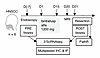 Figure 1
Study overview and patient characteristics. Study scheme and associated analysis on tissue samples.
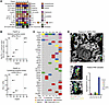 Figure 2
Patient characteristics. (A) Characteristics and outcome for each of the 6 patients. c, clinical categorization; yp, post-neoajuvant therapy pathological categorization. (B) PathR versus PD-L1 immune positive score (IPS; top) and combined positive score (CPS; bottom) for each patient. NR, nonresponder. (C) Genomic alterations and TMB. (D) Multiplex IHC staining images highlighting immune cell aggregates in the PRE biopsy of the MPR (Pt07). Bottom right: quantification of aggregates per stromal area in all patients’ PRE biopsies. Scale bars: 100 μm.
Pre- (PRE) and posttreatment (POST) biopsies were collected by endoscopy in a similar manner to optimize comparability for biological responses. As defined in the clinical trial, the treatment efficacy endpoint was assessed by PathR on the tumor resection. Patients with specimens showing tumor cell necrosis and/or giant cell/histolytic reaction to keratinous debris in ≥ 10% and ≥ 50% of tumor area were considered partial responders (PRs) and major partial responders (MPRs), respectively. These thresholds are adapted to such short neoadjuvant regimens, with the 10% threshold selected based on a phase II trial evaluating short neoadjuvant pembrolizumab in HNSCC (24). Three patients (Pt02, Pt04, and Pt05) were PRs (3/6 patients, 50%), and 1 (Pt07) was MPR, with PathR = 54% (1/6 patients, 17%). Two patients (Pt03 and Pt08) were nonresponders (2/6 patients, 33%). Detailed clinical characteristics are summarized in Figure 2A, Supplemental Figure 1A, and Supplemental Table 1, and representative hematoxylin, eosin, and saffron images across the spectrum of patient responses are in Supplemental Figure 1B (supplemental material available online with this article; https://doi.org/10.1172/JCI198718DS1). We next evaluated the association between PD-L1 expression on tumor or immune cells and PathR (Figure 2B, Supplemental Figure 1C, and Supplemental Table 2). PD-L1 staining in PRE tumor biopsies showed a combined positive score (defined as number of PD-L1+ cells/number of viable tumor cells) ranging from 0 to 30, which showed no positive correlation with PathR (Spearman’s r = 0.20, P = 0.714). When considering immune cells only (immune positive score, number of PD-L1+ immune cells/number of viable tumor cells), ratios ranged from 0 to 13 and were positively correlated with PathR (Spearman’s r = 0.90, P = 0.028).
To ensure we were capturing clear biological features associated with response, we focused on the case with a very distinct response (MPR case, Pt07, >50% PathR). Next-generation sequencing (NGS) showed that the major responder had the most detected oncogene alterations and the highest tumor mutational burden (TMB) (Figure 2C), although all patients were still classified as “TMB low” relative to a previously established dataset (25). We identified deleterious mutations of NSD1, RB1, and CDKN2A (p16) in this patient that explained the accumulation of p16 in cancer cells in the absence of HPV (Figure 2A) (26). Another specificity of the Pt07 PRE biopsy was a higher number of immune aggregates in the stroma, composed of T cells, B cells, and DC-LAMP+ DCs and surrounded by plasma cells (Figure 2D), as detected by multiplex IHC. Notably, the stromal densities of these immune cells were strongly increased in this patient after BA treatment (Supplemental Figure 1D). Pt07 CD68+ macrophages also massively infiltrated the stroma after treatment, especially the zones surrounding necrotic tumor cells (Supplemental Figure 1E). Together with the high PD-L1 immune positive score observed for Pt07, these results could suggest a higher antitumor immunity potential prior to treatment for the MPRs, likely unleashed by the PD-L1/TGF-β inhibition.
BA treatment reduces TGF-β signaling, with the strongest impact on mesenchymal cells. To dissect the biological impact of BA treatment on cancer cells and the TME, we analyzed the 6 paired biopsies by multiplex imaging and 5 pairs by scRNA-seq (1 POST scRNA-seq run [Pt05] failed). scRNA-seq analysis identified 13 clusters of major cell types (see Methods; Figure 3A, Supplemental Figure 2A, and Supplemental Table 3); all major cell types were well represented in all baseline samples (Supplemental Figure 2B). To identify broad changes induced by BA administration, irrespective of response or patient specificity, we first performed a global PRE/POST comparison by performing pseudobulk analysis by patient, cell type, and PRE or POST status and used DESeq2 to compute differentially expressed genes (DEGs) for each patient and cell type (Figure 3, B and C). Overall, we found 60 significantly upregulated DEGs and 96 significantly downregulated DEGs in the POST samples compared with the PRE samples [adjusted P < 0.05; |log2(fold-change)| > 0.5; Figure 3B, Supplemental Figure 3, and Supplemental Table 4]. The top 20 upregulated and downregulated genes are depicted in Figure 3B. Upregulated genes included multiple chemokines (CXCL8, CXCL9, and CXCL10), as well as IFNG, which encodes IFN-γ. The downregulated genes were enriched for TGF-β–inducible genes, with multiple matrix-associated components (e.g., COL1A1, COL3A1, COL11A1, MMP2, POSTN, and ASPN) (Figure 3B, Supplemental Figure 3, and Supplemental Table 4). DEGs were analyzed within each cluster to investigate their cellular origin and to identify the cell types most affected by BA treatment (Figure 3C and Supplemental Table 4). Figure 3C shows the top DEGs from Figure 3B within each cell type before and after treatment (log average expression). As expected, IFNG was mainly expressed and upregulated in CD8+ T cells and in NK cells. All cell types showed an upregulation of IFN-γ–inducible genes (e.g., CXCL9, IDO1, MMP12, WARS, and GBP1), with monocytes/macrophages and DCs exhibiting the strongest signature of IFN-γ activation (Figure 3C). The downregulated matrix-related genes mostly came from mesenchymal cells, especially CAFs and pericytes (Figure 3C), in line with their higher expression levels of TGF-β coreceptors (Supplemental Figure 2, C and D). These results confirmed that TGF-β signaling was reduced in the TME after BA treatment and that mostly stromal cells were affected.
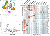 Figure 3
BA treatment reduces TGF-β signaling, with the strongest impact on mesenchymal cells. (A) scRNA-seq of 87,645 cells from n = 6 PRE and n = 5 POST samples after QC filtering projected on a UMAP and clustering of the main cell types. (B) Volcano plot showing the results of DEG analysis between PRE and POST samples from 5 paired patients (n = 5). Pseudobulk profiles were generated by aggregating cells within each patient and cell type (cluster). Red dots represent upregulated genes, and blue dots represent downregulated genes. The top 20 up- and downregulated genes are depicted (all DEGs are listed in Supplemental Table 4). (C) Heatmap showing the gene expression level of the top 20 up- and downregulated DEGs from the analysis in B, shown for each cell type in pre- and posttreatment conditions, respectively. Mo/Mac, monocytes and macrophages. Analysis performed with the n = 5 paired PRE/POST samples. Values represent log10-transformed average expression levels (a difference of 1 corresponds to a 10-fold difference in expression).
To further investigate whether the decrease in TGF-β signaling could be attributable to the aPD-L1 moiety of BA, we performed the same analysis with a publicly available scRNA-seq dataset from patients with HNSCC who had been treated only with aPD-L1 durvalumab (Supplemental Figure 4) (27). In aPD-L1–treated patient data, we found a similar increase of IFN-γ–activated monocytes/macrophages and DCs with upregulated CXCL9 and IDO1, yet no decreased expression of collagens or other TGF-β target genes was observed (Supplemental Figure 4, A and B). Taken together, these results highlight the antifibrotic effect of blocking TGF-β signaling, which was recapitulated in the BA-treated patients in our study.
BA treatment induces a strong remodeling of the stromal architecture. Given the strong TGF-β blocking effect observed on mesenchymal cells, we sought to investigate changes in the stromal architecture. Masson’s trichrome staining revealed drastic posttreatment modifications of global collagen fibers (Figure 4A); we therefore thought to stain and quantify specific types of collagens in all PRE and POST samples. This included the major matrix component collagen I, as well as collagen XII, recently identified as part of a fibrillar barrier associated with T cell exclusion in human lung cancer (6) and collagen IV coverage (Figure 4, B and C). We found reduced collagen I (P = 0.062) and collagen XII (P = 0.062) in 5 of 6 patients (Figure 4, B and C). In contrast, collagen IV, which is more specifically expressed by pericytes and endothelial cells (Supplemental Figure 2A), showed no global reduction (P = 0.844; Figure 4C).
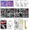 Figure 4
BA induced a strong remodeling of the stromal architecture. (A) Representative image of Masson’s trichrome staining highlighting decrease in collagen fiber content (blue) after treatment. Scale bars: 500 μm. (B) Collagen I staining of each PRE and POST biopsy. Scale bars: 200 μm. (C) Quantification of the surface covered by collagens I, XII, and IV. Wilcoxon’s paired test, P values: collagen I = 0.062, collagen XII = 0.062, and collagen IV = 0.844. (D) Left: example of tumor and stroma classifier on Pt02 (QuPath analysis). Right: quantification of the area covered by the stromal compartment within the whole tumor lesion for all paired samples. Wilcoxon’s paired test, P value = 0.156. Scale bars: 500 μm. (E) MERFISH spatial transcriptomics on Pt04 PRE and POST biopsies. Cells were segmented and unbiasedly clustered. Left: visualization of the resulting cancer cell cluster (dark gray), endothelial cell cluster (green), and CAF cluster (yellow). Right: cell density quantification. Scale bar: 500 μm.
We noticed not only a strong reduction in fibers but also a deep impact on the stroma, with changes in its proportion within the tumor lesion. By applying a stroma and tumor classifier on each PRE and POST sample, we observed a markedly reduced stromal area in a subset of patients, especially Pt02 and Pt04 (Figure 4D). To further investigate the cellular changes underlying this stromal loss, we additionally performed spatial transcriptomic analysis (MERSCOPE technology; see Methods) on the paired PRE and POST samples for Pt04 (Figure 4E). Based on literature showing CAFs as major ECM producers in the TME, and our results showing strong transcriptomic changes in CAFs after BA treatment (Figure 3C), we hypothesized that fibroblasts would be the main impacted cell population. The gene panel was designed to distinguish the main cell types (see Methods; Supplemental Table 8), allowing their quantification after segmentation and clustering. This analysis confirmed the strong decrease in CAFs after BA treatment in Pt04, without reduction in endothelial cell density (Figure 4E).
Given that Pt08 behaved differently with an increased proportion of stroma in the whole lesion (Figure 4D, right), we performed the same spatial transcriptomic analysis on Pt08 PRE and POST samples. However, the MERSCOPE analysis revealed a 4-fold reduction in CAFs after BA treatment (Supplemental Figure 5A). The increased stromal area might be explained by a massive infiltration of immune cells observed in the POST biopsy for this patient (Supplemental Figure 5B), who exhibited bleeding after treatment (Supplemental Table 14).
To our knowledge, our results are the first demonstration that TGF-β– and stromal-oriented drugs can have a rapid and drastic impact on fibroblasts and tumor architecture in cancer patients.
BA induces a systematic decrease in LRRC15+ activated CAFs. To investigate whether several subtypes/states of fibroblasts in the PRE lesions were differentially impacted by the treatment, we further analyzed the scRNA-seq dataset from all patients. Subclustering of the CAFs and pericytes revealed 1 clearly distinct pericyte cluster (expressing MCAM, RGS5, and COX4I2) and 2 CAF subsets (Figure 5A and Supplemental Table 5). We observed 1 CAF subcluster, denoted CD10+/IL24+ CAF, that expressed MME (CD10) as well as genes encoding various cytokines and chemokines (IL24, CXCL1, IL6, and CXCL6), resembling inflammatory CAFs (iCAFs) described across multiple cancer types (28, 29). The second fibroblast cluster, denoted LRRC15+ CAF, expressed a similar transcriptional profile as myofibroblastic CAFs (myCAFs) (15, 28), with the activation marker LRRC15 and ECM-related genes (CTHRC1, COL11A1, and MMP11). Proportion analysis within the total CAF clusters showed a global reduction in the LRRC15+ CAFs (Figure 5B). To confirm this, we quantified LRRC15+ and CD10+ CAF abundance through coverage of the stroma in PRE and POST samples (Figure 5, C and D). This showed a significant (P = 0.031, paired Wilcoxon’s test) reduction of the LRRC15+ myCAFs in all 6 patients, while CD10+ iCAF coverage (quantified as CD10+CD45–) of the stroma was not significantly modified (Figure 5D).
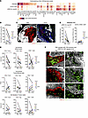 Figure 5
BA induced a systematic decrease in LRRC15+-activated CAFs and an increase in intratumor CD8 T cell infiltration in immune-excluded samples with CAF capsule. (A) Heatmap of the top 15 DEGs of CAF/pericyte subclusters identified via scRNA-seq. (B) Percentage of LRRC15+ CAFs among all CAFs within each scRNA-seq sample (pericytes are not counted as CAFs). Wilcoxon’s paired test, P value = 0.125 (Pt05 POST scRNA-seq run failed). (C) Representative images of PRE and POST biopsies stained for LRRC15, CD10, CD45, and pan-keratin. Scale bars: 100 µm. (D) Stromal area coverage of LRRC15+ (left) and CD10+CD45– (right) CAFs as quantified for each biopsy. Wilcoxon’s paired test, P values: LRRC15+ myCAF = 0.031; CD10+ iCAF = 0.156. (E) Quantification of CD8+ T cell, CD4+ T cell, and Treg infiltration. Wilcoxon’s paired test. P values for whole-lesion density: CD4+ T cells = 0.031, CD4+ Treg = 0.218, and CD8+ T cells = 0.313. P values for tumor/stroma ratio: CD4+ T cells = 0.438, CD4+ Treg = 0.219, and CD8+ T cells = 0.438). P values for density in tumor nests: CD4+ T cells = 0.563, CD4+ Treg = 0.219, and CD8+ T cells = 0.999. (F) ORION images from Pt04, Pt07, and Pt08 presenting LRRC15+ CAF capsule architecture and CD8+ T cell exclusion pretreatment. Right: posttreatment images showing loss of LRRC15 CAFs and increase in CD8+ intratumoral infiltration. Scale bars: 100 μm.
We then performed additional multiplex IHC analyses incorporating markers of apoptosis (cleaved caspase-3) and cell cycle arrest/senescence (p21, p16, and γH2AX). These analyses revealed the presence of cleaved caspase-3+LRRC15+ CAFs in POST biopsies (Supplemental Figure 5C), consistent with ongoing apoptosis. CAFs did not express γH2AX or p16, yet a fraction of residual LRRC15+ CAFs expressed p21 (Supplemental Figure 5D). p21 staining was absent in CAFs prior to treatment, suggesting some phenotypic modulation among the fibroblasts that persist after therapy. When considered together with the overall reduction in CAF abundance (Figure 4E, Figure 5, C and D, and Supplemental Figure 5A), these findings support a model in which treatment induces predominant depletion of LRRC15+ CAFs, along with some phenotypic modulation in residual cells.
To investigate the stromal loss and activated CAF changes in a validation cohort, immunofluorescence (IF) staining was performed on 6 PRE and POST samples from an NIH patient cohort treated with a similar regimen (window-of-opportunity trial, preoperative D1-D14 BA injections in patients with HPV– HNSCC) (23). This confirmed the significant (P = 0.031, paired Wilcoxon’s test) decrease in LRRC15+ CAFs for all 6 additional patients (Supplemental Figure 6, A and B) as well as a marked reduction in the whole stromal compartment in 4 of 6 patients (Supplemental Figure 6, C and D).
Altogether, these results obtained in 2 independent cohorts point toward a dominant CAF depleting effect of BA rather than a simple fibroblast polarization shift toward a less activated CAF state.
BA treatment induces changes in CD4+ and CD8+ T cell infiltration. We previously showed in human lung carcinoma tumors that activated CAFs can form a dense fibrillar capsule around tumor islets, associated with CD3+ and CD8+ T cell marginalization in the stroma (6). Given the stromal disruption observed (Figure 4), we analyzed T cell subset infiltration after BA treatment. While we found an overall reduction in T cell whole-lesion density, especially for conventional (significant decrease) and Treg CD4+ T cell density (Figure 5E, top row), T cell distribution in tumor and stroma was changed (Figure 5E, middle and bottom rows, and Figure 5F). We observed an increased tumor/stroma CD8+ T cell density ratio in 5 patients out of 6. Analyzing CAF topological arrangement, we observed that 3 PRE biopsies (Pt04, Pt07, and Pt08) displayed an architecture of multiple layers of LRRC15+ CAFs lining tumor cell islets, which disappeared after treatment (Figure 5F). These 3 tumors showed an increased CD8+ T cell infiltration, as seen through both tumor/stroma ratio and tumor nest density, revealing a more homogeneous distribution of CD8+ T cells after treatment (Figure 5F). These CD8+ T cell results were confirmed in the NIH validation cohort (Supplemental Figure 6, E and F), where the 3 patients (NIH Pt02, Pt10, and Pt12) with an LRRC15+ CAF capsule and CD8+ T cell exclusion before BA treatment showed a higher CD8+ T cell intratumor penetration (tumor/stroma ratio and tumor nest density) after treatment.
We used scRNA-seq data to further investigate the changes in the CD4+ and CD8+ T cell subsets, which were annotated using ProjecTILs reference maps (Supplemental Tables 6 and 7) (30). We identified exhausted (CD8.TEX) and progenitor-exhausted (CD8.TPEX) CD8+ T cells as the major producers of IFN-γ (Supplemental Figure 7, A and B, and Supplemental Table 7) (30). While CD8.TPEX cells made up a negligible fraction of total CD8+ T cells, CD8.TEX cells were the most enriched subset among CD8+ T cells in all samples (Supplemental Figure 7D). Notably, the average expression of IFN-γ by CD8.TEX increased in all patients after treatment, emphasizing the reinvigorating capacity of BA (Supplemental Figure 7E).
Together with our prior studies (4, 6), these results suggest that PD-L1/TGF-β blockade therapy, even over a short term, can alter the CD8+ T cell–restricting CAF barrier.
Maintenance of LRRC15+ CAFs around a subset of BA-resistant tumor cells in the major responder patient. To extend our knowledge on the biological impact of BA on the TME upon response, the whole surgical specimen of the MPR (Pt07, PathR 54%) was analyzed through both multiplexed imaging and spatial transcriptomics (Figure 6). As previously mentioned, the Pt07 PRE biopsy had the most detected oncogene alterations and the highest TMB, a high PD-L1 immune positive score, and a higher number of immune aggregates (Figure 2, B–D). The tumor harbored LRRC15+ CAF layers surrounding tumor cells, along with a CD8+ T cell exclusion, that were both counteracted upon treatment (Figure 5, C–F).
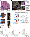 Figure 6
Maintenance of LRRC15+ CAFs around a subset of distinct cancer cells in the major responder patient. (A) Hematoxylin, eosin, and saffron image of the MPR’s resection (Pt07, PathR 54%), with a representative zoom showing dense fibers surrounding resistant tumor cells. Scale bars: 500 μm, 250 μm (zoom). (B) Multiplex IHC image of the MPR’s resection revealing a large area of major cell death with T cell and macrophage infiltration and a minor area of LRRC15+ CAF capsule maintenance and immune exclusion. Right: zooms of each area, with illustrative sketches. Scale bars: 500 μm. (C) Merscope and Xenium transcriptomic analysis of the MPR posttreatment resection. Cells were segmented and unbiasedly clustered. Visualization of the 2 resulting cancer cell clusters: 1 (blue) in the CAF (red) maintenance area and 1 (orange) in the cell death area. (D) Top 10 DEGs between the 2 cancer cell clusters as well as stemness/epithelial–mesenchymal transition–related DEGs. (E) Reclustering of all scRNA-seq cancer cells of patient 7 (both pre- and posttreatment cancer cells) unbiasedly gave 2 clusters. Left: enrichment of the top 10 Merscope and Xenium signatures (Figure 6D) in the 2 cancer cell subclusters. Right: volcano plot of DEGs between cluster A (resistant-like) and cluster B cancer cells from the scRNA-seq dataset. (F) Ligand-receptor interaction analysis based on the scRNA-seq data (all Pt07 cells included) and the CellChat framework, showing the only 3 signaling pathways identified between resistant-like cancer cells (sender) and fibroblasts (receiver).
Most of the MPR tumor resection posttreatment consisted of regions of major tumor cell death (Figure 6A) with a global infiltration of T cells into the stroma and tumor cell islets (Figure 6B). A minor area was marked by intact cancer cells (hereafter, BA-resistant cells) exempt from immune infiltration (Figure 6, A and B). In this region, we observed capsule maintenance of densely layered LRRC15+ CAFs (Figure 6B). Given that BA treatment induced a massive reduction in LRRC15+ CAFs in all patients (Figure 5, C and D), we sought to investigate the specificities of this resistant tumor region. The resistant cells stained positively for pan-keratin (albeit weakly), podoplanin (PDPN), and MCAM, but not for vimentin (Supplemental Figure 8A). A pathologist confirmed that these tumor cells were morphologically poorly differentiated (Figure 6A, zoom). Note that such poorly differentiated cancer cells (e.g., PDPN+, MCAM+, and KERlo) were already observed in the PRE biopsy of this patient, indicating their presence before treatment (Supplemental Figure 8B).
To dissect the potential molecular mechanisms underlying LRRC15+ CAF maintenance, we further characterized the differences between the BA-resistant cancer cells and those in areas of major cell death. We performed MERFISH (multiplexed error-robust fluorescence in situ hybridization) spatial transcriptomics with a customized 300-gene panel (Supplemental Table 8) and Xenium spatial transcriptomics with a predesigned 289-gene panel (Supplemental Table 9) on the MPR surgical specimen. Both techniques provided similar results. Unsupervised clustering identified 2 distinct clusters of cancer cells, corresponding to the tumor cells in the area of CAF maintenance and in the region of cell death (Figure 6C). Based on DEG analyses, the BA-resistant cancer cells were characterized by higher expression of stemness-related transcription factors (e.g., SNAI2, SOX9, LGR5, LGR6, and ETV5) and mesenchyme-associated genes (e.g., MMP2, CLDN11, and NID1), as well as lower levels of genes associated with keratinocyte differentiation (e.g., CDH1 and KLF4) (Figure 6D, Supplemental Figure 8C, Supplemental Tables 10 and 11). Despite the greater presence of LRRC15+ CAFs around them, they expressed lower levels of TGFB1 than their BA-sensitive counterparts (Supplemental Figure 8C). Finally, BA-resistant cancer cells expressed both FGF3 and FGFR1, suggesting an autocrine signaling loop (Supplemental Figure 8C). Although at a lower level than on the BA-resistant cancer cells, FGFR1 was also detected on the surrounding LRRC15+ CAFs (Supplemental Figure 8C), indicating potential crosstalk between the 2 cell types.
The spatial transcriptomic analysis allowed us to identify tumor cells in resistant and sensitive areas. We next analyzed the scRNA-seq data from Pt07 cancer cells (Figure 6E), which provided no spatial information yet deeper information on the transcriptomic profile. We identified 2 clusters of cells that we assigned to resistant- and sensitive-like cancer cells, as they were enriched in the expression of the signatures identified by spatial transcriptomics (Figure 6E, left). scRNA-seq DEG analyses identified similar highly expressed genes compared with Merfish/Xenium data, with higher expression of differentiation genes (e.g., KRT19 and FOXA1) in sensitive-like cells and increased levels of stemness/epithelial–mesenchymal transition–related genes (e.g., HOXA9, WNT6, PODXL, and LGR5) in the resistant ones (Figure 6E, right, and Supplemental Table 12). We further pursued scRNA-seq analysis to investigate crosstalk from cancer cells to fibroblasts potentially driving LRRC15+ CAF maintenance. Using CellChat (31) and filtering for interactions specific for resistant-like tumor cells and CAFs, we identified 3 predicted communication networks, including FGF signaling (FGF3/FGF19 [cancer cells]–FGFR1 [CAFs]), in line with our results from spatial transcriptomics (Supplemental Figure 8C); PDGF signaling (PDGFA [cancer cells]–PDGFRA/B [CAFs]); and GAS signaling (GAS6 [cancer cells]–AXL [CAFs]) (Figure 6F and Supplemental Table 13). These ligand-receptor pairs were specifically highly expressed by resistant-like cancer cells and CAFs, but not sensitive-like cancer cells. Together, these data showed that TGF-β blockade does not systematically affect LRRC15+ CAFs; here, their specific localization around highly distinct, poorly differentiated tumor cells suggests that tumor-derived signals may drive the preservation of the LRRC15+ CAF capsule despite BA treatment.


Copyright © 2026 American Society for Clinical Investigation
ISSN: 0021-9738 (print), 1558-8238 (online)