Advertisement
Research ArticleNeuroscienceOncology
Open Access | ![]() 10.1172/JCI179395
10.1172/JCI179395
Disruption of ataxia telangiectasia–mutated kinase enhances radiation therapy efficacy in spatially directed diffuse midline glioma models
Avani Mangoli,1 Vennesa Valentine,2 Spencer M. Maingi,2 Sophie R. Wu,2 Harrison Q. Liu,2 Michael Aksu,3 Vaibhav Jain,3 Bronwen E. Foreman,2 Joshua A. Regal,2 Loren B. Weidenhammer,2 Connor E. Stewart,2 Maria E. Guerra Garcia,2 Emily Hocke,2 Karen Abramson,3 Tal Falick Michaeli,1 Nerissa T. Williams,2 Lixia Luo,2 Megan Romero,4 Katherine Deland,2 Samantha Gadd,5 Eita Uchida,5 Laura Attardi,6 Kouki Abe,5 Rintaro Hashizume,5 David M. Ashley,1 Oren J. Becher,4 David G. Kirsch,7 Simon G. Gregory,3 and Zachary J. Reitman2
1The Preston Robert Tisch Brain Tumor Center,
2Department of Radiation Oncology, and
3The Preston Robert Tisch Brain Tumor Center Omics Program, Duke University, Durham, North Carolina, USA.
4Department of Pediatric Hematology Oncology, Mount Sinai Kravis Children’s Hospital, New York, New York, USA.
5Department of Pediatrics, University of Alabama at Birmingham, Birmingham, Alabama, USA.
6Departments of Radiation Oncology and Genetics, Stanford University School of Medicine, Stanford, California, USA.
7Princess Margaret Cancer Centre, University of Toronto, Toronto, Ontario, Canada.
Address correspondence to: Zachary J. Reitman, Duke University, Department of Radiation, 30 Duke Medicine Circle, Box 3085, Durham, North Carolina 27710, USA. Phone: 919.668.7336; Email: zjr@duke.edu. Or to: Simon G. Gregory, 300 N. Duke Street, DUMC 104775, Durham, North Carolina 27701, USA. Phone: 919.684.0726; Email: simon.gregory@duke.edu.
Authorship note: AM and VV are co–first authors.
Find articles by Mangoli, A. in: PubMed | Google Scholar
1The Preston Robert Tisch Brain Tumor Center,
2Department of Radiation Oncology, and
3The Preston Robert Tisch Brain Tumor Center Omics Program, Duke University, Durham, North Carolina, USA.
4Department of Pediatric Hematology Oncology, Mount Sinai Kravis Children’s Hospital, New York, New York, USA.
5Department of Pediatrics, University of Alabama at Birmingham, Birmingham, Alabama, USA.
6Departments of Radiation Oncology and Genetics, Stanford University School of Medicine, Stanford, California, USA.
7Princess Margaret Cancer Centre, University of Toronto, Toronto, Ontario, Canada.
Address correspondence to: Zachary J. Reitman, Duke University, Department of Radiation, 30 Duke Medicine Circle, Box 3085, Durham, North Carolina 27710, USA. Phone: 919.668.7336; Email: zjr@duke.edu. Or to: Simon G. Gregory, 300 N. Duke Street, DUMC 104775, Durham, North Carolina 27701, USA. Phone: 919.684.0726; Email: simon.gregory@duke.edu.
Authorship note: AM and VV are co–first authors.
Find articles by Valentine, V. in: PubMed | Google Scholar
1The Preston Robert Tisch Brain Tumor Center,
2Department of Radiation Oncology, and
3The Preston Robert Tisch Brain Tumor Center Omics Program, Duke University, Durham, North Carolina, USA.
4Department of Pediatric Hematology Oncology, Mount Sinai Kravis Children’s Hospital, New York, New York, USA.
5Department of Pediatrics, University of Alabama at Birmingham, Birmingham, Alabama, USA.
6Departments of Radiation Oncology and Genetics, Stanford University School of Medicine, Stanford, California, USA.
7Princess Margaret Cancer Centre, University of Toronto, Toronto, Ontario, Canada.
Address correspondence to: Zachary J. Reitman, Duke University, Department of Radiation, 30 Duke Medicine Circle, Box 3085, Durham, North Carolina 27710, USA. Phone: 919.668.7336; Email: zjr@duke.edu. Or to: Simon G. Gregory, 300 N. Duke Street, DUMC 104775, Durham, North Carolina 27701, USA. Phone: 919.684.0726; Email: simon.gregory@duke.edu.
Authorship note: AM and VV are co–first authors.
Find articles by Maingi, S. in: PubMed | Google Scholar
1The Preston Robert Tisch Brain Tumor Center,
2Department of Radiation Oncology, and
3The Preston Robert Tisch Brain Tumor Center Omics Program, Duke University, Durham, North Carolina, USA.
4Department of Pediatric Hematology Oncology, Mount Sinai Kravis Children’s Hospital, New York, New York, USA.
5Department of Pediatrics, University of Alabama at Birmingham, Birmingham, Alabama, USA.
6Departments of Radiation Oncology and Genetics, Stanford University School of Medicine, Stanford, California, USA.
7Princess Margaret Cancer Centre, University of Toronto, Toronto, Ontario, Canada.
Address correspondence to: Zachary J. Reitman, Duke University, Department of Radiation, 30 Duke Medicine Circle, Box 3085, Durham, North Carolina 27710, USA. Phone: 919.668.7336; Email: zjr@duke.edu. Or to: Simon G. Gregory, 300 N. Duke Street, DUMC 104775, Durham, North Carolina 27701, USA. Phone: 919.684.0726; Email: simon.gregory@duke.edu.
Authorship note: AM and VV are co–first authors.
Find articles by Wu, S. in: PubMed | Google Scholar
1The Preston Robert Tisch Brain Tumor Center,
2Department of Radiation Oncology, and
3The Preston Robert Tisch Brain Tumor Center Omics Program, Duke University, Durham, North Carolina, USA.
4Department of Pediatric Hematology Oncology, Mount Sinai Kravis Children’s Hospital, New York, New York, USA.
5Department of Pediatrics, University of Alabama at Birmingham, Birmingham, Alabama, USA.
6Departments of Radiation Oncology and Genetics, Stanford University School of Medicine, Stanford, California, USA.
7Princess Margaret Cancer Centre, University of Toronto, Toronto, Ontario, Canada.
Address correspondence to: Zachary J. Reitman, Duke University, Department of Radiation, 30 Duke Medicine Circle, Box 3085, Durham, North Carolina 27710, USA. Phone: 919.668.7336; Email: zjr@duke.edu. Or to: Simon G. Gregory, 300 N. Duke Street, DUMC 104775, Durham, North Carolina 27701, USA. Phone: 919.684.0726; Email: simon.gregory@duke.edu.
Authorship note: AM and VV are co–first authors.
Find articles by Liu, H. in: PubMed | Google Scholar
1The Preston Robert Tisch Brain Tumor Center,
2Department of Radiation Oncology, and
3The Preston Robert Tisch Brain Tumor Center Omics Program, Duke University, Durham, North Carolina, USA.
4Department of Pediatric Hematology Oncology, Mount Sinai Kravis Children’s Hospital, New York, New York, USA.
5Department of Pediatrics, University of Alabama at Birmingham, Birmingham, Alabama, USA.
6Departments of Radiation Oncology and Genetics, Stanford University School of Medicine, Stanford, California, USA.
7Princess Margaret Cancer Centre, University of Toronto, Toronto, Ontario, Canada.
Address correspondence to: Zachary J. Reitman, Duke University, Department of Radiation, 30 Duke Medicine Circle, Box 3085, Durham, North Carolina 27710, USA. Phone: 919.668.7336; Email: zjr@duke.edu. Or to: Simon G. Gregory, 300 N. Duke Street, DUMC 104775, Durham, North Carolina 27701, USA. Phone: 919.684.0726; Email: simon.gregory@duke.edu.
Authorship note: AM and VV are co–first authors.
Find articles by Aksu, M. in: PubMed | Google Scholar
1The Preston Robert Tisch Brain Tumor Center,
2Department of Radiation Oncology, and
3The Preston Robert Tisch Brain Tumor Center Omics Program, Duke University, Durham, North Carolina, USA.
4Department of Pediatric Hematology Oncology, Mount Sinai Kravis Children’s Hospital, New York, New York, USA.
5Department of Pediatrics, University of Alabama at Birmingham, Birmingham, Alabama, USA.
6Departments of Radiation Oncology and Genetics, Stanford University School of Medicine, Stanford, California, USA.
7Princess Margaret Cancer Centre, University of Toronto, Toronto, Ontario, Canada.
Address correspondence to: Zachary J. Reitman, Duke University, Department of Radiation, 30 Duke Medicine Circle, Box 3085, Durham, North Carolina 27710, USA. Phone: 919.668.7336; Email: zjr@duke.edu. Or to: Simon G. Gregory, 300 N. Duke Street, DUMC 104775, Durham, North Carolina 27701, USA. Phone: 919.684.0726; Email: simon.gregory@duke.edu.
Authorship note: AM and VV are co–first authors.
Find articles by
Jain, V.
in:
PubMed
|
Google Scholar
|

1The Preston Robert Tisch Brain Tumor Center,
2Department of Radiation Oncology, and
3The Preston Robert Tisch Brain Tumor Center Omics Program, Duke University, Durham, North Carolina, USA.
4Department of Pediatric Hematology Oncology, Mount Sinai Kravis Children’s Hospital, New York, New York, USA.
5Department of Pediatrics, University of Alabama at Birmingham, Birmingham, Alabama, USA.
6Departments of Radiation Oncology and Genetics, Stanford University School of Medicine, Stanford, California, USA.
7Princess Margaret Cancer Centre, University of Toronto, Toronto, Ontario, Canada.
Address correspondence to: Zachary J. Reitman, Duke University, Department of Radiation, 30 Duke Medicine Circle, Box 3085, Durham, North Carolina 27710, USA. Phone: 919.668.7336; Email: zjr@duke.edu. Or to: Simon G. Gregory, 300 N. Duke Street, DUMC 104775, Durham, North Carolina 27701, USA. Phone: 919.684.0726; Email: simon.gregory@duke.edu.
Authorship note: AM and VV are co–first authors.
Find articles by Foreman, B. in: PubMed | Google Scholar
1The Preston Robert Tisch Brain Tumor Center,
2Department of Radiation Oncology, and
3The Preston Robert Tisch Brain Tumor Center Omics Program, Duke University, Durham, North Carolina, USA.
4Department of Pediatric Hematology Oncology, Mount Sinai Kravis Children’s Hospital, New York, New York, USA.
5Department of Pediatrics, University of Alabama at Birmingham, Birmingham, Alabama, USA.
6Departments of Radiation Oncology and Genetics, Stanford University School of Medicine, Stanford, California, USA.
7Princess Margaret Cancer Centre, University of Toronto, Toronto, Ontario, Canada.
Address correspondence to: Zachary J. Reitman, Duke University, Department of Radiation, 30 Duke Medicine Circle, Box 3085, Durham, North Carolina 27710, USA. Phone: 919.668.7336; Email: zjr@duke.edu. Or to: Simon G. Gregory, 300 N. Duke Street, DUMC 104775, Durham, North Carolina 27701, USA. Phone: 919.684.0726; Email: simon.gregory@duke.edu.
Authorship note: AM and VV are co–first authors.
Find articles by Regal, J. in: PubMed | Google Scholar
1The Preston Robert Tisch Brain Tumor Center,
2Department of Radiation Oncology, and
3The Preston Robert Tisch Brain Tumor Center Omics Program, Duke University, Durham, North Carolina, USA.
4Department of Pediatric Hematology Oncology, Mount Sinai Kravis Children’s Hospital, New York, New York, USA.
5Department of Pediatrics, University of Alabama at Birmingham, Birmingham, Alabama, USA.
6Departments of Radiation Oncology and Genetics, Stanford University School of Medicine, Stanford, California, USA.
7Princess Margaret Cancer Centre, University of Toronto, Toronto, Ontario, Canada.
Address correspondence to: Zachary J. Reitman, Duke University, Department of Radiation, 30 Duke Medicine Circle, Box 3085, Durham, North Carolina 27710, USA. Phone: 919.668.7336; Email: zjr@duke.edu. Or to: Simon G. Gregory, 300 N. Duke Street, DUMC 104775, Durham, North Carolina 27701, USA. Phone: 919.684.0726; Email: simon.gregory@duke.edu.
Authorship note: AM and VV are co–first authors.
Find articles by Weidenhammer, L. in: PubMed | Google Scholar
1The Preston Robert Tisch Brain Tumor Center,
2Department of Radiation Oncology, and
3The Preston Robert Tisch Brain Tumor Center Omics Program, Duke University, Durham, North Carolina, USA.
4Department of Pediatric Hematology Oncology, Mount Sinai Kravis Children’s Hospital, New York, New York, USA.
5Department of Pediatrics, University of Alabama at Birmingham, Birmingham, Alabama, USA.
6Departments of Radiation Oncology and Genetics, Stanford University School of Medicine, Stanford, California, USA.
7Princess Margaret Cancer Centre, University of Toronto, Toronto, Ontario, Canada.
Address correspondence to: Zachary J. Reitman, Duke University, Department of Radiation, 30 Duke Medicine Circle, Box 3085, Durham, North Carolina 27710, USA. Phone: 919.668.7336; Email: zjr@duke.edu. Or to: Simon G. Gregory, 300 N. Duke Street, DUMC 104775, Durham, North Carolina 27701, USA. Phone: 919.684.0726; Email: simon.gregory@duke.edu.
Authorship note: AM and VV are co–first authors.
Find articles by Stewart, C. in: PubMed | Google Scholar
1The Preston Robert Tisch Brain Tumor Center,
2Department of Radiation Oncology, and
3The Preston Robert Tisch Brain Tumor Center Omics Program, Duke University, Durham, North Carolina, USA.
4Department of Pediatric Hematology Oncology, Mount Sinai Kravis Children’s Hospital, New York, New York, USA.
5Department of Pediatrics, University of Alabama at Birmingham, Birmingham, Alabama, USA.
6Departments of Radiation Oncology and Genetics, Stanford University School of Medicine, Stanford, California, USA.
7Princess Margaret Cancer Centre, University of Toronto, Toronto, Ontario, Canada.
Address correspondence to: Zachary J. Reitman, Duke University, Department of Radiation, 30 Duke Medicine Circle, Box 3085, Durham, North Carolina 27710, USA. Phone: 919.668.7336; Email: zjr@duke.edu. Or to: Simon G. Gregory, 300 N. Duke Street, DUMC 104775, Durham, North Carolina 27701, USA. Phone: 919.684.0726; Email: simon.gregory@duke.edu.
Authorship note: AM and VV are co–first authors.
Find articles by Guerra Garcia, M. in: PubMed | Google Scholar
1The Preston Robert Tisch Brain Tumor Center,
2Department of Radiation Oncology, and
3The Preston Robert Tisch Brain Tumor Center Omics Program, Duke University, Durham, North Carolina, USA.
4Department of Pediatric Hematology Oncology, Mount Sinai Kravis Children’s Hospital, New York, New York, USA.
5Department of Pediatrics, University of Alabama at Birmingham, Birmingham, Alabama, USA.
6Departments of Radiation Oncology and Genetics, Stanford University School of Medicine, Stanford, California, USA.
7Princess Margaret Cancer Centre, University of Toronto, Toronto, Ontario, Canada.
Address correspondence to: Zachary J. Reitman, Duke University, Department of Radiation, 30 Duke Medicine Circle, Box 3085, Durham, North Carolina 27710, USA. Phone: 919.668.7336; Email: zjr@duke.edu. Or to: Simon G. Gregory, 300 N. Duke Street, DUMC 104775, Durham, North Carolina 27701, USA. Phone: 919.684.0726; Email: simon.gregory@duke.edu.
Authorship note: AM and VV are co–first authors.
Find articles by Hocke, E. in: PubMed | Google Scholar
1The Preston Robert Tisch Brain Tumor Center,
2Department of Radiation Oncology, and
3The Preston Robert Tisch Brain Tumor Center Omics Program, Duke University, Durham, North Carolina, USA.
4Department of Pediatric Hematology Oncology, Mount Sinai Kravis Children’s Hospital, New York, New York, USA.
5Department of Pediatrics, University of Alabama at Birmingham, Birmingham, Alabama, USA.
6Departments of Radiation Oncology and Genetics, Stanford University School of Medicine, Stanford, California, USA.
7Princess Margaret Cancer Centre, University of Toronto, Toronto, Ontario, Canada.
Address correspondence to: Zachary J. Reitman, Duke University, Department of Radiation, 30 Duke Medicine Circle, Box 3085, Durham, North Carolina 27710, USA. Phone: 919.668.7336; Email: zjr@duke.edu. Or to: Simon G. Gregory, 300 N. Duke Street, DUMC 104775, Durham, North Carolina 27701, USA. Phone: 919.684.0726; Email: simon.gregory@duke.edu.
Authorship note: AM and VV are co–first authors.
Find articles by Abramson, K. in: PubMed | Google Scholar
1The Preston Robert Tisch Brain Tumor Center,
2Department of Radiation Oncology, and
3The Preston Robert Tisch Brain Tumor Center Omics Program, Duke University, Durham, North Carolina, USA.
4Department of Pediatric Hematology Oncology, Mount Sinai Kravis Children’s Hospital, New York, New York, USA.
5Department of Pediatrics, University of Alabama at Birmingham, Birmingham, Alabama, USA.
6Departments of Radiation Oncology and Genetics, Stanford University School of Medicine, Stanford, California, USA.
7Princess Margaret Cancer Centre, University of Toronto, Toronto, Ontario, Canada.
Address correspondence to: Zachary J. Reitman, Duke University, Department of Radiation, 30 Duke Medicine Circle, Box 3085, Durham, North Carolina 27710, USA. Phone: 919.668.7336; Email: zjr@duke.edu. Or to: Simon G. Gregory, 300 N. Duke Street, DUMC 104775, Durham, North Carolina 27701, USA. Phone: 919.684.0726; Email: simon.gregory@duke.edu.
Authorship note: AM and VV are co–first authors.
Find articles by Michaeli, T. in: PubMed | Google Scholar
1The Preston Robert Tisch Brain Tumor Center,
2Department of Radiation Oncology, and
3The Preston Robert Tisch Brain Tumor Center Omics Program, Duke University, Durham, North Carolina, USA.
4Department of Pediatric Hematology Oncology, Mount Sinai Kravis Children’s Hospital, New York, New York, USA.
5Department of Pediatrics, University of Alabama at Birmingham, Birmingham, Alabama, USA.
6Departments of Radiation Oncology and Genetics, Stanford University School of Medicine, Stanford, California, USA.
7Princess Margaret Cancer Centre, University of Toronto, Toronto, Ontario, Canada.
Address correspondence to: Zachary J. Reitman, Duke University, Department of Radiation, 30 Duke Medicine Circle, Box 3085, Durham, North Carolina 27710, USA. Phone: 919.668.7336; Email: zjr@duke.edu. Or to: Simon G. Gregory, 300 N. Duke Street, DUMC 104775, Durham, North Carolina 27701, USA. Phone: 919.684.0726; Email: simon.gregory@duke.edu.
Authorship note: AM and VV are co–first authors.
Find articles by Williams, N. in: PubMed | Google Scholar
1The Preston Robert Tisch Brain Tumor Center,
2Department of Radiation Oncology, and
3The Preston Robert Tisch Brain Tumor Center Omics Program, Duke University, Durham, North Carolina, USA.
4Department of Pediatric Hematology Oncology, Mount Sinai Kravis Children’s Hospital, New York, New York, USA.
5Department of Pediatrics, University of Alabama at Birmingham, Birmingham, Alabama, USA.
6Departments of Radiation Oncology and Genetics, Stanford University School of Medicine, Stanford, California, USA.
7Princess Margaret Cancer Centre, University of Toronto, Toronto, Ontario, Canada.
Address correspondence to: Zachary J. Reitman, Duke University, Department of Radiation, 30 Duke Medicine Circle, Box 3085, Durham, North Carolina 27710, USA. Phone: 919.668.7336; Email: zjr@duke.edu. Or to: Simon G. Gregory, 300 N. Duke Street, DUMC 104775, Durham, North Carolina 27701, USA. Phone: 919.684.0726; Email: simon.gregory@duke.edu.
Authorship note: AM and VV are co–first authors.
Find articles by Luo, L. in: PubMed | Google Scholar
1The Preston Robert Tisch Brain Tumor Center,
2Department of Radiation Oncology, and
3The Preston Robert Tisch Brain Tumor Center Omics Program, Duke University, Durham, North Carolina, USA.
4Department of Pediatric Hematology Oncology, Mount Sinai Kravis Children’s Hospital, New York, New York, USA.
5Department of Pediatrics, University of Alabama at Birmingham, Birmingham, Alabama, USA.
6Departments of Radiation Oncology and Genetics, Stanford University School of Medicine, Stanford, California, USA.
7Princess Margaret Cancer Centre, University of Toronto, Toronto, Ontario, Canada.
Address correspondence to: Zachary J. Reitman, Duke University, Department of Radiation, 30 Duke Medicine Circle, Box 3085, Durham, North Carolina 27710, USA. Phone: 919.668.7336; Email: zjr@duke.edu. Or to: Simon G. Gregory, 300 N. Duke Street, DUMC 104775, Durham, North Carolina 27701, USA. Phone: 919.684.0726; Email: simon.gregory@duke.edu.
Authorship note: AM and VV are co–first authors.
Find articles by Romero, M. in: PubMed | Google Scholar
1The Preston Robert Tisch Brain Tumor Center,
2Department of Radiation Oncology, and
3The Preston Robert Tisch Brain Tumor Center Omics Program, Duke University, Durham, North Carolina, USA.
4Department of Pediatric Hematology Oncology, Mount Sinai Kravis Children’s Hospital, New York, New York, USA.
5Department of Pediatrics, University of Alabama at Birmingham, Birmingham, Alabama, USA.
6Departments of Radiation Oncology and Genetics, Stanford University School of Medicine, Stanford, California, USA.
7Princess Margaret Cancer Centre, University of Toronto, Toronto, Ontario, Canada.
Address correspondence to: Zachary J. Reitman, Duke University, Department of Radiation, 30 Duke Medicine Circle, Box 3085, Durham, North Carolina 27710, USA. Phone: 919.668.7336; Email: zjr@duke.edu. Or to: Simon G. Gregory, 300 N. Duke Street, DUMC 104775, Durham, North Carolina 27701, USA. Phone: 919.684.0726; Email: simon.gregory@duke.edu.
Authorship note: AM and VV are co–first authors.
Find articles by Deland, K. in: PubMed | Google Scholar
1The Preston Robert Tisch Brain Tumor Center,
2Department of Radiation Oncology, and
3The Preston Robert Tisch Brain Tumor Center Omics Program, Duke University, Durham, North Carolina, USA.
4Department of Pediatric Hematology Oncology, Mount Sinai Kravis Children’s Hospital, New York, New York, USA.
5Department of Pediatrics, University of Alabama at Birmingham, Birmingham, Alabama, USA.
6Departments of Radiation Oncology and Genetics, Stanford University School of Medicine, Stanford, California, USA.
7Princess Margaret Cancer Centre, University of Toronto, Toronto, Ontario, Canada.
Address correspondence to: Zachary J. Reitman, Duke University, Department of Radiation, 30 Duke Medicine Circle, Box 3085, Durham, North Carolina 27710, USA. Phone: 919.668.7336; Email: zjr@duke.edu. Or to: Simon G. Gregory, 300 N. Duke Street, DUMC 104775, Durham, North Carolina 27701, USA. Phone: 919.684.0726; Email: simon.gregory@duke.edu.
Authorship note: AM and VV are co–first authors.
Find articles by Gadd, S. in: PubMed | Google Scholar
1The Preston Robert Tisch Brain Tumor Center,
2Department of Radiation Oncology, and
3The Preston Robert Tisch Brain Tumor Center Omics Program, Duke University, Durham, North Carolina, USA.
4Department of Pediatric Hematology Oncology, Mount Sinai Kravis Children’s Hospital, New York, New York, USA.
5Department of Pediatrics, University of Alabama at Birmingham, Birmingham, Alabama, USA.
6Departments of Radiation Oncology and Genetics, Stanford University School of Medicine, Stanford, California, USA.
7Princess Margaret Cancer Centre, University of Toronto, Toronto, Ontario, Canada.
Address correspondence to: Zachary J. Reitman, Duke University, Department of Radiation, 30 Duke Medicine Circle, Box 3085, Durham, North Carolina 27710, USA. Phone: 919.668.7336; Email: zjr@duke.edu. Or to: Simon G. Gregory, 300 N. Duke Street, DUMC 104775, Durham, North Carolina 27701, USA. Phone: 919.684.0726; Email: simon.gregory@duke.edu.
Authorship note: AM and VV are co–first authors.
Find articles by Uchida, E. in: PubMed | Google Scholar
1The Preston Robert Tisch Brain Tumor Center,
2Department of Radiation Oncology, and
3The Preston Robert Tisch Brain Tumor Center Omics Program, Duke University, Durham, North Carolina, USA.
4Department of Pediatric Hematology Oncology, Mount Sinai Kravis Children’s Hospital, New York, New York, USA.
5Department of Pediatrics, University of Alabama at Birmingham, Birmingham, Alabama, USA.
6Departments of Radiation Oncology and Genetics, Stanford University School of Medicine, Stanford, California, USA.
7Princess Margaret Cancer Centre, University of Toronto, Toronto, Ontario, Canada.
Address correspondence to: Zachary J. Reitman, Duke University, Department of Radiation, 30 Duke Medicine Circle, Box 3085, Durham, North Carolina 27710, USA. Phone: 919.668.7336; Email: zjr@duke.edu. Or to: Simon G. Gregory, 300 N. Duke Street, DUMC 104775, Durham, North Carolina 27701, USA. Phone: 919.684.0726; Email: simon.gregory@duke.edu.
Authorship note: AM and VV are co–first authors.
Find articles by Attardi, L. in: PubMed | Google Scholar
1The Preston Robert Tisch Brain Tumor Center,
2Department of Radiation Oncology, and
3The Preston Robert Tisch Brain Tumor Center Omics Program, Duke University, Durham, North Carolina, USA.
4Department of Pediatric Hematology Oncology, Mount Sinai Kravis Children’s Hospital, New York, New York, USA.
5Department of Pediatrics, University of Alabama at Birmingham, Birmingham, Alabama, USA.
6Departments of Radiation Oncology and Genetics, Stanford University School of Medicine, Stanford, California, USA.
7Princess Margaret Cancer Centre, University of Toronto, Toronto, Ontario, Canada.
Address correspondence to: Zachary J. Reitman, Duke University, Department of Radiation, 30 Duke Medicine Circle, Box 3085, Durham, North Carolina 27710, USA. Phone: 919.668.7336; Email: zjr@duke.edu. Or to: Simon G. Gregory, 300 N. Duke Street, DUMC 104775, Durham, North Carolina 27701, USA. Phone: 919.684.0726; Email: simon.gregory@duke.edu.
Authorship note: AM and VV are co–first authors.
Find articles by Abe, K. in: PubMed | Google Scholar
1The Preston Robert Tisch Brain Tumor Center,
2Department of Radiation Oncology, and
3The Preston Robert Tisch Brain Tumor Center Omics Program, Duke University, Durham, North Carolina, USA.
4Department of Pediatric Hematology Oncology, Mount Sinai Kravis Children’s Hospital, New York, New York, USA.
5Department of Pediatrics, University of Alabama at Birmingham, Birmingham, Alabama, USA.
6Departments of Radiation Oncology and Genetics, Stanford University School of Medicine, Stanford, California, USA.
7Princess Margaret Cancer Centre, University of Toronto, Toronto, Ontario, Canada.
Address correspondence to: Zachary J. Reitman, Duke University, Department of Radiation, 30 Duke Medicine Circle, Box 3085, Durham, North Carolina 27710, USA. Phone: 919.668.7336; Email: zjr@duke.edu. Or to: Simon G. Gregory, 300 N. Duke Street, DUMC 104775, Durham, North Carolina 27701, USA. Phone: 919.684.0726; Email: simon.gregory@duke.edu.
Authorship note: AM and VV are co–first authors.
Find articles by
Hashizume, R.
in:
PubMed
|
Google Scholar
|
1The Preston Robert Tisch Brain Tumor Center,
2Department of Radiation Oncology, and
3The Preston Robert Tisch Brain Tumor Center Omics Program, Duke University, Durham, North Carolina, USA.
4Department of Pediatric Hematology Oncology, Mount Sinai Kravis Children’s Hospital, New York, New York, USA.
5Department of Pediatrics, University of Alabama at Birmingham, Birmingham, Alabama, USA.
6Departments of Radiation Oncology and Genetics, Stanford University School of Medicine, Stanford, California, USA.
7Princess Margaret Cancer Centre, University of Toronto, Toronto, Ontario, Canada.
Address correspondence to: Zachary J. Reitman, Duke University, Department of Radiation, 30 Duke Medicine Circle, Box 3085, Durham, North Carolina 27710, USA. Phone: 919.668.7336; Email: zjr@duke.edu. Or to: Simon G. Gregory, 300 N. Duke Street, DUMC 104775, Durham, North Carolina 27701, USA. Phone: 919.684.0726; Email: simon.gregory@duke.edu.
Authorship note: AM and VV are co–first authors.
Find articles by Ashley, D. in: PubMed | Google Scholar
1The Preston Robert Tisch Brain Tumor Center,
2Department of Radiation Oncology, and
3The Preston Robert Tisch Brain Tumor Center Omics Program, Duke University, Durham, North Carolina, USA.
4Department of Pediatric Hematology Oncology, Mount Sinai Kravis Children’s Hospital, New York, New York, USA.
5Department of Pediatrics, University of Alabama at Birmingham, Birmingham, Alabama, USA.
6Departments of Radiation Oncology and Genetics, Stanford University School of Medicine, Stanford, California, USA.
7Princess Margaret Cancer Centre, University of Toronto, Toronto, Ontario, Canada.
Address correspondence to: Zachary J. Reitman, Duke University, Department of Radiation, 30 Duke Medicine Circle, Box 3085, Durham, North Carolina 27710, USA. Phone: 919.668.7336; Email: zjr@duke.edu. Or to: Simon G. Gregory, 300 N. Duke Street, DUMC 104775, Durham, North Carolina 27701, USA. Phone: 919.684.0726; Email: simon.gregory@duke.edu.
Authorship note: AM and VV are co–first authors.
Find articles by Becher, O. in: PubMed | Google Scholar
1The Preston Robert Tisch Brain Tumor Center,
2Department of Radiation Oncology, and
3The Preston Robert Tisch Brain Tumor Center Omics Program, Duke University, Durham, North Carolina, USA.
4Department of Pediatric Hematology Oncology, Mount Sinai Kravis Children’s Hospital, New York, New York, USA.
5Department of Pediatrics, University of Alabama at Birmingham, Birmingham, Alabama, USA.
6Departments of Radiation Oncology and Genetics, Stanford University School of Medicine, Stanford, California, USA.
7Princess Margaret Cancer Centre, University of Toronto, Toronto, Ontario, Canada.
Address correspondence to: Zachary J. Reitman, Duke University, Department of Radiation, 30 Duke Medicine Circle, Box 3085, Durham, North Carolina 27710, USA. Phone: 919.668.7336; Email: zjr@duke.edu. Or to: Simon G. Gregory, 300 N. Duke Street, DUMC 104775, Durham, North Carolina 27701, USA. Phone: 919.684.0726; Email: simon.gregory@duke.edu.
Authorship note: AM and VV are co–first authors.
Find articles by
Kirsch, D.
in:
PubMed
|
Google Scholar
|
1The Preston Robert Tisch Brain Tumor Center,
2Department of Radiation Oncology, and
3The Preston Robert Tisch Brain Tumor Center Omics Program, Duke University, Durham, North Carolina, USA.
4Department of Pediatric Hematology Oncology, Mount Sinai Kravis Children’s Hospital, New York, New York, USA.
5Department of Pediatrics, University of Alabama at Birmingham, Birmingham, Alabama, USA.
6Departments of Radiation Oncology and Genetics, Stanford University School of Medicine, Stanford, California, USA.
7Princess Margaret Cancer Centre, University of Toronto, Toronto, Ontario, Canada.
Address correspondence to: Zachary J. Reitman, Duke University, Department of Radiation, 30 Duke Medicine Circle, Box 3085, Durham, North Carolina 27710, USA. Phone: 919.668.7336; Email: zjr@duke.edu. Or to: Simon G. Gregory, 300 N. Duke Street, DUMC 104775, Durham, North Carolina 27701, USA. Phone: 919.684.0726; Email: simon.gregory@duke.edu.
Authorship note: AM and VV are co–first authors.
Find articles by
Gregory, S.
in:
PubMed
|
Google Scholar
|
1The Preston Robert Tisch Brain Tumor Center,
2Department of Radiation Oncology, and
3The Preston Robert Tisch Brain Tumor Center Omics Program, Duke University, Durham, North Carolina, USA.
4Department of Pediatric Hematology Oncology, Mount Sinai Kravis Children’s Hospital, New York, New York, USA.
5Department of Pediatrics, University of Alabama at Birmingham, Birmingham, Alabama, USA.
6Departments of Radiation Oncology and Genetics, Stanford University School of Medicine, Stanford, California, USA.
7Princess Margaret Cancer Centre, University of Toronto, Toronto, Ontario, Canada.
Address correspondence to: Zachary J. Reitman, Duke University, Department of Radiation, 30 Duke Medicine Circle, Box 3085, Durham, North Carolina 27710, USA. Phone: 919.668.7336; Email: zjr@duke.edu. Or to: Simon G. Gregory, 300 N. Duke Street, DUMC 104775, Durham, North Carolina 27701, USA. Phone: 919.684.0726; Email: simon.gregory@duke.edu.
Authorship note: AM and VV are co–first authors.
Find articles by Reitman, Z. in: PubMed | Google Scholar
Published April 17, 2025 - More info
J Clin Invest. 2025;135(12):e179395. https://doi.org/10.1172/JCI179395.
© 2025 Mangoli et al. This work is licensed under the Creative Commons Attribution 4.0 International License. To view a copy of this license, visit http://creativecommons.org/licenses/by/4.0/.
Received: January 18, 2024; Accepted: April 15, 2025

-
Results
Conditional p53 loss and H3.3K27M expression in retrovirus-induced mouse DMGs. To express H3.3K27M from the endogenous H3f3a locus in retrovirus-induced primary mouse gliomas, we used a H3f3aLSL-K27M-Tag allele that expresses H3.3K27M in the presence of Cre recombinase (10). To incorporate the H3f3aLSL-K27M-Tag allele into the replication-competent avian sarcoma-leukosis virus (ASLV) long terminal repeat (LTR) with splice acceptor (RCAS/tv-a) retrovirus system, mice were bred with NestinTVA mice to allow RCAS retroviruses to specifically transduce TVA+ Nestin–expressing neural stem cells. To investigate the deletion of p53 specific to tumors, we crossbred a p53 variant in which critical exons were flanked by loxP sites (floxed), allowing for functional deletion of p53 in the presence of Cre recombinase. We first introduced retroviruses into NestinTVA p53fl/fl H3f3aLSL-K27M-Tag/+ mice (hereafter referred to as nPH mice) and compared them with matched mice lacking the H3f3aLSL-K27M-Tag allele (hereafter referred to as nP mice) (Figure 1A). We induced DMGs by injecting mice with RCAS retroviruses expressing Cre recombinase, firefly luciferase, and the oncogene platelet-derived growth factor–ligand β (PDGFB) and monitored them for tumor formation by in vivo imaging. Using luciferase-based bioluminescence imaging to detect tumors, we determined that there was no difference in time to tumor formation in H3f3aLSL-K27M-Tag/+ mice compared with matched mice lacking the H3f3aLSL-K27M-Tag allele (Figure 1B). To investigate the effects of Atm deletion in these tumors, we also generated NestinTVA p53fl/fl H3f3aLSL-K27M-Tag/+ Atmfl/fl mice (hereafter referred to as nPHAfl/fl mice) and littermate controls with intact Atm in their tumors of genotype NestinTVA p53fl/fl H3f3aLSL-K27M-Tag/+ Atmfl/+ (hereafter referred to as nPHAfl/+ mice) (Figure 1, C–E, see description of Atm loss results below). Tumors exhibiting hypercellularity and diffuse infiltration of the nearby normal brain on H&E formed within 4–8 weeks with high penetrance (Figure 1F). We detected HA expression indicating the presence of the HA tag on both H3.3K27M and PDGF-β constructs (Figure 1G). As expected, p53 was not detected in p53fl/fl tumors by IHC (Figure 1H). Histone 3 lysine 27 trimethylation was significantly decreased by IHC in H3f3aLSL-K27M-Tag/+ tumors compared with controls (mean, 50.49 % [nP] vs. 5.757 % [nPH] of cells staining positive, P < 0.001; Figure 1I and Supplemental Figure 1; supplemental material available online with this article; https://doi.org/10.1172/JCI179395DS1), indicating that H3.3K27M could functionally deplete H3K27me3 as predicted (19). Differentially methylated features between nP and K27M-bearing tumors showed hyper- and hypomethylated features within promoters (Supplemental Figure 2A) and enhancers (Supplemental Figure 2B) Additional analysis showed a difference in the percentage of methylation within hypomethylated tiles and de novo tiles in K27M tumors compared with nP and normal murine tissue (Supplemental Figure 2C and D). Differentially methylated genes yielded from the hypomethylated genomic regions were most enriched for processes involving neuronal development and differentiation, suggesting developmental properties for promoter and enhancer tiles (Supplemental Figure 2E). This finding is consistent with the DNA methylation state of other tissues of these specific tiles and with the role of K27M in regulating oncogenic and developmental processes (20). Sequence motif analysis identified differential methylation of motifs associated with the transcription factors Hoxd13 and Hoxa11 (Supplemental Figure 2F), which are known to be involved in hindbrain development. Ki67 was elevated in more than 50% of tumor cells regardless of H3.3K27M status (Figure 1J). Anti-FLAG IHC confirmed the presence of the FLAG tag on the H3.3K27M construct (Figure 1K). FLAG IHC demonstrated that H3.3K27M-Tag+ cells diffusely infiltrated from a hypercellular tumor core into the brain parenchyma, suggesting the diffuse, infiltrative biology seen in human DMG. These results demonstrate that RCAS/tv-a and a conditional H3f3aLSL-K27M-Tag allele can be combined to target K27M to the H3f3a gene in time, lineage, and space to generate primary mouse DMGs that recapitulate human disease.
 Figure 1
Atm loss improves radiosensitivity of primary murine DMBs generated using a conditional H3.3K27M allele. (A) Representation of mouse genotypes used to generate primary mouse DMGs with p53 loss (NestinTVA p53fl/fl [nP]) and mouse DMGs with p53 loss and H3.3K27M (NestinTVA p53fl/fl H3f3aloxP-Stop-loxP-K27M-Tag/+ [nPH]) with or without the conditional H3.3K27M allele. Mice also contained 1 intact and 1 floxed allele of Atm (Atmfl/+, not shown). (B) Dot plot showing the time to tumor formation between nPH and nP mice without any statistical significance. Welch’s t test was used to determine statistical significance. (C) Schematic showing nPHAfl/+ (Atmfl/+) and nPHAfl/fl (Atmfl/fl) within the RCAS/tv-a retrovirus and conditional H3K27M allele. (D) Time to tumor formation showing no statistical difference between nPHAfl/fl and nPHAfl/+ mice. Welch’s t test was used to determine statistical significance. (E) Overall survival of nPHAfl/fl and nPHAfl/+ mice following administration of 3 daily fractions of 10 Gy image-guided focal brain irradiation, with significantly longer median survival of nPHAfl/fl mice. P = 0.03, by Mantel-Cox (log-rank) test. (F) Whole-mount and H&E images of tumor cells from nP, nPH, nPHAfl/+, and nPHAfl/fl (top to bottom) mice exhibiting hypercellularity and infiltration of normal brain tissue. (G) IHC for HA expression indicating the presence of the PDGF-β HA tag. (H) IHC for p53. (I) IHC for histone 3 lysine 27 trimethylation (H3K27me3). (J) IHC displaying the Ki67 proliferation of tumor cells. (K) Anti-FLAG IHC confirmed the presence of the FLAG-HA tag. Scale bars: 100 μm (for all H&E and IHC images in F–K); 1,000 μm (for whole-mount images in F)
Atm loss radiosensitizes primary p53-null/H3.3K27M DMGs. Targeting ATM kinase has emerged as a potential strategy to increase the efficacy of standard-of-care RT for brain tumors (5, 6, 17). We sought to determine whether disruption of ATM could radiosensitize primary mouse DMGs with p53 and H3.3K27M alterations. Previously, we established that H3f3a-WT brainstem gliomas lacking Atm in tumor cells were radiosensitized compared with littermate controls with a functional Atm allele in their tumors (5). However, these mice lack H3.3K27M, which disrupts the G1-to-S cell-cycle checkpoint (13) and may thereby affect the downstream effects of ATM deficiency (17). We hypothesized that Atm inactivation in the presence of the H3.3K27M allele would also radiosensitize tumors. To test this, we assessed the tumor-free survival of nPHAfl/fl mice and compared their survival rates with those of control mice with intact ATM in their tumors of genotype nPHAfl/+ (Figure 1C). We found no difference in tumor-free survival between nPHAfl/fl and nPHAfl/+ mice in the absence of irradiation (Figure 1D). To test whether Atm deletion radiosensitizes p53-null/H3.3K27M DMGs, we delivered 3 daily fractions of 10 Gy focal brain irradiation to mice using the Small Animal Radiation Research Platform (SARRP). nPHAfl/fl mice had significantly longer median survival than did nPHAfl/+ mice (P = 0.03, Mantel-Cox [log-rank test], Figure 1E). Thus, Atm deletion in tumor cells enhanced the efficacy of focal brain irradiation for primary p53-null/H3.3K27M DMGs. H&E staining confirmed tumor cell presence and infiltration (Figure 1F), followed by IHC confirmation of HA expression (Figure 1G), p53 loss (Figure 1H), and the presence of H3.3K27M (Figure 1I), Ki67 (Figure 1J), and anti-FLAG antibodies (Figure 1K). These results show that Atm disruption enhanced the efficacy of RT for primary mouse DMGs that had p53 loss and the H3.3K27M mutation.
In situ multiplexed microscopy reveals cell-cycle and semaphorin pathway changes after irradiation and Atm disruption. To explore the mechanisms underlying radiation efficacy and resistance, we performed spatially resolved gene expression analyses of primary mouse DMGs. Our previous work identified key differences in the response to irradiation and Atm loss between the neoplastic and vascular compartments within primary mouse tumors (12). To distinguish compartment-specific changes in gene expression, such as vascular and immune cells in specific regions of the tumor and nontumor brain, we needed to profile expression changes at single-cell resolution and in a spatially resolved manner. To achieve such a resolution, we used the 10x Genomics Xenium ISS platform to profile primary p53-null/H3.3K27M mouse DMGs. We examined DMG-bearing mice treated or not with focal brain irradiation (10 Gy × 3), with or without tumor Atm loss, as depicted in Figure 2A. We examined 5 μm mid-sagittal sections of formalin-fixed, paraffin-embedded (FFPE) tumor-bearing brains. We supplemented 10x Genomics’ standard mouse brain content with a custom panel containing padlock probes, resulting in 298 brain- and DMG-specific mRNA transcript assays (Supplemental Table 1). Individual cells were detected by nuclear DAPI staining, and cell boundaries were defined by in silico segmentation (see Methods). This yielded 790,374 individual cells across the 4 tumor-bearing brains. Next, we clustered cells on the basis of their transcriptional profiles and compared cell-type composition between the samples. Uniform manifold approximation and projection (UMAP) (21) was performed for reduction, projection, and harmony integration of differentiated normal and neoplastic brain cells into 20 and 29 clusters per specimen, respectively (Figure 2B and Supplemental Figure 3). Examination of differentially expressed marker genes in each cluster identified neoplastic and normal cells including GABAergic interneurons marked by Gad1 and Gad2; microglia marked by P2ry12, Lyz2, and C1qa; and endothelial cells marked by Cd34, Fn1, and Adgrl4 (Supplemental Figure 4). We used canonical cell-type markers and label transfer–based methods to collapse cell clusters into 10 cell archetypes (neoplastic, endothelial, neuronal, astrocytic, oligodendrocytic, microglial, T lymphocytic, etc.) that could be directly compared across specimens (Supplemental Table 2 and Supplemental Figure 5). This analysis revealed mass-like tumors with infiltrating edges recapitulating diffuse glioma biology (Figure 2C). Notably, an Atm-null post-irradiation tumor was smaller and involuted, which was suggestive of rapid treatment response. The tumor core, periphery, and nontumor areas were contoured using these data to allow comparisons between matching cell types and locations after irradiation or Atm loss (Figure 2C).
 Figure 2
Spatial clustering in primary mouse DMGs treated with focal brain irradiation or tumoral Atm deletion. (A) Schematic of DMG-bearing mice were subjected or not to focal brain irradiation and ISS. All mice were of the genotype NestinTVA p53fl/fl H3f3aloxP-Stop-loxP-K27M-Tag/+ with either Atm-intact (Atmfl/+) or Atm-null (Atmfl/fl) tumors. (B) Harmony integration showing clustering of 4 tumor-bearing mice with the H3f3aloxP-Stop-loxP-K27M-Tag/+ genotype with either Atm-intact (Atmfl/+) or Atm-null (Atmfl/fl) tumors. (C) Spatial clustering of cells into 10 cell archetypes based on label transfer in 4 tumor-bearing mouse brains (bottom color panel), H&E images of whole brain (left), and distribution of cells within normal brain, tumor periphery, and tumor core annotated in bar graph (right). Top row indicates Atm intact with and without irradiation. Bottom row indicates Atm null with and without irradiation. Color legend on the bottom corresponds to individual cell type noted on bar graph.
We used the spatially resolved expression data to identify differentially expressed genes among neoplastic cells within the tumor cores. We first localized the tumor core within the full-brain sagittal sections using the canonical DMG neoplastic cell markers Olig1, Olig2, and Pdgfra (Figure 3A). As expected, we could not detect p53 in the neoplastic cells within the tumor core in the Tp53fl/fl model, whereas low baseline levels could be detected in non-neoplastic cell types (Figure 3B). Similarly, Atm transcripts were nearly undetectable in neoplastic cells from Atm-null tumors (mean fold-change –0.636, P < 0.0001 vs. Atm-intact tumor; Supplemental Figure 6). To identify transcripts that may be differentially expressed after irradiation and/or Atm loss, we interrogated differentially expressed genes in neoplastic cells after focal brain irradiation in Atm intact tumors (Supplemental Table 3) and Atm-null tumors (Supplemental Table 4). Cyclin-dependent kinase 1a (Cdkn1a), which encodes p21, a potent regulator of cell-cycle progression at G1, was the most differentially expressed gene after focal brain irradiation among Atm intact tumors (log-fold change 0.8, P = 0.0001, by Wilcoxon test, Figure 3C). Cdkn1a was still upregulated, albeit to a lesser degree, after focal brain irradiation among Atm-null tumors (log-fold change 0.6, P = 5.46 × 10⁻⁸, by Wilcoxon test, Figure 3D). Conversely, transcription factors such as Sox8 and Sox9, which are associated with developmental cell states, were substantially downregulated after irradiation in Atm-intact tumors, whereas Sox2, Sox4, Pdgfra, and Olig2 were associated with early glial differentiation were all substantially downregulated after irradiation in Atm-null tumors. These results identify the differential expression of cell-cycle regulators and cell-fate–regulating transcription factors after irradiation in a primary DMG mouse model.
 Figure 3
Differentially expressed genes and neighborhood analysis of primary mouse DMGs with tumoral Atm loss and/or focal irradiation. (A) Spatial identification of tumors by expression of Pdgfra, Olig1, and Olig2 in all conditions (top to bottom): Atm-intact, Atm-intact with irradiation, Atm-null, Atm-null with irradiation. (B) Spatial identification of p53 loss in all tumor conditions: Atm-intact without and with irradiation (top row, left to right). Atm-null without and with irradiation (bottom row, left to right). (C) Key differentially expressed genes in Atm-intact neoplastic tumor cells treated with and without focal brain irradiation. The log2 fold change and P values for all genes are indicated in Supplemental Table 3. (D) Key differentially expressed genes in Atm-null neoplastic tumor cells treated with and without focal brain irradiation. The log2 fold change and P value for all genes are indicated in Supplemental Table 4. (E) Co-occurrence plot of Atm-intact (nPHAfl/+) tumor showing the number compared with the distance of various cell types in relation to neoplastic cells. (F) Co-occurrence plot of Atm-intact (nPHAfl/+) tumor with irradiation showing the number compared with the distance of various cell types in relation to neoplastic cells. (G) Co-occurrence plot of Atm-null (nPHAfl/fl) tumor showing the number compared with the distance of various cell types in relation to neoplastic cells. (H) Co-occurrence plot of Atm-null (nPHAfl/fl) tumor with irradiation showing the number compared with the distance of various cell types in relation to neoplastic cells. Red arrow indicates increased frequency of immune cells compared with neoplastic cells. Color legend for E–H is on the right side panel. OPC, oligodendrocyte precursor cell; TLC, T lymphocyte. Neighborhood enrichment and co-occurrence analyses were conducted on the entire slide. All unlabeled cells were removed for analysis.
Irradiation and Atm loss were associated with changes in the expression of semaphorin genes specifically, semaphorin 6A (Sema6a) and semaphorin 3D (Sema3d) which have been implicated in the tumor cell proliferation and survival in glioma mouse models and in glioblastomas (22, 23). After irradiation in Atm-intact tumors, Sema3d was significantly increased (log-fold change 1.13, P = 0), suggesting that RT may influence proliferation within the neoplastic core. After irradiation of Atm-null tumors, Sema6a was significantly decreased (log-fold change –0.40, P = 6.59 × 10⁻¹⁵). We utilized single-nucleus RNA-Seq (snRNA-Seq) data from additional primary murine models derived via in utero electroporation approaches to validate semaphorin, p21, and endothelial cell interactions in orthogonal models (Supplemental Figure 7) (9). However, our single-cell spatial transcriptomics provided additional mechanistic insight indicating that after radiotherapy, specific semaphorin genes are altered in neoplastic cells that might play a critical role in glioma biology.
Neighborhood analysis shows altered immune-neoplastic interactions after treatment. Targeting ATM combined with irradiation can bridge innate and adaptive immune processes in extracranial cancers (24, 25). This led us to interrogate the spatial relationship between neoplastic cells and the immune microenvironment. We examined whether the proximity between neoplastic cells and normal cells varied across irradiated or Atm-null tumors. Neighborhood analysis quantified the spatial proximity between different cell types and was used to estimate the mean distance between the neoplastic cells and other cell types (Supplemental Figure 8). These data identified an increased proximity of neoplastic cells and immune cells, such as antigen-presenting cells (APCs) and microglia, after Atm loss and after treatment with irradiation, which was especially pronounced in the irradiated Atm-null tumor. Colocalization analysis between neoplastic cells and other cell types confirmed that microglia and APCs were most enriched within 0–500 μm (Figure 3, E–H), and these cell types were most colocalized in the irradiated Atm-null tumor (Figure 3H).
Ligand-receptor analysis reveals endothelial cell communication. Next, cell-cell and cell-ligand-receptor interactions in primary mouse DMGs established that endothelial cells had the highest frequency of interactions (Supplemental Figure 9 and Supplemental Table 5). We evaluated statistically significant ligand-receptor interactions (P < 0.05) among the tumors and identified the interaction between the endothelium, microglia, and neoplastic cells with decreased Col1a2-CD93 receptor interaction after Atm loss and irradiation (Supplemental Figure 9). CD93 plays a role in tumor-associated vasculature (26), and changes in Col1a2 expression have been observed after radiotherapy in other cancers (27). These results provide insight into the changes in endothelial cell interactions after tumor irradiation. After irradiation of Atm-intact tumors, the cell-ligand interaction of Sema3a:NRP2 between neoplastic cells and microglia decreased. This interaction has been noted to affect glioma cell migration (28), implying a potential alteration in migration with irradiation. We observed the opposite effect in Atm-null tumors after irradiation (Supplemental Figure 9). Thus, ligand-receptor analysis of ISS data suggests that glioma-linked collagen and semaphorin interactions can be examined in primary DMG mouse models.
Pharmacologic ATM inhibition deregulates DNA damage responses and improves survival. To validate these findings, we confirmed that pharmacological inhibition of ATM could radiosensitize patient-derived models of DMG. To do so, we tested whether the brain-penetrant ATM inhibitor AZD1390 (18), combined with focal brain irradiation, could similarly improve the survival of a patient-derived xenograft model of the H3.3K27M-mutant and p53-mutant diffuse midline glioma SF8628 (29–32), which lacks a functional ATM mutation (Supplemental Figure 10 and Supplemental Table 6). The combination of AZD1390 and irradiation significantly extended the median survival of mice compared with either treatment alone (Figure 4A). We tested an Atm-intact genetically engineered model with a combination of AZD1930 and irradiation, which led to a trend for extended median survival compared with irradiation alone (median 29 days vs. 10 days, P = 0.1, log-rank test; Supplemental Figure 11). These results confirmed that pharmacologic or genetic targeting of ATM can radiosensitize multiple types of in vivo DMG models.
 Figure 4
Pharmacologic inhibition and DNA damage response signaling in primary mouse DMGs with tumoral Atm loss and/or focal irradiation. (A) Overall survival of mice bearing patient-derived SF8628 DMG xenografts were treated with 20 mg/kg AZD1390 for 2 weeks (ATM inhibitor [ATMi], 5 days per week for 2 weeks) and/or focal brain irradiation (RT, 2 Gy for 3 days per week for a total dose of 12 Gy). (B) Western blot of SF8628 followed by AZD1390 treatment with and without RT (0 hours, 1 hour, 3 hours, 6 hours, 24 hours). (C) Gross dissection and images of the brain with tumor were captured. Representative IHC images of tumor cells from NestinTVA p53fl/fl PDGF-β + H3.3K27M + Cre (PKC) mice treated with vehicle (V) or the ATM inhibitor drug (D) AZD1390, with or without 10 Gy irradiation, for the following (top to bottom): H&E, p-KAP1, total KAP1, and γH2AX. Imaged with Zeiss Axio imager. Original magnification, ×20, insets ×40. (D) PDGF-β + H3.3K27M + p53fl/fl (n = 5 per treatment group) cells stained for p-KAP1 revealed increased p-KAP1 expression in samples treated with 1 dose of 10 Gy and showed significance in vehicle-treated samples (PKC + V +/– RT). P = 0.0079, by Mann-Whitney U test. This expression was significantly reduced in drug-treated tumors subjected to RT compared with vehicle-treated tumors subjected to RT, suggesting the ATM inhibitor AZD1390 sensitized the DIPG tumor–bearing mice to irradiation. *P = 0.0159 and **P = 0.0079, by Mann-Whitney U test. (E) PDGF-β + H3.3K27M + p53fl/fl tumor cells stained for total KAP1 show unchanged levels across all treatment groups (n = 5 per treatment group). (F) PDGF-β + H3.3K27M + p53fl/fl tumor cells stained with yH2AX demonstrated increased expression in samples treated with 1 dose of 10 Gy RT when compared with their respective non-RT-treated samples (n = 5 per treatment group). **P = 0.0079, by Mann-Whitney U test for the vehicle-treated groups and *P = 0.0317, by Mann-Whitney U test for drug-treated groups.
We investigated the DNA damage response in the SF8628 line by performing Western blotting in cells treated with AZD1390 with or without irradiation, which showed increased expression of GH2AX up to 24 hours after irradiation (Figure 4B and Supplemental Figure 12). To further interrogate the effects of irradiation after treatment with AZD1390, we treated Nestin-Tva Cre p53fl/fl PDGF-β H3.3K27M mice (13) with vehicle or drug, along with 10 Gy irradiation and harvested mouse brain tumors 1 hour after irradiation. AZD1390 effectively inhibited ATM, as indicated by significantly reduced phosphorylated KAP1 (p-KAP1) expression in the treated group when compared with expression levels in the control group (P = 0.01, Mann-Whitney U test) and increased GH2AX expression (P = 0.03, Mann-Whitney U test) in tumor-bearing mice (Figure 4, C–F). The differential change in GH2AX expression in tumor-bearing mice treated with AZD1390 when compared with RT alone indicated a synergistic effect.
Atm radiosensitizes Cdkn1a-null primary murine DMGs. Next, we dissected the specific functions of p53 that may affect the radiosensitivity of mouse DMG. Our primary models of DMG indicated that the presence of functional p53 was a key determinant of whether tumors were radiosensitized by Atm loss. For instance, primary p53-null/H3.3K27M tumors and primary p53-null/H3f3a-WT tumors were radiosensitized by Atm loss (Figure 1E and ref. 5, respectively). Conversely, p53 WT primary DMG models driven by inhibitor of cyclin-dependent kinase 4a (Ink4A) and alternative reading frame (ARF) or phosphatase and tensin homolog (PTEN) loss were not radiosensitized by Atm loss (5, 11). However, it is unknown whether the loss of p53 transcriptional activation and/or loss of other p53 functions enables radiosensitization by Atm loss. Our ISS data identified increased Cdkn1a expression in neoplastic cells after radiation. Since Cdkn1a (encoding p21) is a major transcriptional target of p53 (33), we hypothesized that the loss of Cdkn1a function downstream of p53 may be a key determinant of whether Atm loss can radiosensitize primary DMGs. To test whether Cdkn1a loss allows primary mouse DMGs to be radiosensitized by Atm loss, we examined our model of WT p53 DMGs driven by Ink4A/ARF loss, which was not radiosensitized by Atm loss (NestinTVA Ink4A/ARFfl/fl) (5). To test whether p21 loss could radiosensitize these mice when Atm was lost, we bred mice with constitutive p21 loss into this genotype (NestinTVA p21–/– Ink4A/ARFfl/fl Atmfl/fl, referred to hereafter as nlp21Afl/fl mice). We tested the effects of tumor-specific Atm loss by comparing these mice with their littermate controls with intact Atm with the genotype NestinTVA p21–/– Ink4A/ARFfl/fl Atmfl/fl (nIp21Afl/+) (Figure 5A). The time to tumor formation was similar regardless of the presence of intact Atm (Figure 5B). Surprisingly, p21-null mice bearing tumors with Atm deletion had shorter survival following fractionated focal brain irradiation compared with their littermate controls with intact Atm in the tumors (P < 0.03, by log-rank test; Figure 5C). We confirmed p21 loss using IHC (Figure 5, D–E). We found that p21-null tumors with and without Atm loss had similar proliferation indices, as assessed by Ki67 staining (Figure 5F). TUNEL staining of irradiated tumors showed that Atm loss was associated with significantly increased TUNEL staining (P < 0.05, Figure 5, G and H), suggesting that tumors lacking both Atm and Cdkn1a were primed for apoptosis. These results show that functional Cdkn1a was not a key mediator of radiosensitization by Atm loss in the primary mouse model of DMG.
background.") Figure 5
Figure 5Effect of tumor-specific Atm loss in primary DMGs on a Cdkn1a-null (p21–/–)background. (A) Overview of the p21–/– genotypes analyzed. (B) Tumor-free survival of nIp21A mice with and without intact Atm using the log-rank test. (C) Post-focal brain irradiation survival of nIp21A mice with and without intact Atm indicating a statistically significant survival benefit in nlp21Afl/+ mice (P < 0.05, by log-rank test). (D) IHC showing p21 expression in nIp21A mouse brains. NestinTVA Ptenfl/fl Atmfl/+ (nPtenA) tumor–bearing brain generated with identical RCAS viruses shown as a control. (E) Plot indicating the percentage of tumor cells that stained positive for p21 compared with the total cell count. (F) IHC images of staining for Ki67 showing proliferation for nlp21Afl/+ and nIp21Afl/fl. (G) TUNEL staining of tumor-bearing brains of nIp21A mice with and without intact Atm in tumors collected 1 hour after focal brain RT. (H) Quantification of TUNEL staining in nlp21Afl/+ mice. *P < 0.05, by unpaired t test. Scale bars: 2 mm (D top row) 50 μm, (D bottom row, F, and G).
A p53 transactivation domain mutant retains its tumor suppressor function in mouse DMG. Since Atm loss could not radiosensitize Cdkn1a-null DMGs, we reasoned that regulation of p53 transcriptional targets other than Cdkn1a may cause radioresistance in WT p53, Atm-null DMGs. To investigate this possibility, we leveraged the conditional loxP-Stop-loxP-p5325,26 allele (p53LSL-25,26) (34). In the presence of Cre recombinase, this allele expresses p5325,26, a p53 mutant that is severely compromised for the transactivation of most p53 target genes and cannot induce G1 arrest or apoptosis in response to acute DNA damage (34). Interestingly, p5325,26 retains tumor suppressor activity in lung tumors (34), however it is unknown whether it retains tumor suppressor activity in brain tumors. We first determined whether p5325,26 retains tumor suppressor activity in DMG. To test this hypothesis, we compared littermate mice with either p53LSL-25,26/fl or p53fl/fl mice. All mice harbored NestinTVA and were injected with Cre, luciferase, and PDGF-β retrovirus constructs as described above (Figure 6A). We noted a marked delay in tumor presentation in the p53 LSL-25,26/fl group compared with that in the p53fl/fl controls (Figure 6, B and C). Immunohistochemical analysis revealed heterogeneous p53 expression in the p53LSL-25,26/fl group and apparently absent p53 expression in the p53fl/fl group (Figure 6, D and E). Thus, a p53 mutant with severely compromised transactivation activity retained its tumor suppressor activity in primary mouse brainstem gliomas. These results indicate that p53 transactivation function is dispensable for p53 tumor suppression in DMG.
 Figure 6
Tumor formation in mice expressing a p53 transactivation domain 1 mutant. (A) Schematic for conditional the p53 transactivation domain 1 mutant and mouse genotypes for expression of a p53 transactivation domain 1 mutant. (B) Tumor-free survival in the np53(25,26) group compared with the np53 control group based on a log-rank test. (C) Time to tumor presentation in the p5325,26/fl group compared with the p53fl/fl control group (Wilcoxon test). (D) IHC for anti-HA staining in the p53 and p53(25,26) groups. Scale bars: 50 μm. (E) IHC for p53 expression in the p53 (scale bar: 100 μm) and p53(25,26) (scale bar: 50 μm) groups.
Atm loss does not radiosensitize mouse DMGs lacking a functional p53 transactivation domain. We next sought to determine whether Atm loss could radiosensitize DMGs lacking p53 transcriptional activity but retaining other nontranscriptional functions of p53. We previously showed that Atm loss did not radiosensitize brainstem gliomas driven by Ink4A and ARF loss, however Atm loss modestly radiosensitized brainstem gliomas with both Ink4A and ARF loss and p53 loss (5). We reasoned that if loss of p53 transactivation domain function is the determinant of radiosensitization by Atm loss, then brainstem gliomas with both Ink4A and ARF loss and expression of a transactivation-deficient p5325,26 allele would be radiosensitized by Atm loss. To test if mouse DMGs with p5325,26 and Ink4A and ARF loss were radiosensitized by Atm loss, we bred mice of the NestinTVA p53LSL-25,26/fl Ink4A/ARFfl/fl Atmfl/fl genotype. To test the effects of Atm loss, we compared these mice with their littermate controls with the same genotype except an intact Atm allele (NestinTVA p53LSL-25,26/fl Ink4A/ARFfl/fl Atmfl/+) (Figure 7A). We noted similar time to tumor formation in both models (Figure 7D). Atm loss was associated with differential staining of p-ATM and p-KAP1 after focal brain irradiation (Figure 7, B and C), confirming the loss of ATM functional activity. After subjecting the mice to fractionated focal brain irradiation, no difference in overall survival was appreciated (Figure 7E). These results indicate that the transactivation-independent functions of p53 may be the primary determinants of whether mouse DMGs can be radiosensitized by Atm loss.
 Figure 7
Effect of Atm loss on survival after fractionated focal brain irradiation in mouse DMGs expressing a p53 transactivation domain 1 mutant. (A) Schematic showing the p53LSL(25,26) allele and genotypes for NestinTVA p53LSL(25,26)/fl Ink4A/ARFfl/fl mice with either Atmfl/fl or Atmfl/+. (B) IHC images showing p-Atm in Atmfl/+ and Atmfl/fl tumors. Scale bars: 50 μm. (C) IHC images showing p-KAP1 expression in Atmfl/+ and Atmfl/fl tumors. Scale bars: 50 μm. (D) Time to tumor formation in NestinTVA p53LSL(25,26)/fl Ink4A/ARFfl/fl mice with either Atmfl/fl or Atmfl/+ (dot plot). NS, by Wilcoxon test. (E) Overall survival following fractionated brain irradiation in mouse DMGs expressing a p53 transactivation domain 1 mutation with or without Atm loss. The P value is based on a log-rank test.


background.")


-
Discussion
Here, we describe the generation of primary mouse DMGs based on recent advances in murine genetic engineering including the conditional H3.3K27M allele and the RCAS/tv-a retrovirus platform (10, 13, 14). We used this model to show that the genetic loss of Atm, an important target for drugs that have entered clinical trials for patients with brain tumors (18), radiosensitizes primary DMG models. Our results in p53-null/H3.3K27M mouse DMGs were similar to those reported previously for p53-null mouse brainstem gliomas (5), in which the sole difference was the presence of H3.3K27M expression from the endogenous H3f3a locus in the neoplastic tumor cells in our current model. In addition, we generated several unique genetically engineered mouse models with differential responses based on genotype (highlighted in Table 1), which suggests that H3.3K27M is not a primary determinant of the ability to target ATM to enhance the efficacy of RT in primary mouse DMG models (Supplemental Figure 13).
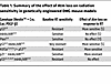 Table 1
Summary of the effect of Atm loss on radiation sensitivity in genetically engineered DMG mouse models
Our results from genetic experiments in primary mouse models indicate that p53 is a key determinant of the ability of DMGs to be radiosensitized by Atm loss. Almost all p53-altered primary mouse models were radioresistant and radiosensitized by Atm loss, including (a) a model driven by p53 loss with WT H3f3a (5); (b) a model driven by both p53 loss and loss of Ink4A/ARF(5); and (c) the H3.3K27M/TP53 mutant model reported here (Figure 1E). In contrast, Atm loss is unable to radiosensitize primary p53-WT brainstem glioma mouse models, including models driven by Ink4A and ARF loss (5) and models driven by Pten loss (11). Notably, a recent study comprehensively found that pharmacological ATM inhibition radiosensitizes both p53-mutant and WT p53 patient-derived models of DMG and pediatric high-grade glioma (6). Also, H3.3K27M may enhance ATM signaling, increasing radiosensitivity with and without ATM inhibition (35). Together, these data suggest that the mutational status of p53, H3.3K27M, and other alterations should be tested in correlative analyses in future clinical trials of ATM inhibitors in patients with DMG.
Our ISS data provide what we believe to be the first high-resolution transcriptional analysis at high gene plexy (~300 gene targets) in a mouse tumor model, which is critical to defining the model’s tumor vasculature and neoplastic compartments that play distinct roles in the therapeutic response (12). Future work will leverage these data to interrogate tumor immune and vascular microenvironment alterations induced by irradiation and Atm loss, which may guide the rational design of combinations of RT, ATM inhibitors, and therapies targeting the immune system or vasculature.
This work has several limitations. H3.3K27M did not decrease tumor latency in our system, as has been observed in other experimental systems (36). This may be due to the highly restricted manner of H3.3K27M induction in our system (i.e., from the endogeneous H3f3a locus and only in spatially and lineage-restricted cells) and/or the use of a relatively strong PDGF-β co-driver alteration that could mask more subtle H3.3K27M driver phenotypes in our system. Also, the presence of an HA tag on both HA–PDGF-β and H3.3K27M-FLAG-HA constructs precludes specific identification of PDGF-β in the nPH system. Finally, we observed a trend toward improved overall survival with ATM inhibition in an Atm-intact genetic mouse model. We hypothesize that the heterogeneity in tumor latency and the timing of treatment delivery may have conferred a significant survival benefit that was difficult to detect compared with the genetic loss of Atm and compared with the pharmacological xenograft experiment. Future studies could interrogate ATM inhibition effects in Atm-null models to discern on-target versus off-target effects of the ATM inhibitor.
The current work implicates transactivation-independent mechanisms by which p53 mediates radioresistance in Atm-null tumors. Our ISS data show that irradiation elicited overexpression of Cdkn1a, a key downstream target of p53 that mediates cell senescence and G1-to-S checkpoint arrest, in p53-null tumors, indicating p53-independent mechanisms of Cdkn1a expression (37). This finding led us to dissect the contribution of p53 transactivation functions that regulate p21 expression to radiosensitization in Atm-null DMGs. While Atm loss radiosensitizes tumors lacking p53, we found that Atm loss could not radiosensitize tumors containing a p5325,26 allele deficient in p53 transactivation function. Similarly, tumors lacking Cdkn1a (p21) could not be radiosensitized by Atm loss. Our findings highlight the importance of carefully considering p21 status in clinical trials involving Atm inhibition, given the complex role of p21 in tumor growth and the microenvironment (38). Strikingly, we found that Atm loss made tumors more radioresistant in mice that lacked Cdkn1a and that this loss was associated with increased apoptosis. These findings implicate the transactivation-independent function of p53 as a key determinant of radiosensitivity in Atm-null tumors. Future work will dissect the transactivation-independent functions of p53, such as the promotion of apoptosis through mitochondrial membrane permeabilization, direct repression of transcription, and/or direct interaction with complexes that detect DNA lesions (39, 40). Our data provide genetic and mechanistic insights that build upon studies of pharmacological ATM inhibition in patient-derived xenograft models (6). In addition, our work shows that ATM inhibition improves the response to irradiation, leading to extended survival. Further studies are needed to determine the transactivation-independent mechanisms of p53 and ATM-directed therapies and their effect on overcoming resistance to RT in patients with H3.3K27M-mutant DMG.


Copyright © 2026 American Society for Clinical Investigation
ISSN: 0021-9738 (print), 1558-8238 (online)