Go to JCI Insight
  • About
  • Editors
  • Consulting Editors
  • For authors
  • Publication ethics
  • Alerts
  • Advertising
  • Job board
  • Subscribe
  • Contact
  • Current issue
  • Past issues
  • By specialty
    • COVID-19
    • Cardiology
    • Gastroenterology
    • Immunology
    • Metabolism
    • Nephrology
    • Neuroscience
    • Oncology
    • Pulmonology
    • Vascular biology
    • All ...
  • Videos
    • Conversations with Giants in Medicine
    • Author's Takes
  • Reviews
    • View all reviews ...
    • Lung inflammatory injury and tissue repair (Jul 2023)
    • Immune Environment in Glioblastoma (Feb 2023)
    • Korsmeyer Award 25th Anniversary Collection (Jan 2023)
    • Aging (Jul 2022)
    • Next-Generation Sequencing in Medicine (Jun 2022)
    • New Therapeutic Targets in Cardiovascular Diseases (Mar 2022)
    • Immunometabolism (Jan 2022)
    • View all review series ...
  • Viewpoint
  • Collections
    • In-Press Preview
    • Commentaries
    • Research letters
    • Letters to the editor
    • Editorials
    • Viewpoint
    • Top read articles
  • Clinical Medicine
  • JCI This Month
    • Current issue
    • Past issues

  • Current issue
  • Past issues
  • Specialties
  • Reviews
  • Review series
  • Conversations with Giants in Medicine
  • Author's Takes
  • In-Press Preview
  • Commentaries
  • Research letters
  • Letters to the editor
  • Editorials
  • Viewpoint
  • Top read articles
  • About
  • Editors
  • Consulting Editors
  • For authors
  • Publication ethics
  • Alerts
  • Advertising
  • Job board
  • Subscribe
  • Contact
Tracking the clonal origin of lethal prostate cancer
Michael C. Haffner, … , William G. Nelson, Srinivasan Yegnasubramanian
Published October 25, 2013
Citation Information: J Clin Invest. 2013;123(11):4918-4922. https://doi.org/10.1172/JCI70354.
View: Text | PDF
Brief Report

Tracking the clonal origin of lethal prostate cancer

  • Text
  • PDF
Abstract

Recent controversies surrounding prostate cancer overtreatment emphasize the critical need to delineate the molecular features associated with progression to lethal metastatic disease. Here, we have used whole-genome sequencing and molecular pathological analyses to characterize the lethal cell clone in a patient who died of prostate cancer. We tracked the evolution of the lethal cell clone from the primary cancer to metastases through samples collected during disease progression and at the time of death. Surprisingly, these analyses revealed that the lethal clone arose from a small, relatively low-grade cancer focus in the primary tumor, and not from the bulk, higher-grade primary cancer or from a lymph node metastasis resected at prostatectomy. Despite being limited to one case, these findings highlight the potential importance of developing and implementing molecular prognostic and predictive markers, such as alterations of tumor suppressor proteins PTEN or p53, to augment current pathological evaluation and delineate clonal heterogeneity. Furthermore, this case illustrates the potential need in precision medicine to longitudinally sample metastatic lesions to capture the evolving constellation of alterations during progression. Similar comprehensive studies of additional prostate cancer cases are warranted to understand the extent to which these issues may challenge prostate cancer clinical management.

Authors

Michael C. Haffner, Timothy Mosbruger, David M. Esopi, Helen Fedor, Christopher M. Heaphy, David A. Walker, Nkosi Adejola, Meltem Gürel, Jessica Hicks, Alan K. Meeker, Marc K. Halushka, Jonathan W. Simons, William B. Isaacs, Angelo M. De Marzo, William G. Nelson, Srinivasan Yegnasubramanian

×

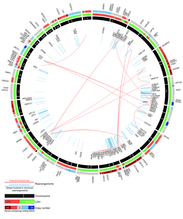
Figure 1

Common genomic consensus alterations found in 3 distant metastases (M5, M38, and M40) by whole-genome sequencing are plotted.

Options: View larger image (or click on image) Download as PowerPoint
Common genomic consensus alterations found in 3 distant metastases (M5, ...
Genes with nonsynonymous single nucleotide variants (SNVs) and indels in coding sequences are indicated in the outermost circle. Copy number alterations are color-coded and shown in the adjacent circle. Regions of high copy number gain (>4; dark red), and significant copy number loss (<0.5; blue) are indicated. LOH of individual regions (orange) is indicated in the middle circle. Arcs in the interior connect regions of structural rearrangements (blue, intrachromosomal; orange, interchromosomal), and associated genes are shown in the inner circle.

Copyright © 2023 American Society for Clinical Investigation
ISSN: 0021-9738 (print), 1558-8238 (online)

Sign up for email alerts