Advertisement
Commentary Free access | 10.1172/JCI69568
Illuminating regeneration: noninvasive imaging of disease progression in muscular dystrophy
Jennifer R. Levy and Kevin P. Campbell
Howard Hughes Medical Institute, Department of Molecular Physiology and Biophysics, Department of Neurology, Department of Internal Medicine, Roy J. and Lucille A. Carver College of Medicine, The University of Iowa, Iowa City, Iowa, USA.
Address correspondence to: Kevin P. Campbell, Howard Hughes Medical Institute, Department of Molecular Biology and Biophysics, The University of Iowa College of Medicine, 4283 CBRB, 285 Newton Road, Iowa City, Iowa, 52242-1101, USA. Phone: 319.335.7867; Fax: 319.335.6957; E-mail: kevin-campbell@uiowa.edu.
Find articles by Levy, J. in: PubMed | Google Scholar
Howard Hughes Medical Institute, Department of Molecular Physiology and Biophysics, Department of Neurology, Department of Internal Medicine, Roy J. and Lucille A. Carver College of Medicine, The University of Iowa, Iowa City, Iowa, USA.
Address correspondence to: Kevin P. Campbell, Howard Hughes Medical Institute, Department of Molecular Biology and Biophysics, The University of Iowa College of Medicine, 4283 CBRB, 285 Newton Road, Iowa City, Iowa, 52242-1101, USA. Phone: 319.335.7867; Fax: 319.335.6957; E-mail: kevin-campbell@uiowa.edu.
Find articles by Campbell, K. in: PubMed | Google Scholar
Published April 24, 2013 - More info
J Clin Invest. 2013;123(5):1931–1934. https://doi.org/10.1172/JCI69568.
© 2013 The American Society for Clinical Investigation
Related article:
Abstract
Muscular dystrophies are a class of disorders that cause progressive muscle wasting. A major hurdle for discovering treatments for the muscular dystrophies is a lack of reliable assays to monitor disease progression in animal models. We have developed a novel mouse model to assess disease activity noninvasively in mice with muscular dystrophies. These mice express an inducible luciferase reporter gene in muscle stem cells. In dystrophic mice, muscle stem cells activate and proliferate in response to muscle degeneration, resulting in an increase in the level of luciferase expression, which can be monitored by noninvasive, bioluminescence imaging. We applied this noninvasive imaging to assess disease activity in a mouse model of the human disease limb girdle muscular dystrophy 2B (LGMD2B), caused by a mutation in the dysferlin gene. We monitored the natural history and disease progression in these dysferlin-deficient mice up to 18 months of age and were able to detect disease activity prior to the appearance of any overt disease manifestation by histopathological analyses. Disease activity was reflected by changes in luciferase activity over time, and disease burden was reflected by cumulative luciferase activity, which paralleled disease progression as determined by histopathological analysis. The ability to monitor disease activity noninvasively in mouse models of muscular dystrophy will be invaluable for the assessment of disease progression and the effectiveness of therapeutic interventions.
Authors
Katie K. Maguire, Leland Lim, Sedona Speedy, Thomas A. Rando
-
Abstract
Muscular dystrophies are characterized by progressive muscle weakness and wasting. Among the key obstacles to the development of therapies is the absence of an assay to monitor disease progression in live animals. In this issue of the JCI, Maguire and colleagues use noninvasive bioluminescence imaging to monitor luciferase activity in mice expressing an inducible luciferase reporter gene in satellite cells. These cells proliferate in response to degeneration, therefore increasing the level of luciferase expression in dystrophic muscle.
-
Introduction
Skeletal muscle has a robust regenerative capacity, with rapid reestablishment of full strength, even after severe damage to the tissue. Regeneration is mediated by muscle stem cells, called satellite cells. In response to muscle damage, satellite cells proliferate, differentiate into myoblasts, and fuse into myotubes, which act to repair damaged muscle. In muscular dystrophies, continuous muscle degeneration is accompanied by regeneration of muscle fibers mediated by satellite cell progeny (1).
Currently, the standard method for evaluating disease progression in muscular dystrophy animal models is muscle histopathology. This approach is labor intensive, as it involves the removal and processing of the tissue of interest, imaging of the slides, and analysis of the images. Furthermore, the invasiveness of this approach does not permit consecutive sampling, hindering the ability to evaluate the course of a disease or success of a therapeutic strategy. Other methods for evaluating muscle disease include behavior testing and force testing of the dissected muscle, although the specificity of the results obtained from these tests can often be difficult to assess. High levels of serum biomarkers, such as serum creatine kinase, can be indicative of muscle damage, but levels depend on muscle mass and can be widely variable over time in individual dystrophic mice (2).
Perhaps the best candidate technology for studying muscle disease in live animals is MRI, which can reveal the permeability of muscle fibers correlating with disease severity (3). While MRI is noninvasive, it is more expensive and less widely available than bioluminescence imaging systems in animal research laboratories.
-
A “regeneration reporter” mouse strain
The first group to use bioluminescence imaging to reveal satellite cell proliferation was Sacco et al., who transplanted a single luciferase-expressing satellite cell into the tibialis anterior (TA) muscle of NOD/SCID mice that were depleted of endogenous satellite cells by irradiation (4). They observed that a single luciferase-expressing satellite cell is capable of self renewal after transplantation. Further, they found a substantial increase in satellite cell proliferation, as indicated by increased bioluminescence values, in response to muscle tissue damage by notexin.
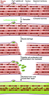
In this issue, Maguire et al. (5) utilized the Pax7CreER/LuSEAP mouse first generated by Nishijo et al. (6) to develop a mouse model that could be used to monitor muscle regeneration in response to disease and injury. This mouse expresses a Cre-dependent firefly luciferase gene and an estrogen-responsive Cre-recombinase under the control of the Pax7 locus. Because satellite cells are the only muscle cells in the adult that express Pax7, these mice express the bioreporter following tamoxifen treatment specifically in satellite cells. This expression is maintained as satellite cell progeny proliferate and differentiate into myotubes to repair the muscle, and expression of luciferase can be monitored by noninvasive, bioluminescence imaging (Figure 1). Nishijo et al. previously utilized the Pax7CreER/LuSEAP mice to characterize satellite cell kinetics in growing postnatal muscle and found a doubling of the signal every 3.93 weeks from adolescence to young adulthood (6). Maguire et al. use the Pax7CreER/LuSEAP mouse to assess satellite cell activation and proliferation as an indicator of regeneration. They first injured the TA muscles with cardiotoxin and compared the luciferase signal between the injured and uninjured limbs. Luciferase activity increased following injury, and immunohistochemical studies confirmed that luciferase-expressing cells were present in the injured TA as nascent myofibers and myotubes (5). This suggests that the noninvasive measurement of luciferase activity in the Pax7CreER/LuSEAP strain is an accurate measure of the proliferative activity of satellite cells and therefore has potential to be used to measure muscle regeneration in muscular dystrophy models as well.
 Figure 1
A “regeneration reporter” mouse strain. Pax7-positive muscle satellite cells express luciferase (indicated by green shading) following tamoxifen injection. Upon damage to muscle by either cardiotoxin injury or disease, satellite cells differentiate, and the progeny also express luciferase. Satellite cell progeny proliferate and differentiate into myotubes to repair the muscle, increasing the luciferase signal that can be detected by a bioluminescence imager.
-
Using luciferase to monitor disease progression
A key feature of dystrophic muscle is the degeneration of mature muscle fibers and the subsequent fusion of satellite cell–derived myoblasts to form myotubes, which mature into fibers. This degeneration/regeneration is apparent by muscle histopathology, where regenerating fibers can be identified by their centrally located nuclei. Maguire and colleagues hypothesized that the Pax7CreER/LuSEAP strain could be used to develop a noninvasive and quantifiable measure of regeneration activity in dystrophic mice. To test this, they crossed this reporter strain with a dysferlin-deficient (Dysf–/–) mouse model of limb girdle muscular dystrophy 2B (LGMD2B) (7, 8). Dysferlin is a transmembrane protein involved in calcium-mediated plasma membrane repair (9). Like human LGMD2B patients, Dysf–/– mice develop a slowly progressive muscular dystrophy, mainly in proximal limb muscles. Muscles from Dysf–/– mice exhibit centrally nucleated fibers, necrotic fibers, fat deposition, and inflammatory cell infiltrates (10).
The authors injected 2-month-old Dysf–/–/Pax7CreER/LuSEAP mice with tamoxifen to induce luciferase expression in satellite cells and found luciferase expression increased over time in the hind limb muscles of dysferlin-deficient, but not wild-type, mice. They found luciferase activity mainly in the proximal limb muscles as early as 3 months of age, with some involvement in the distal muscles starting at 6 months, and increasing luciferase activity in both proximal and distal muscle groups up to 18 months of age. Further examination of these mice showed a correlation between intensity of luciferase signal (as determined by bioluminescent imaging), number of luciferase positive fibers (as determined by immunohistochemistry), and extent of histopathology, including centrally nucleated fibers and fibers that express embryonic myosin heavy chain, a marker for newly regenerated myofibers (5). This validates luciferase imaging as an alternative to conventional measures of disease progression in muscular dystrophy.
There are several immediate advantages to using bioluminescent imaging to determine the extent of disease in muscular dystrophy models. First, the method is less labor intensive and more quantitative than classic histopathology. Second, measurements for individual mice are consistent and show patterns of luciferase activity that parallel the entire cohort. This suggests that a bioluminescence scheme for quantifying regeneration activity could be useful in therapeutic studies, in which each mouse could be used as its own pre- and posttreatment control. A third advantage to this system is the apparent increased sensitivity compared with conventional measures. The Dysf–/– mouse has been previously characterized as having a slowly progressive muscular dystrophy by 6 months of age, followed by rapid disease progression (7, 8). This is similar to other dysferlin-deficient LGMD2B mouse models (the naturally occurring A/J mice (11) and a targeted dysferlin knockout (12) both show active myopathy at approximately 6 to 8 months). Excitingly, the current paper finds a significant increase in luciferase expression in Dysf–/– mice as early as 3 months. This suggests that researchers may now have the opportunity to study the early pathophysiology of dysferlin deficiency and test the effectiveness of early therapeutic interventions.
One major limit to a bioluminescence-based reporter of regeneration activity is that it is light based. Therefore, luciferase signal is influenced by hair, skin pigmentation, thickness of skin and fat, and depth of tissue. This is particularly unfortunate in the case of the diaphragm, which is severely affected in several muscular dystrophies (especially Duchenne, ref. 13). Due to the depth of the diaphragm and the amount of tissue through which light must travel to image it, this method would not be particularly conducive to evaluating regeneration in the diaphragm. For researchers interested in regeneration activity in limb muscles, however, a bioluminescence strategy appears to have potential as a reliable and sensitive technique. Another disadvantage to this technique is the limited potential for an equivalent diagnostic tool for human patients. While therapeutic studies can be designed on animal models using luciferase activity as an indicator of regeneration activity, subsequent testing in humans will need to utilize a different technique, such as MRI imaging, to measure outcome. Finally, regeneration, while closely linked, may not be directly proportional to disease severity. Several mouse models have been generated that have regenerative defects (for examples, see ref. 14). Breeding these mice with the Pax7CreER/LuSEAP reporter line may give an incomplete picture of disease progression. Researchers must keep this caveat in mind when utilizing this technique on mouse models where the satellite cell regenerative capacity has not yet been characterized.
-
Future applications
Maguire and colleagues have demonstrated that bioluminescence imaging of satellite cells is an accurate representation of regeneration activity in a mouse model of LGMD2B. By crossing the Pax7CreER/LuSEAP mouse to other muscular dystrophy models (the mdx model of Duchenne, for example), this strategy can easily be used to study muscle degeneration and regeneration in any number of muscle diseases. Further, the Pax7CreER/LuSEAP mouse can be used to examine satellite cell proliferation in models of atrophy and hypertrophy (hind limb suspension and external loading, respectively), as well as other neuromuscular disorders. In the case of LGMD2B, the authors found that luciferase activity was significantly increased compared with wild-type controls at ages as early as 3 months. Bioluminescence imaging may also reveal early time points at which a difference can be detected in other models of muscle disease, which could provide insights into disease mechanism and alter the timing in which researchers consider applying therapies.
The Pax7CreER/LuSEAP mouse expresses luciferase in differentiated satellite cells, regardless of fate (myogenic, adipogenic, or fibrogenic). In the future, it would be interesting to utilize key transcription factors in order to design a reporter that could differentiate between self-renewed satellite cells and differentiated satellite cell progeny.
Compared to classical histology, which requires substantial effort in the dissection, processing, imaging, and analysis of each tissue, bioluminescence imaging is high throughput and quantitative. In addition, it is noninvasive and therefore it is ideal for studies in which each animal, and even each muscle, can be used as its own control. This technology is certainly an avenue investigators should consider when designing future studies of regeneration activity and therapeutic intervention in muscular disease.
-
Acknowledgments
J.R. Levy is supported by a Muscular Dystrophy Association Development grant (MDA200826). K.P. Campbell is an investigator of the Howard Hughes Medical Institute.
Address correspondence to: Kevin P. Campbell, Howard Hughes Medical Institute, Department of Molecular Biology and Biophysics, The University of Iowa College of Medicine, 4283 CBRB, 285 Newton Road, Iowa City, Iowa, 52242-1101, USA. Phone: 319.335.7867; Fax: 319.335.6957; E-mail: kevin-campbell@uiowa.edu.
-
Footnotes
Conflict of interest: The authors have declared that no conflict of interest exists.
Reference information: J Clin Invest. 2013;123(5):1931–1934. doi:10.1172/JCI69568.
See the related article at Assessment of disease activity in muscular dystrophies by noninvasive imaging.
-
References
- Yin H, Price F, Rudnicki MA. Satellite cells and the muscle stem cell niche. Physiol Rev. 2013;93(1):23–67.
- Lieberman JS, Taylor RG, Fowler WM. Serum creatine phosphokinase variations in dystrophic mice. Exp Neurol. 1981;73(3):716–724.
- McIntosh L, Granberg KE, Briere KM, Anderson JE. Nuclear magnetic resonance spectroscopy study of muscle growth, mdx dystrophy and glucocorticoid treatments: correlation with repair. NMR Biomed. 1998;11(1):1–10.
- Sacco A, Doyonnas R, Kraft P, Vitorovic S, Blau HM. Self-renewal and expansion of single transplanted muscle stem cells. Nature. 2008;456(7221):502–506.
- Maguire KK, Lim L, Speedy S, Rando TA. Assessment of disease activity in muscular dystrophies by noninvasive imaging. J Clin Invest. 2013;123(5):2298–2305.
- Nishijo K, et al. Biomarker system for studying muscle, stem cells, and cancer in vivo. FASEB J. 2009;23(8):2681–2690.
- Bittner RE, et al. Dysferlin deletion in SJL mice (SJL-Dysf) defines a natural model for limb girdle muscular dystrophy 2B. Nat Genet. 1999;23(2):141–142.
- Weller AH, Magliato SA, Bell KP, Rosenberg NL. Spontaneous myopathy in the SJL/J mouse: pathology and strength loss. Muscle Nerve. 1997;20(1):72–82.
- Han R, Campbell KP. Dysferlin and muscle membrane repair. Curr Opin Cell Biol. 2007;19(4):409–416.
- Hornsey MA, Laval SH, Barresi R, Lochmüller H, Bushby K. Muscular dystrophy in dysferlin-deficient mouse models. Neuromuscul Disord. 2013;23(5):377–387.View this article via: PubMed Google Scholar
- Ho M, et al. Disruption of muscle membrane and phenotype divergence in two novel mouse models of dysferlin deficiency. Hum Mol Genet. 2004;13(18):1999–2010.
- Bansal D, et al. Defective membrane repair in dysferlin-deficient muscular dystrophy. Nature. 2003;423(6936):168–172.
- Stedman HH, et al. The mdx mouse diaphragm reproduces the degenerative changes of Duchenne muscular dystrophy. Nature. 1991;352(6335):536–539.
- Wallace GQ, McNally EM. Mechanisms of muscle degeneration, regeneration, and repair in the muscular dystrophies. Annu Rev Physiol. 2009;71:37–57.
-
Version history
- Version 1 (April 24, 2013): No description
- Version 2 (May 1, 2013): No description


Copyright © 2025 American Society for Clinical Investigation
ISSN: 0021-9738 (print), 1558-8238 (online)

