Issue published April 1, 2016 Previous issue | Next issue
- Volume 126, Issue 4
Go to section:
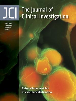
On the cover: Extracellular vesicles in vascular calcification

-
Review Series
×
Abstract
Intercellular signaling via extracellular vesicles (EVs) is an underappreciated modality of cell-cell crosstalk that enables cells to convey packages of complex instructions to specific recipient cells. EVs transmit these instructions through their cargoes of multiple proteins, nucleic acids, and specialized lipids, which are derived from their cells of origin and allow for combinatorial effects upon recipient cells. This Review series brings together the recent progress in our understanding of EV signaling in physiological and pathophysiological conditions, highlighting how certain EVs, particularly exosomes, can promote or regulate infections, host immune responses, development, and various diseases — notably cancer. Given the diverse nature of EVs and their abilities to profoundly modulate host cells, this series puts particular emphasis on the clinical applications of EVs as therapeutics and as diagnostic biomarkers.
Authors
Jonathan M. Pitt, Guido Kroemer, Laurence Zitvogel
×Abstract
New biomarkers are needed to improve the diagnosis of prostate cancer. Similarly to healthy cells, prostate epithelial cancer cells produce extracellular vesicles (prostasomes) that can be isolated from seminal fluid, urine, and blood. Prostasomes contain ubiquitously expressed and prostate-specific membrane and cytosolic proteins, as well as RNA. Both quantitative and qualitative changes in protein, mRNA, long noncoding RNA, and microRNA composition of extracellular vesicles isolated from prostate cancer patients have been reported. In general, however, the identified extracellular vesicle–associated single-marker molecules or combinations of marker molecules require confirmation in large cohorts of patients to validate their specificity and sensitivity as prostate cancer markers. Complications include variable factors such as prostate manipulation and urine flux, as well as masking by ubiquitously expressed free molecules and extracellular vesicles from tissues other than the prostate. Herein, we propose that the most promising methods include comprehensive combinational screening for (mutant) RNA in prostasomes that are immunoisolated with antibodies targeting prostate-specific epitopes.
Authors
Carla Zijlstra, Willem Stoorvogel
×Abstract
Two broad categories of extracellular vesicles (EVs), exosomes and shed microvesicles (sMVs), which differ in size distribution as well as protein and RNA profiles, have been described. EVs are known to play key roles in cell-cell communication, acting proximally as well as systemically. This Review discusses the nature of EV subtypes, strategies for isolating EVs from both cell-culture media and body fluids, and procedures for quantifying EVs. We also discuss proteins selectively enriched in exosomes and sMVs that have the potential for use as markers to discriminate between EV subtypes, as well as various applications of EVs in clinical diagnosis.
Authors
Rong Xu, David W. Greening, Hong-Jian Zhu, Nobuhiro Takahashi, Richard J. Simpson
×Abstract
Numerous studies have shown that non–cell-autonomous regulation of cancer cells is an important aspect of tumorigenesis. Cancer cells need to communicate with stromal cells by humoral factors such as VEGF, FGFs, and Wnt in order to survive. Recently, extracellular vesicles (EVs) have also been shown to be involved in cell-cell communication between cancer cells and the surrounding microenvironment and to be important for the development of cancer. In addition, these EVs contain small noncoding RNAs, including microRNAs (miRNAs), which contribute to the malignancy of cancer cells. Here, we provide an overview of current research on EVs, especially miRNAs in EVs. We also propose strategies to treat cancers by targeting EVs around cancer cells.
Authors
Nobuyoshi Kosaka, Yusuke Yoshioka, Yu Fujita, Takahiro Ochiya
×Abstract
Almost all cell types release extracellular vesicles (EVs), which are derived either from multivesicular bodies or from the plasma membrane. EVs contain a subset of proteins, lipids, and nucleic acids from the cell from which they are derived. EV factors, particularly small RNAs such as miRNAs, likely play important roles in cell-to-cell communication both locally and systemically. Most of the functions associated with EVs are in the regulation of immune responses to pathogens and cancer, as well as in regulating autoimmunity. This Review will focus on the different modes of immune regulation, both direct and indirect, by EVs. The therapeutic utility of EVs for the regulation of immune responses will also be discussed.
Authors
Paul D. Robbins, Akaitz Dorronsoro, Cori N. Booker
×Abstract
Exosomes and other extracellular microvesicles (ExMVs) have important functions in intercellular communication and regulation. During the course of infection, these vesicles can convey pathogen molecules that serve as antigens or agonists of innate immune receptors to induce host defense and immunity, or that serve as regulators of host defense and mediators of immune evasion. These molecules may include proteins, nucleic acids, lipids, and carbohydrates. Pathogen molecules may be disseminated by incorporation into vesicles that are created and shed by host cells, or they may be incorporated into vesicles shed from microbial cells. Involvement of ExMVs in the induction of immunity and host defense is widespread among many pathogens, whereas their involvement in immune evasion mechanisms is prominent among pathogens that establish chronic infection and is found in some that cause acute infection. Because of their immunogenicity and enrichment of pathogen molecules, exosomes may also have potential in vaccine preparations and as diagnostic markers. Additionally, the ability of exosomes to deliver molecules to recipient cells raises the possibility of their use for drug/therapy delivery. Thus, ExMVs play a major role in the pathogenesis of infection and provide exciting potential for the development of novel diagnostic and therapeutic approaches.
Authors
Jeffrey S. Schorey, Clifford V. Harding
×Abstract
Stroke is one of the leading causes of death and disability worldwide. Stroke recovery is orchestrated by a set of highly interactive processes that involve the neurovascular unit and neural stem cells. Emerging data suggest that exosomes play an important role in intercellular communication by transferring exosomal protein and RNA cargo between source and target cells in the brain. Here, we review these advances and their impact on promoting coupled brain remodeling processes after stroke. The use of exosomes for therapeutic applications in stroke is also highlighted.
Authors
Zheng Gang Zhang, Michael Chopp
×Abstract
Extracellular vesicles (EVs, including exosomes) are implicated in many aspects of nervous system development and function, including regulation of synaptic communication, synaptic strength, and nerve regeneration. They mediate the transfer of packets of information in the form of nonsecreted proteins and DNA/RNA protected within a membrane compartment. EVs are essential for the packaging and transport of many cell-fate proteins during development as well as many neurotoxic misfolded proteins during pathogenesis. This form of communication provides another dimension of cellular crosstalk, with the ability to assemble a “kit” of directional instructions made up of different molecular entities and address it to specific recipient cells. This multidimensional form of communication has special significance in the nervous system. How EVs help to orchestrate the wiring of the brain while allowing for plasticity associated with learning and memory and contribute to regeneration and degeneration are all under investigation. Because they carry specific disease-related RNAs and proteins, practical applications of EVs include potential uses as biomarkers and therapeutics. This Review describes our current understanding of EVs and serves as a springboard for future advances, which may reveal new important mechanisms by which EVs in coordinate brain and body function and dysfunction.
Authors
Valentina Zappulli, Kristina Pagh Friis, Zachary Fitzpatrick, Casey A. Maguire, Xandra O. Breakefield
×Abstract
Humans circulate quadrillions of exosomes at all times. Exosomes are a class of extracellular vesicles released by all cells, with a size range of 40–150 nm and a lipid bilayer membrane. Exosomes contain DNA, RNA, and proteins. Exosomes likely remove excess and/or unnecessary constituents from the cells, functioning like garbage bags, although their precise physiological role remains unknown. Additionally, exosomes may mediate specific cell-to-cell communication and activate signaling pathways in cells they fuse or interact with. Exosomes are detected in the tumor microenvironment, and emerging evidence suggests that they play a role in facilitating tumorigenesis by regulating angiogenesis, immunity, and metastasis. Circulating exosomes can be used as liquid biopsies and noninvasive biomarkers for early detection, diagnosis, and treatment of cancer patients.
Authors
Raghu Kalluri
×Abstract
Tumor-derived exosomes (TEX) are harbingers of tumor-induced immune suppression: they carry immunosuppressive molecules and factors known to interfere with immune cell functions. By delivering suppressive cargos consisting of proteins similar to those in parent tumor cells to immune cells, TEX directly or indirectly influence the development, maturation, and antitumor activities of immune cells. TEX also deliver genomic DNA, mRNA, and microRNAs to immune cells, thereby reprogramming functions of responder cells to promote tumor progression. TEX carrying tumor-associated antigens can interfere with antitumor immunotherapies. TEX also have the potential to serve as noninvasive biomarkers of tumor progression. In the tumor microenvironment, TEX may be involved in operating numerous signaling pathways responsible for the downregulation of antitumor immunity.
Authors
Theresa L. Whiteside
×Abstract
DC-derived exosomes (Dex) are nanometer-sized membrane vesicles that are secreted by the sentinel antigen-presenting cells of the immune system: DCs. Like DCs, the molecular composition of Dex includes surface expression of functional MHC-peptide complexes, costimulatory molecules, and other components that interact with immune cells. Dex have the potential to facilitate immune cell–dependent tumor rejection and have distinct advantages over cell-based immunotherapies involving DCs. Accordingly, Dex-based phase I and II clinical trials have been conducted in advanced malignancies, showing the feasibility and safety of the approach, as well as the propensity of these nanovesicles to mediate T and NK cell–based immune responses in patients. This Review will evaluate the interactions of Dex with immune cells, their clinical progress, and the future of Dex immunotherapy for cancer.
Authors
Jonathan M. Pitt, Fabrice André, Sebastian Amigorena, Jean-Charles Soria, Alexander Eggermont, Guido Kroemer, Laurence Zitvogel


-
Commentaries
×
Abstract
Precise epigenetic modifications in stem cells control developmental programs and cell fate decisions. In particular, the addition or removal of trimethylation of histone 3 lysine 27 (H3K27me3) at lineage-specific genes has been linked to the repression of gene expression, and a precise balance of methyltransferases and demethylases within cells determines H3K27me3 levels. The demethylase UTX is essential for development and tissue homeostasis; however, a role for UTX in stem cell–mediated tissue regeneration is unknown. In this issue of the
JCI , Dilworth and colleagues reveal that UTX and its demethylase activity are required in the muscle stem cell lineage for muscle regeneration in response to injury. Specifically, UTX mediates the removal of H3K27me3 in the promoter of the transcription factor myogenin, which regulates myogenic differentiation. The results of this study provide important insight into the contribution of epigenetic regulation in stem cell–mediated regeneration of adult tissues.Authors
Ling Liu, Thomas A. Rando
×Abstract
Exon skipping uses antisense oligonucleotides (ASOs) to alter transcript splicing for the purpose of rescuing or modulating protein expression. In this issue of the
JCI , Lee and colleagues developed and evaluated an ASO-dependent method for treating certain molecularly defined diseases associated with alterations in lamin A/C (LMNA ) splicing. Exon skipping by ASOs is gaining traction as a therapeutic strategy, and the use of ASOs is now being applied to bypass mutations and generate modified but functional proteins for an array of genetic disorders.Authors
Elizabeth M. McNally, Eugene J. Wyatt
×Abstract
Genetic alterations are known drivers of autoimmune disease; however, there is a much higher incidence of autoimmunity in women, implicating sex-specific factors in disease development. The autoimmune regulator (AIRE) gene contributes to the maintenance of central tolerance, and complete loss of AIRE function results in the development of autoimmune polyendocrinopathy syndrome type 1. In this issue of the JCI, Dragin and colleagues demonstrate that AIRE expression is downregulated in females as the result of estrogen-mediated alterations at the AIRE promoter. The association between estrogen and reduction of AIRE may at least partially account for the elevated incidence of autoimmune disease in women and has potential implications for sex hormone therapy.
Authors
Pearl Bakhru, Maureen A. Su
×Abstract
The invasion of cancer cells around and into nerves is associated with increased cancer aggression and poor patient outcome. As this perineural invasion increases disease severity, a better understanding of how the process is regulated may help in the development of therapeutics to target neuronal involvement in cancer. In this issue of the JCI, Deborde and colleagues show that direct contact between Schwann cells and cancer cells promotes cancer cell dissociation, migration, and invasion. Moreover, their data specifically suggest NCAM1 as an important molecular mediator of this Schwann cell–directed regulation of cancer cells in perineural invasion. The results of this study provide new insight into the cellular and molecular mechanisms of perineural invasion.
Authors
Salma H. Azam, Chad V. Pecot
×Abstract
Influenza infection can cause acute respiratory distress syndrome (ARDS), leading to poor disease outcome with high mortality. One of the driving features in the pathogenesis of ARDS is the accumulation of fluid in the alveoli, which causes severe pulmonary edema and impaired oxygen uptake. In this issue of the
JCI , Peteranderl and colleagues define a paracrine communication between macrophages and type II alveolar epithelial cells during influenza infection where IFNα induces macrophage secretion of TRAIL that causes endocytosis of Na,K-ATPase by the alveolar epithelium. This reduction of Na,K-ATPase expression decreases alveolar fluid clearance, which in turn leads to pulmonary edema. Inhibition of the TRAIL signaling pathway has been shown to improve lung injury after influenza infection, and future studies will be needed to determine if blocking this pathway is a viable option in the treatment of ARDS.Authors
Rena Brauer, Peter Chen
×Abstract
Regulatory T cells (Tregs) modulate the function of a variety of immune cells and are critical for maintaining self-tolerance and preventing the development of autoimmune disease. Due to their ability to suppress effector T cells, Tregs have been increasingly explored for clinical use to suppress alloresponses. While this approach has been promising in preclinical models and early clinical trials, widespread clinical use of Tregs has been limited by the low number of these cells in the periphery and the unknown frequency of allo-responsive Tregs. In this issue of the
JCI , MacDonald and colleagues transduced human Tregs with a chimeric antigen receptor (CAR) that targets the HLA class I molecule A2. These CAR-expressing T cells were readily activated via CAR stimulation and exerted potent immunosuppressive effects when stimulated in vitro. In a murine model of hematopoietic stem cell transplantation, CAR-modified Tregs were more effective in preventing the development of graft-versus–host disease compared with polyclonal Tregs. The results of this study lay the groundwork for the further evaluation of CAR-expressing Tregs in the prevention or treatment of transplant complications.Authors
Matthias Edinger


-
Research Articles
×
Abstract
Controlled and site-specific regulation of growth factor signaling remains a major challenge for current antiangiogenic therapies, as these antiangiogenic agents target normal vasculature as well tumor vasculature. In this article, we identified the prion-like protein doppel as a potential therapeutic target for tumor angiogenesis. We investigated the interactions between doppel and VEGFR2 and evaluated whether blocking the doppel/VEGFR2 axis suppresses the process of angiogenesis. We discovered that tumor endothelial cells (TECs), but not normal ECs, express doppel; tumors from patients and mouse xenografts expressed doppel in their vasculatures. Induced doppel overexpression in ECs enhanced vascularization, whereas doppel constitutively colocalized and complexed with VEGFR2 in TECs. Doppel inhibition depleted VEGFR2 from the cell membrane, subsequently inducing the internalization and degradation of VEGFR2 and thereby attenuating VEGFR2 signaling. We also synthesized an orally active glycosaminoglycan (LHbisD4) that specifically binds with doppel. We determined that LHbisD4 concentrates over the tumor site and that genetic loss of doppel in TECs decreases LHbisD4 binding and targeting both in vitro and in vivo. Moreover, LHbisD4 eliminated VEGFR2 from the cell membrane, prevented VEGF binding in TECs, and suppressed tumor growth. Together, our results demonstrate that blocking doppel can control VEGF signaling in TECs and selectively inhibit tumor angiogenesis.
Authors
Taslim A. Al-Hilal, Seung Woo Chung, Jeong Uk Choi, Farzana Alam, Jooho Park, Seong Who Kim, Sang Yoon Kim, Fakhrul Ahsan, In-San Kim, Youngro Byun
×Abstract
Philadelphia chromosome–like acute lymphoblastic leukemia (Ph-like ALL) is a high-risk ALL commonly associated with alterations that affect the tyrosine kinase pathway, tumor suppressors, and lymphoid transcription factors. Loss-of-function mutations in the gene-encoding adaptor protein LNK (also known as SH2B3) are found in Ph-like ALLs; however, it is not clear how LNK regulates normal B cell development or promotes leukemogenesis. Here, we have shown that combined loss of
Lnk and tumor suppressorsTp53 orInk4a /Arf in mice triggers a highly aggressive and transplantable precursor B-ALL.Tp53–/–Lnk–/– B-ALLs displayed similar gene expression profiles to human Ph-like B-ALLs, supporting use of this model for preclinical and molecular studies. PreleukemicTp53–/–Lnk–/– pro-B progenitors were hypersensitive to IL-7, exhibited marked self-renewal in vitro and in vivo, and were able to initiate B-ALL in transplant recipients. Mechanistically, we demonstrated that LNK regulates pro-B progenitor homeostasis by attenuating IL-7–stimuated JAK/STAT5 signaling via a direct interaction with phosphorylated JAK3. Moreover, JAK inhibitors were effective in prolonging survival of mice transplanted withLnk–/–Tp53–/– leukemia. Additionally, synergistic administration of PI3K/mTOR and JAK inhibitors further abrogated leukemia development. Hence, our results suggest that LNK suppresses IL-7R/JAK/STAT signaling to restrict pro-/pre-B progenitor expansion and leukemia development, providing a pathogenic mechanism and a potential therapeutic approach for B-ALLs withLNK mutations.Authors
Ying Cheng, Kudakwashe Chikwava, Chao Wu, Haibing Zhang, Anchit Bhagat, Dehua Pei, John K. Choi, Wei Tong
×Abstract
Abdominal aortic aneurysm (AAA) is a major cause of morbidity and mortality; however, the mechanisms that are involved in disease initiation and progression are incompletely understood. Extracellular matrix proteins play an integral role in modulating vascular homeostasis in health and disease. Here, we determined that the expression of the matricellular protein CCN3 is strongly reduced in rodent AAA models, including angiotensin II–induced AAA and elastase perfusion–stimulated AAA. CCN3 levels were also reduced in human AAA biopsies compared with those in controls. In murine models of induced AAA, germline deletion of
Ccn3 resulted in severe phenotypes characterized by elastin fragmentation, vessel dilation, vascular inflammation, dissection, heightened ROS generation, and smooth muscle cell loss. Conversely, overexpression of CCN3 mitigated both elastase- and angiotensin II–induced AAA formation in mice. BM transplantation experiments suggested that the AAA phenotype of CCN3-deficient mice is intrinsic to the vasculature, as AAA was not exacerbated in WT animals that received CCN3-deficient BM and WT BM did not reduce AAA severity in CCN3-deficient mice. Genetic and pharmacological approaches implicated the ERK1/2 pathway as a critical regulator of CCN3-dependent AAA development. Together, these results demonstrate that CCN3 is a nodal regulator in AAA biology and identify CCN3 as a potential therapeutic target for vascular disease.Authors
Chao Zhang, Dustin van der Voort, Hong Shi, Rongli Zhang, Yulan Qing, Shuichi Hiraoka, Minoru Takemoto, Koutaro Yokote, Joseph V. Moxon, Paul Norman, Laure Rittié, Helena Kuivaniemi, G. Brandon Atkins, Stanton L. Gerson, Guo-Ping Shi, Jonathan Golledge, Nianguo Dong, Bernard Perbal, Domenick A. Prosdocimo, Zhiyong Lin
×Abstract
Regulation of STAT3 activation is critical for normal and malignant hematopoietic cell proliferation. Here, we have reported that the endogenous transmembrane protein upstream-of-mTORC2 (UT2) negatively regulates activation of STAT3. Specifically, we determined that UT2 interacts directly with GP130 and inhibits phosphorylation of STAT3 on tyrosine 705 (STAT3Y705). This reduces cytokine signaling including IL6 that is implicated in multiple myeloma and other hematopoietic malignancies. Modulation of UT2 resulted in inverse effects on animal survival in myeloma models. Samples from multiple myeloma patients also revealed a decreased copy number of
UT2 and decreased expression ofUT2 in genomic and transcriptomic analyses, respectively. Together, these studies identify a transmembrane protein that functions to negatively regulate cytokine signaling through GP130 and pSTAT3Y705 and is molecularly and mechanistically distinct from the suppressors of cytokine signaling (SOCS) family of genes. Moreover, this work provides evidence that perturbations of this activation-dampening molecule participate in hematologic malignancies and may serve as a key determinant of multiple myeloma pathophysiology. UT2 is a negative regulator shared across STAT3 and mTORC2 signaling cascades, functioning as a tumor suppressor in hematologic malignancies driven by those pathways.Authors
Dongjun Lee, Ying-Hua Wang, Demetrios Kalaitzidis, Janani Ramachandran, Homare Eda, David B. Sykes, Noopur Raje, David T. Scadden
×Abstract
Acute and chronic tissue injury results in the generation of a myriad of environmental cues that macrophages respond to by changing their phenotype and function. This phenotypic regulation is critical for controlling tissue inflammation and resolution. Here, we have identified the adaptor protein disabled homolog 2 (DAB2) as a regulator of phenotypic switching in macrophages.
Dab2 expression was upregulated in M2 macrophages and suppressed in M1 macrophages isolated from both mice and humans, and genetic deletion ofDab2 predisposed macrophages to adopt a proinflammatory M1 phenotype. In mice with myeloid cell–specific deletion ofDab2 (Dab2fl/fl Lysm-Cre ), treatment with sublethal doses of LPS resulted in increased proinflammatory gene expression and macrophage activation. Moreover, chronic high-fat feeding exacerbated adipose tissue inflammation, M1 polarization of adipose tissue macrophages, and the development of insulin resistance in DAB2-deficient animals compared with controls. Mutational analyses revealed that DAB2 interacts with TNF receptor–associated factor 6 (TRAF6) and attenuates IκB kinase β–dependent (IKKβ-dependent) phosphorylation of Ser536 in the transactivation domain of NF-κB p65. Together, these findings reveal that DAB2 is critical for controlling inflammatory signaling during phenotypic polarization of macrophages and suggest that manipulation of DAB2 expression and function may hold therapeutic potential for the treatment of acute and chronic inflammatory disorders.Authors
Samantha E. Adamson, Rachael Griffiths, Radim Moravec, Subramanian Senthivinayagam, Garren Montgomery, Wenshu Chen, Jenny Han, Poonam R. Sharma, Garrett R. Mullins, Stacey A. Gorski, Jonathan A. Cooper, Alexandra Kadl, Kyle Enfield, Thomas J. Braciale, Thurl E. Harris, Norbert Leitinger
×Abstract
Vascular calcification is a common feature of major cardiovascular diseases. Extracellular vesicles participate in the formation of microcalcifications that are implicated in atherosclerotic plaque rupture; however, the mechanisms that regulate formation of calcifying extracellular vesicles remain obscure. Here, we have demonstrated that sortilin is a key regulator of smooth muscle cell (SMC) calcification via its recruitment to extracellular vesicles. Sortilin localized to calcifying vessels in human and mouse atheromata and participated in formation of microcalcifications in SMC culture. Sortilin regulated the loading of the calcification protein tissue nonspecific alkaline phosphatase (TNAP) into extracellular vesicles, thereby conferring its calcification potential. Furthermore, SMC calcification required Rab11-dependent trafficking and FAM20C/casein kinase 2–dependent C-terminal phosphorylation of sortilin. In a murine model,
Sort1 -deficiency reduced arterial calcification but did not affect bone mineralization. Additionally, transfer of sortilin-deficient BM cells to irradiated atherosclerotic mice did not affect vascular calcification, indicating a primary role of SMC-derived sortilin. Together, the results of this study identify sortilin phosphorylation as a potential therapeutic target for ectopic calcification/microcalcification and may clarify the mechanism that underlies the genetic association between theSORT1 gene locus and coronary artery calcification.Authors
Claudia Goettsch, Joshua D. Hutcheson, Masanori Aikawa, Hiroshi Iwata, Tan Pham, Anders Nykjaer, Mads Kjolby, Maximillian Rogers, Thomas Michel, Manabu Shibasaki, Sumihiko Hagita, Rafael Kramann, Daniel J. Rader, Peter Libby, Sasha A. Singh, Elena Aikawa
×Abstract
Alloreactive donor T cells are the driving force in the induction of graft-versus-host disease (GVHD), yet little is known about T cell metabolism in response to alloantigens after hematopoietic cell transplantation (HCT). Here, we have demonstrated that donor T cells undergo metabolic reprograming after allogeneic HCT. Specifically, we employed a murine allogeneic BM transplant model and determined that T cells switch from fatty acid β-oxidation (FAO) and pyruvate oxidation via the tricarboxylic (TCA) cycle to aerobic glycolysis, thereby increasing dependence upon glutaminolysis and the pentose phosphate pathway. Glycolysis was required for optimal function of alloantigen-activated T cells and induction of GVHD, as inhibition of glycolysis by targeting mTORC1 or 6-phosphofructo-2-kinase/fructose-2,6-biphosphatase 3 (PFKFB3) ameliorated GVHD mortality and morbidity. Together, our results indicate that donor T cells use glycolysis as the predominant metabolic process after allogeneic HCT and suggest that glycolysis has potential as a therapeutic target for the control of GVHD.
Authors
Hung D. Nguyen, Shilpak Chatterjee, Kelley M.K. Haarberg, Yongxia Wu, David Bastian, Jessica Heinrichs, Jianing Fu, Anusara Daenthanasanmak, Steven Schutt, Sharad Shrestha, Chen Liu, Honglin Wang, Hongbo Chi, Shikhar Mehrotra, Xue-Zhong Yu
×Abstract
Macrophages have long been considered to contribute to HIV infection of the CNS; however, a recent study has contradicted this early work and suggests that myeloid cells are not an in vivo source of virus production. Here, we addressed the role of macrophages in HIV infection by first analyzing monocytes isolated from viremic patients and patients undergoing antiretroviral treatment. We were unable to find viral DNA or viral outgrowth in monocytes isolated from peripheral blood. To determine whether tissue macrophages are productively infected, we used 3 different but complementary humanized mouse models. Two of these models (bone marrow/liver/thymus [BLT] mice and T cell–only mice [ToM]) have been previously described, and the third model was generated by reconstituting immunodeficient mice with human CD34+ hematopoietic stem cells that were devoid of human T cells (myeloid-only mice [MoM]) to specifically evaluate HIV replication in this population. Using MoM, we demonstrated that macrophages can sustain HIV replication in the absence of T cells; HIV-infected macrophages are distributed in various tissues including the brain; replication-competent virus can be rescued ex vivo from infected macrophages; and infected macrophages can establish de novo infection. Together, these results demonstrate that macrophages represent a genuine target for HIV infection in vivo that can sustain and transmit infection.
Authors
Jenna B. Honeycutt, Angela Wahl, Caroline Baker, Rae Ann Spagnuolo, John Foster, Oksana Zakharova, Stephen Wietgrefe, Carolina Caro-Vegas, Victoria Madden, Garrett Sharpe, Ashley T. Haase, Joseph J. Eron, J. Victor Garcia
×Abstract
Most skin cancers develop as the result of UV light–induced DNA damage; however, a substantial number of cases appear to occur independently of UV damage. A causal link between UV-independent skin cancers and chronic inflammation has been suspected, although the precise mechanism underlying this association is unclear. Here, we have proposed that activation-induced cytidine deaminase (AID, encoded by
AICDA ) links chronic inflammation and skin cancer. We demonstrated that Tg mice expressing AID in the skin spontaneously developed skin squamous cell carcinoma withHras andTrp53 mutations. Furthermore, genetic deletion ofAicda reduced tumor incidence in a murine model of chemical-induced skin carcinogenesis. AID was expressed in human primary keratinocytes in an inflammatory stimulus–dependent manner and was detectable in human skin cancers. Together, the results of this study indicate that inflammation-induced AID expression promotes skin cancer development independently of UV damage and suggest AID as a potential target for skin cancer therapeutics.Authors
Taichiro Nonaka, Yoshinobu Toda, Hiroshi Hiai, Munehiro Uemura, Motonobu Nakamura, Norio Yamamoto, Ryo Asato, Yukari Hattori, Kazuhisa Bessho, Nagahiro Minato, Kazuo Kinoshita
×Abstract
Chronic mucosal inflammation is associated with a greater risk of gastric cancer (GC) and, therefore, requires tight control by suppressive counter mechanisms. Gastrokine-2 (GKN2) belongs to a family of secreted proteins expressed within normal gastric mucosal cells. GKN2 expression is frequently lost during GC progression, suggesting an inhibitory role; however, a causal link remains unsubstantiated. Here, we developed
Gkn2 knockout and transgenic overexpressing mice to investigate the functional impact of GKN2 loss in GC pathogenesis. In mouse models of GC, decreased GKN2 expression correlated with gastric pathology that paralleled human GC progression. At baseline,Gkn2 knockout mice exhibited defective gastric epithelial differentiation but not malignant progression. Conversely,Gkn2 knockout in the IL-11/STAT3-dependentgp130 F/F GC model caused tumorigenesis of the proximal stomach. Additionally, gastric immunopathology was accelerated inHelicobacter pylori –infectedGkn2 knockout mice and was associated with augmented T helper cell type 1 (Th1) but not Th17 immunity. Heightened Th1 responses inGkn2 knockout mice were linked to deregulated mucosal innate immunity and impaired myeloid-derived suppressor cell activation. Finally, transgenic overexpression of human gastrokines (GKNs) attenuated gastric tumor growth ingp130 F/F mice. Together, these results reveal an antiinflammatory role for GKN2, provide in vivo evidence that links GKN2 loss to GC pathogenesis, and suggest GKN restoration as a strategy to restrain GC progression.Authors
Trevelyan R. Menheniott, Louise O’Connor, Yok Teng Chionh, Jan Däbritz, Michelle Scurr, Benjamin N. Rollo, Garrett Z. Ng, Shelley Jacobs, Angelique Catubig, Bayzar Kurklu, Stephen Mercer, Toshinari Minamoto, David E. Ong, Richard L. Ferrero, James G. Fox, Timothy C. Wang, Philip Sutton, Louise M. Judd, Andrew S. Giraud
×Abstract
The phosphatidylinositol 3-kinase (PI3K) signaling pathway is central to the action of insulin and many growth factors. Heterozygous mutations in the gene encoding the p85α regulatory subunit of PI3K (
PIK3R1 ) have been identified in patients with SHORT syndrome — a disorder characterized by short stature, partial lipodystrophy, and insulin resistance. Here, we evaluated whether SHORT syndrome–associatedPIK3R1 mutations account for the pathophysiology that underlies the abnormalities by generating knockin mice that are heterozygous for thePik3r1 Arg649Trp mutation, which is homologous to the mutation found in the majority of affected individuals. Similar to the patients, mutant mice exhibited a reduction in body weight and length, partial lipodystrophy, and systemic insulin resistance. These derangements were associated with a reduced capacity of insulin and other growth factors to activate PI3K in liver, muscle, and fat; marked insulin resistance in liver and fat of mutation-harboring animals; and insulin resistance in vitro in cells derived from these mice. In addition, mutant mice displayed defective insulin secretion and GLP-1 action on islets in vivo and in vitro. These data demonstrate the ability of this heterozygous mutation to alter PI3K activity in vivo and the central role of PI3K in insulin/growth factor action, adipocyte function, and glucose metabolism.Authors
Jonathon N. Winnay, Marie H. Solheim, Ercument Dirice, Masaji Sakaguchi, Hye-Lim Noh, Hee Joon Kang, Hirokazu Takahashi, Kishan K. Chudasama, Jason K. Kim, Anders Molven, C. Ronald Kahn, Pål R. Njølstad
×Abstract
Adoptive immunotherapy with regulatory T cells (Tregs) is a promising treatment for allograft rejection and graft-versus-host disease (GVHD). Emerging data indicate that, compared with polyclonal Tregs, disease-relevant antigen-specific Tregs may have numerous advantages, such as a need for fewer cells and reduced risk of nonspecific immune suppression. Current methods to generate alloantigen-specific Tregs rely on expansion with allogeneic antigen-presenting cells, which requires access to donor and recipient cells and multiple MHC mismatches. The successful use of chimeric antigen receptors (CARs) for the generation of antigen-specific effector T cells suggests that a similar approach could be used to generate alloantigen-specific Tregs. Here, we have described the creation of an HLA-A2–specific CAR (A2-CAR) and its application in the generation of alloantigen-specific human Tregs. In vitro, A2-CAR–expressing Tregs maintained their expected phenotype and suppressive function before, during, and after A2-CAR–mediated stimulation. In mouse models, human A2-CAR–expressing Tregs were superior to Tregs expressing an irrelevant CAR at preventing xenogeneic GVHD caused by HLA-A2+ T cells. Together, our results demonstrate that use of CAR technology to generate potent, functional, and stable alloantigen-specific human Tregs markedly enhances their therapeutic potential in transplantation and sets the stage for using this approach for making antigen-specific Tregs for therapy of multiple diseases.
Authors
Katherine G. MacDonald, Romy E. Hoeppli, Qing Huang, Jana Gillies, Dan S. Luciani, Paul C. Orban, Raewyn Broady, Megan K. Levings
×Abstract
The adult kidney plays a central role in erythropoiesis and is the main source of erythropoietin (EPO), an oxygen-sensitive glycoprotein that is essential for red blood cell production. Decreases of renal pO2 promote hypoxia-inducible factor 2–mediated (HIF-2–mediated) induction of EPO in peritubular interstitial fibroblast-like cells, which serve as the cellular site of EPO synthesis in the kidney. It is not clear whether HIF signaling in other renal cell types also contributes to the regulation of EPO production. Here, we used a genetic approach in mice to investigate the role of renal epithelial HIF in erythropoiesis. Specifically, we found that HIF activation in the proximal nephron via induced inactivation of the von Hippel–Lindau tumor suppressor, which targets the HIF-α subunit for proteasomal degradation, led to rapid development of hypoproliferative anemia that was associated with a reduction in the number of EPO-producing renal interstitial cells. Moreover, suppression of renal EPO production was associated with increased glucose uptake, enhanced glycolysis, reduced mitochondrial mass, diminished O2 consumption, and elevated renal tissue pO2. Our genetic analysis suggests that tubulointerstitial cellular crosstalk modulates renal EPO production under conditions of epithelial HIF activation in the kidney.
Authors
Navid M. Farsijani, Qingdu Liu, Hanako Kobayashi, Olena Davidoff, Feng Sha, Joachim Fandrey, T. Alp Ikizler, Paul M. O’Connor, Volker H. Haase
×Abstract
Meningioma-1 (MN1) overexpression is frequently observed in patients with acute myeloid leukemia (AML) and is predictive of poor prognosis. In murine models, forced expression of MN1 in hematopoietic progenitors induces an aggressive myeloid leukemia that is strictly dependent on a defined gene expression program in the cell of origin, which includes the homeobox genes
Hoxa9 andMeis1 as key components. Here, we have shown that this program is controlled by two histone methyltransferases, MLL1 and DOT1L, as deletion of eitherMll1 orDot1l in MN1-expressing cells abrogated the cell of origin–derived gene expression program, including the expression ofHoxa cluster genes. In murine models, genetic inactivation of eitherMll1 orDot1l impaired MN1-mediated leukemogenesis. We determined thatHOXA9 andMEIS1 are coexpressed withMN1 in a subset of clinicalMN1hi leukemia, and humanMN1hi/HOXA9hi leukemias were sensitive to pharmacologic inhibition of DOT1L. Together, these data point to DOT1L as a potential therapeutic target inMN1hi AML. In addition, our findings suggest that epigenetic modulation of the interplay between an oncogenic lesion and its cooperating developmental program has therapeutic potential in AML.Authors
Simone S. Riedel, Jessica N. Haladyna, Matthew Bezzant, Brett Stevens, Daniel A. Pollyea, Amit U. Sinha, Scott A. Armstrong, Qi Wei, Roy M. Pollock, Scott R. Daigle, Craig T. Jordan, Patricia Ernst, Tobias Neff, Kathrin M. Bernt
×Abstract
BACKGROUND. Recently, 1-octanol has been shown to have efficacy in treating patients with essential tremor (ET). The primary metabolite of 1-octanol is octanoic acid (OA), which is now thought to be the active substance that mediates tremor suppression. Our aim was to describe the maximum tolerated dose (MTD) of oral OA in patients with ET and assess the pharmacokinetics (PK) and pharmacodynamics (PD) profile of OA.METHODS. The MTD was studied using an open-label, single-ascending 3 + 3 dose–escalation design. Predefined single doses ranged from 8 to 128 mg/kg, with grade 2 adverse events (AEs) defined as dose-limiting toxicity. Tremor was assessed using accelerometry, digital spiral analysis, and a standard clinical rating scale at baseline and up to 600 minutes after intake. Safety assessments and PK sampling were also performed.RESULTS. Dose-limiting toxicity was not reached. The most frequent AE was mild abdominal discomfort. Exposure (AUC) increased linearly with the dose. Secondary efficacy measures suggested a dose-dependent reduction of tremor. Accordingly, a single unified PK/PD model with an effect compartment and sigmoid maximum effect (Emax) response could be built that accounted well for the time profiles of plasma concentrations as well as effects on tremor severity across the 5 dose levels.CONCLUSION. Although our trial did not reach an MTD, a dose-dependent effect was demonstrated in the PK/PD model as well as in secondary efficacy outcomes. Future studies are needed to explore the safety in higher dose ranges and to confirm dose-dependent efficacy in a placebo-controlled design.TRIAL REGISTRATION. Clinicaltrials.gov NCT01468948FUNDING. NINDS Intramural Research Program; TG Therapeutics Inc.Authors
Bernhard Voller, Emily Lines, Gayle McCrossin, Sule Tinaz, Codrin Lungu, George Grimes, Judith Starling, Gopal Potti, Peter Buchwald, Dietrich Haubenberger, Mark Hallett
×Abstract
Advances in the field of cancer immunology, including studies on tumor-infiltrating CD8+ cytotoxic T lymphocytes (CTLs), have led to new immunotherapeutics with proven efficacy against late-stage cancers. However, the antitumor potential of the immune system in targeting early-stage cancers remains uncertain. Here, we demonstrated that both genetic and chemical induction of thymic stromal lymphopoietin (TSLP) at a distant site leads to robust antitumor immunity against spontaneous breast carcinogenesis in mice. Breast tumors exposed to high circulating levels of TSLP were arrested at an early adenoma-like stage and were prevented from advancing to late carcinoma and metastasis. Additionally, CD4+ Th2 cells mediated the antitumor effects of TSLP, challenging the notion that Th2 cells only promote cancer. We also discovered that TSLP is expressed by the breast tumor cells themselves and acts to block breast cancer promotion. Moreover, TSLP-induced immunity also blocked early stages of pancreatic cancer development. Together, our findings demonstrate that TSLP potently induces immunity directed against early stages of breast cancer development without causing inflammation in the normal breast tissue. Moreover, our results highlight a previously unappreciated function of the immune system in controlling the early development of cancer and establish a fundamental role for TSLP and Th2 cells in tumor immunity against early-stage cancers.
Authors
Shadmehr Demehri, Trevor J. Cunningham, Sindhu Manivasagam, Kenneth H. Ngo, Sara Moradi Tuchayi, Rasika Reddy, Melissa A. Meyers, David G. DeNardo, Wayne M. Yokoyama
×Abstract
Fracture nonunions develop in 10%–20% of patients with fractures, resulting in prolonged disability. Current data suggest that bone union during fracture repair is achieved via proliferation and differentiation of skeletal progenitors within periosteal and soft tissues surrounding bone, while bone marrow stromal/stem cells (BMSCs) and other skeletal progenitors may also contribute. The NOTCH signaling pathway is a critical maintenance factor for BMSCs during skeletal development, although the precise role for NOTCH and the requisite nature of BMSCs following fracture is unknown. Here, we evaluated whether NOTCH and/or BMSCs are required for fracture repair by performing nonstabilized and stabilized fractures on NOTCH-deficient mice with targeted deletion of
RBPjk in skeletal progenitors, maturing osteoblasts, and committed chondrocytes. We determined that removal of NOTCH signaling in BMSCs and subsequent depletion of this population result in fracture nonunion, as the fracture repair process was normal in animals harboring either osteoblast- or chondrocyte-specific deletion ofRBPjk . Together, this work provides a genetic model of a fracture nonunion and demonstrates the requirement for NOTCH and BMSCs in fracture repair, irrespective of fracture stability and vascularity.Authors
Cuicui Wang, Jason A. Inzana, Anthony J. Mirando, Yinshi Ren, Zhaoyang Liu, Jie Shen, Regis J. O’Keefe, Hani A. Awad, Matthew J. Hilton
×Abstract
Avian H7N9 influenza viruses are group 2 influenza A viruses that have been identified as the etiologic agent for a current major outbreak that began in China in 2013 and may pose a pandemic threat. Here, we examined the human H7-reactive antibody response in 75 recipients of a monovalent inactivated A/Shanghai/02/2013 H7N9 vaccine. After 2 doses of vaccine, the majority of donors had memory B cells that secreted IgGs specific for H7 HA, with dominant responses against single HA subtypes, although frequencies of H7-reactive B cells ranged widely between donors. We isolated 12 naturally occurring mAbs with low half-maximal effective concentrations for binding, 5 of which possessed neutralizing and HA-inhibiting activities. The 5 neutralizing mAbs exhibited narrow breadth of reactivity with influenza H7 strains. Epitope-mapping studies using neutralization escape mutant analysis, deuterium exchange mass spectrometry, and x-ray crystallography revealed that these neutralizing mAbs bind near the receptor-binding pocket on HA. All 5 neutralizing mAbs possessed low numbers of somatic mutations, suggesting the clones arose from naive B cells. The most potent mAb, H7.167, was tested as a prophylactic treatment in a mouse intranasal virus challenge study, and systemic administration of the mAb markedly reduced viral lung titers.
Authors
Natalie J. Thornburg, Heng Zhang, Sandhya Bangaru, Gopal Sapparapu, Nurgun Kose, Rebecca M. Lampley, Robin G. Bombardi, Yingchun Yu, Stephen Graham, Andre Branchizio, Sandra M. Yoder, Michael T. Rock, C. Buddy Creech, Kathryn M. Edwards, David Lee, Sheng Li, Ian A. Wilson, Adolfo García-Sastre, Randy A. Albrecht, James E. Crowe Jr.
×Abstract
Posttranscriptional control of gene expression is important for defining both normal and pathological cellular phenotypes. In vitro, RNA-binding proteins (RBPs) have recently been shown to play important roles in posttranscriptional regulation; however, the contribution of RBPs to cell specification is not well understood. Here, we determined that the RBP insulin-like growth factor 2 mRNA-binding protein 3 (IGF2BP3) is specifically overexpressed in mixed lineage leukemia–rearranged (MLL-rearranged) B-acute lymphoblastic leukemia (B-ALL), which constitutes a subtype of this malignancy associated with poor prognosis and high risk of relapse. IGF2BP3 was required for the survival of B-ALL cell lines, as knockdown led to decreased proliferation and increased apoptosis. Enforced expression of IGF2BP3 provided murine BM cells with a strong survival advantage, led to proliferation of hematopoietic stem and progenitor cells, and skewed hematopoietic development to the B cell/myeloid lineage. Cross-link immunoprecipitation and high throughput sequencing uncovered the IGF2BP3-regulated transcriptome, which includes oncogenes
MYC andCDK6 as direct targets. IGF2BP3 regulated transcripts via targeting elements within 3′ untranslated regions (3′UTR), and enforced IGF2BP3 expression in mice resulted in enhanced expression ofMyc andCdk6 in BM. Together, our data suggest that IGF2BP3-mediated targeting of oncogenic transcripts may represent a critical pathogenetic mechanism in MLL-rearranged B-ALL and support IGF2BP3 and its cognate RNA-binding partners as potential therapeutic targets in this disease.Authors
Jayanth Kumar Palanichamy, Tiffany M. Tran, Jonathan M. Howard, Jorge R. Contreras, Thilini R. Fernando, Timothy Sterne-Weiler, Sol Katzman, Masoud Toloue, Weihong Yan, Giuseppe Basso, Martina Pigazzi, Jeremy R. Sanford, Dinesh S. Rao
×Abstract
Vanishing white matter (VWM) is a fatal leukodystrophy that is caused by mutations in genes encoding subunits of eukaryotic translation initiation factor 2B (eIF2B). Disease onset and severity are codetermined by genotype. White matter astrocytes and oligodendrocytes are almost exclusively affected; however, the mechanisms of VWM development remain unclear. Here, we used VWM mouse models, patients’ tissue, and cell cultures to investigate whether astrocytes or oligodendrocytes are the primary affected cell type. We generated 2 mouse models with mutations (
Eif2b5Arg191His/Arg191His andEif2b4Arg484Trp/Arg484Trp ) that cause severe VWM in humans and then crossed these strains to develop mice with various mutation combinations. Phenotypic severity was highly variable and dependent on genotype, reproducing the clinical spectrum of human VWM. In all mutant strains, impaired maturation of white matter astrocytes preceded onset and paralleled disease severity and progression. Bergmann glia and retinal Müller cells, nonforebrain astrocytes that have not been associated with VWM, were also affected, and involvement of these cells was confirmed in VWM patients. In coculture, VWM astrocytes secreted factors that inhibited oligodendrocyte maturation, whereas WT astrocytes allowed normal maturation of VWM oligodendrocytes. These studies demonstrate that astrocytes are central in VWM pathomechanisms and constitute potential therapeutic targets. Importantly, astrocytes should also be considered in the pathophysiology of other white matter disorders.Authors
Stephanie Dooves, Marianna Bugiani, Nienke L. Postma, Emiel Polder, Niels Land, Stephen T. Horan, Anne-Lieke F. van Deijk, Aleid van de Kreeke, Gerbren Jacobs, Caroline Vuong, Jan Klooster, Maarten Kamermans, Joke Wortel, Maarten Loos, Lisanne E. Wisse, Gert C. Scheper, Truus E.M. Abbink, Vivi M. Heine, Marjo S. van der Knaap
×Abstract
Autoimmune diseases affect 5% to 8% of the population, and females are more susceptible to these diseases than males. Here, we analyzed human thymic transcriptome and revealed sex-associated differences in the expression of tissue-specific antigens that are controlled by the autoimmune regulator (AIRE), a key factor in central tolerance. We hypothesized that the level of AIRE is linked to sexual dimorphism susceptibility to autoimmune diseases. In human and mouse thymus, females expressed less AIRE (mRNA and protein) than males after puberty. These results were confirmed in purified murine thymic epithelial cells (TECs). We also demonstrated that AIRE expression is related to sexual hormones, as male castration decreased AIRE thymic expression and estrogen receptor α–deficient mice did not show a sex disparity for AIRE expression. Moreover, estrogen treatment resulted in downregulation of AIRE expression in cultured human TECs, human thymic tissue grafted to immunodeficient mice, and murine fetal thymus organ cultures. AIRE levels in human thymus grafted in immunodeficient mice depended upon the sex of the recipient. Estrogen also upregulated the number of methylated CpG sites in the AIRE promoter. Together, our results indicate that in females, estrogen induces epigenetic changes in the AIRE gene, leading to reduced AIRE expression under a threshold that increases female susceptibility to autoimmune diseases.
Authors
Nadine Dragin, Jacky Bismuth, Géraldine Cizeron-Clairac, Maria Grazia Biferi, Claire Berthault, Alain Serraf, Rémi Nottin, David Klatzmann, Ana Cumano, Martine Barkats, Rozen Le Panse, Sonia Berrih-Aknin
×Abstract
Nerves enable cancer progression, as cancers have been shown to extend along nerves through the process of perineural invasion, which carries a poor prognosis. Furthermore, the innervation of some cancers promotes growth and metastases. It remains unclear, however, how nerves mechanistically contribute to cancer progression. Here, we demonstrated that Schwann cells promote cancer invasion through direct cancer cell contact. Histological evaluation of murine and human cancer specimens with perineural invasion uncovered a subpopulation of Schwann cells that associates with cancer cells. Coculture of cancer cells with dorsal root ganglion extracts revealed that Schwann cells direct cancer cells to migrate toward nerves and promote invasion in a contact-dependent manner. Upon contact, Schwann cells induced the formation of cancer cell protrusions in their direction and intercalated between the cancer cells, leading to cancer cell dispersion. The formation of these processes was dependent on Schwann cell expression of neural cell adhesion molecule 1 (NCAM1) and ultimately promoted perineural invasion. Moreover, NCAM1-deficient mice showed decreased neural invasion and less paralysis. Such Schwann cell behavior reflects normal Schwann cell programs that are typically activated in nerve repair but are instead exploited by cancer cells to promote perineural invasion and cancer progression.
Authors
Sylvie Deborde, Tatiana Omelchenko, Anna Lyubchik, Yi Zhou, Shizhi He, William F. McNamara, Natalya Chernichenko, Sei-Young Lee, Fernando Barajas, Chun-Hao Chen, Richard L. Bakst, Efsevia Vakiani, Shuangba He, Alan Hall, Richard J. Wong
×Abstract
The X chromosome–encoded histone demethylase UTX (also known as KDM6A) mediates removal of repressive trimethylation of histone H3 lysine 27 (H3K27me3) to establish transcriptionally permissive chromatin. Loss of UTX in female mice is embryonic lethal. Unexpectedly, male UTX-null mice escape embryonic lethality due to expression of UTY, a paralog that lacks H3K27 demethylase activity, suggesting an enzyme-independent role for UTX in development and thereby challenging the need for active H3K27 demethylation in vivo. However, the requirement for active H3K27 demethylation in stem cell–mediated tissue regeneration remains untested. Here, we employed an inducible mouse KO that specifically ablates
Utx in satellite cells (SCs) and demonstrated that active H3K27 demethylation is necessary for muscle regeneration. Loss of UTX in SCs blocked myofiber regeneration in both male and female mice. Furthermore, we demonstrated that UTX mediates muscle regeneration through its H3K27 demethylase activity, as loss of demethylase activity either by chemical inhibition or knock-in of demethylase-dead UTX resulted in defective muscle repair. Mechanistically, dissection of the muscle regenerative process revealed that the demethylase activity of UTX is required for expression of the transcription factor myogenin, which in turn drives differentiation of muscle progenitors. Thus, we have identified a critical role for the enzymatic activity of UTX in activating muscle-specific gene expression during myofiber regeneration and have revealed a physiological role for active H3K27 demethylation in vivo.Authors
Hervé Faralli, Chaochen Wang, Kiran Nakka, Aissa Benyoucef, Soji Sebastian, Lenan Zhuang, Alphonse Chu, Carmen G. Palii, Chengyu Liu, Brendan Camellato, Marjorie Brand, Kai Ge, F. Jeffrey Dilworth
×Abstract
Influenza A viruses (IAV) can cause lung injury and acute respiratory distress syndrome (ARDS), which is characterized by accumulation of excessive fluid (edema) in the alveolar airspaces and leads to hypoxemia and death if not corrected. Clearance of excess edema fluid is driven mostly by the alveolar epithelial Na,K-ATPase and is crucial for survival of patients with ARDS. We therefore investigated whether IAV infection alters Na,K-ATPase expression and function in alveolar epithelial cells (AECs) and the ability of the lung to clear edema. IAV infection reduced Na,K-ATPase in the plasma membrane of human and murine AECs and in distal lung epithelium of infected mice. Moreover, induced Na,K-ATPase improved alveolar fluid clearance (AFC) in IAV-infected mice. We identified a paracrine cell communication network between infected and noninfected AECs and alveolar macrophages that leads to decreased alveolar epithelial Na,K-ATPase function and plasma membrane abundance and inhibition of AFC. We determined that the IAV-induced reduction of Na,K-ATPase is mediated by a host signaling pathway that involves epithelial type I IFN and an IFN-dependent elevation of macrophage TNF-related apoptosis–inducing ligand (TRAIL). Our data reveal that interruption of this cellular crosstalk improves edema resolution, which is of biologic and clinical importance to patients with IAV-induced lung injury.
Authors
Christin Peteranderl, Luisa Morales-Nebreda, Balachandar Selvakumar, Emilia Lecuona, István Vadász, Rory E. Morty, Carole Schmoldt, Julia Bespalowa, Thorsten Wolff, Stephan Pleschka, Konstantin Mayer, Stefan Gattenloehner, Ludger Fink, Juergen Lohmeyer, Werner Seeger, Jacob I. Sznajder, Gökhan M. Mutlu, G.R. Scott Budinger, Susanne Herold
×Abstract
Lymphatic collecting vessels direct lymph into and from lymph nodes (LNs) and can become hyperpermeable as the result of a previous infection. Enhanced permeability has been implicated in compromised immunity due to reduced flow of lymph and immune cells to LNs, which are the primary site of antigen presentation to T cells. Presently, very little is known about the molecular signals that affect lymphatic collecting vessel permeability. Here, we have shown that lymphatic collecting vessel permeability is controlled by CCR7 and that the chronic hyperpermeability of collecting vessels observed in
Ccr7–/– mice is followed by vessel fibrosis. Reexpression of CCR7 in DCs, however, was sufficient to reverse the development of such fibrosis. IFN regulatory factor 4–positive (IRF4+) DCs constitutively interacted with collecting lymphatics, and selective ablation of this DC subset inCd11c-Cre Irf4fl/fl mice also rendered lymphatic collecting vessels hyperpermeable and fibrotic. Together, our data reveal that CCR7 plays multifaceted roles in regulating collecting vessel permeability and fibrosis, with one of the key players being IRF4-dependent DCs.Authors
Stoyan Ivanov, Joshua P. Scallan, Ki-Wook Kim, Kathrin Werth, Michael W. Johnson, Brian T. Saunders, Peter L. Wang, Emma L. Kuan, Adam C. Straub, Melissa Ouhachi, Erica G. Weinstein, Jesse W. Williams, Carlos Briseño, Marco Colonna, Brant E. Isakson, Emmanuel L. Gautier, Reinhold Förster, Michael J. Davis, Bernd H. Zinselmeyer, Gwendalyn J. Randolph
×Abstract
The alternatively spliced products of
LMNA , lamin C and prelamin A (the precursor to lamin A), are produced in similar amounts in most tissues and have largely redundant functions. This redundancy suggests that diseases, such as Hutchinson-Gilford progeria syndrome (HGPS), that are caused by prelamin A–specific mutations could be treated by shifting the output ofLMNA more toward lamin C. Here, we investigated mechanisms that regulateLMNA mRNA alternative splicing and assessed the feasibility of reducing prelamin A expression in vivo. We identified an exon 11 antisense oligonucleotide (ASO) that increased lamin C production at the expense of prelamin A when transfected into mouse and human fibroblasts. The same ASO also reduced the expression of progerin, the mutant prelamin A protein in HGPS, in fibroblasts derived from patients with HGPS. Mechanistic studies revealed that the exon 11 sequences contain binding sites for serine/arginine-rich splicing factor 2 (SRSF2), andSRSF2 knockdown lowered lamin A production in cells and in murine tissues. Moreover, administration of the exon 11 ASO reduced lamin A expression in wild-type mice and progerin expression in an HGPS mouse model. Together, these studies identify ASO-mediated reduction of prelamin A as a potential strategy to treat prelamin A–specific diseases.Authors
John M. Lee, Chika Nobumori, Yiping Tu, Catherine Choi, Shao H. Yang, Hea-Jin Jung, Timothy A. Vickers, Frank Rigo, C. Frank Bennett, Stephen G. Young, Loren G. Fong


Copyright © 2025 American Society for Clinical Investigation
ISSN: 0021-9738 (print), 1558-8238 (online)