Issue published March 2, 2015 Previous issue | Next issue

- Volume 125, Issue 3
Go to section:
On the cover: Inflammasome response exacerbates parasite infection
The cover image shows bone marrow–derived macrophages infected with Leishmania major (depicted in rainbow colors), with staining for macrophage nuclei (Hoescht, blue), mitochondria (Tom20, gray), and tubulin (purple). On page 1329, Gurung et al. demonstrate that the inflammasome response promotes infection by the parasite L. major by skewing T helper cell polarization. Image credit: Clifford Guy.
-
Review Series
×
Abstract
The enteric nervous system has been studied thus far as an isolated unit. As researchers probe deeper into the function of this system, it is evident that the neural network stretches beyond enteric neurons. It is formed by both intrinsic and extrinsic neurons innervating the gut, enteric glia, and innervated sensory epithelial cells, such as enteroendocrine cells. This Review series summarizes recent knowledge on function and disease of nerves, glia, and sensory epithelial cells of the gut in eight distinctive articles. The timing and growing knowledge for each individual field calls for an appropriate term encompassing the entire system. We call this neuronal ensemble the “gut connectome” and summarize the work from a food sensory perspective.
Authors
Diego V. Bohórquez, Rodger A. Liddle
×Abstract
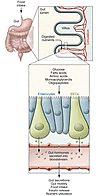
Fat is a vital macronutrient, and its intake is closely monitored by an array of molecular sensors distributed throughout the alimentary canal. In the mouth, dietary fat constituents such as mono- and diunsaturated fatty acids give rise to taste signals that stimulate food intake, in part by enhancing the production of lipid-derived endocannabinoid messengers in the gut. As fat-containing chyme enters the small intestine, it causes the formation of anorexic lipid mediators, such as oleoylethanolamide, which promote satiety. These anatomically and functionally distinct responses may contribute to the homeostatic control and, possibly, the pathological dysregulation of food intake.
Authors
Nicholas V. DiPatrizio, Daniele Piomelli
×Abstract
The enteric nervous system (ENS) is sometimes called the “second brain” because of the diversity of neuronal cell types and complex, integrated circuits that permit the ENS to autonomously regulate many processes in the bowel. Mechanisms supporting ENS development are intricate, with numerous proteins, small molecules, and nutrients that affect ENS morphogenesis and mature function. Damage to the ENS or developmental defects cause vomiting, abdominal pain, constipation, growth failure, and early death. Here, we review molecular mechanisms and cellular processes that govern ENS development, identify areas in which more investigation is needed, and discuss the clinical implications of new basic research.
Authors
Marina Avetisyan, Ellen Merrick Schill, Robert O. Heuckeroth
×Abstract
The enteroendocrine system is the primary sensor of ingested nutrients and is responsible for secreting an array of gut hormones, which modulate multiple physiological responses including gastrointestinal motility and secretion, glucose homeostasis, and appetite. This Review provides an up-to-date synopsis of the molecular mechanisms underlying enteroendocrine nutrient sensing and highlights our current understanding of the neuro-hormonal regulation of gut hormone secretion, including the interaction between the enteroendocrine system and the enteric nervous system. It is hoped that a deeper understanding of how these systems collectively regulate postprandial physiology will further facilitate the development of novel therapeutic strategies.
Authors
Arianna Psichas, Frank Reimann, Fiona M. Gribble
×Abstract
Enteric glia are important components of the enteric nervous system (ENS) and also form an extensive network in the mucosa of the gastrointestinal (GI) tract. Initially regarded as passive support cells, it is now clear that they are actively involved as cellular integrators in the control of motility and epithelial barrier function. Enteric glia form a cellular and molecular bridge between enteric nerves, enteroendocrine cells, immune cells, and epithelial cells, depending on their location. This Review highlights the role of enteric glia in GI motility disorders and in barrier and defense functions of the gut, notably in states of inflammation. It also discusses the involvement of enteric glia in neurological diseases that involve the GI tract.
Authors
Keith A. Sharkey
×Abstract
Tremendous progress has been made in characterizing the bidirectional interactions between the central nervous system, the enteric nervous system, and the gastrointestinal tract. A series of provocative preclinical studies have suggested a prominent role for the gut microbiota in these gut-brain interactions. Based on studies using rodents raised in a germ-free environment, the gut microbiota appears to influence the development of emotional behavior, stress- and pain-modulation systems, and brain neurotransmitter systems. Additionally, microbiota perturbations by probiotics and antibiotics exert modulatory effects on some of these measures in adult animals. Current evidence suggests that multiple mechanisms, including endocrine and neurocrine pathways, may be involved in gut microbiota–to–brain signaling and that the brain can in turn alter microbial composition and behavior via the autonomic nervous system. Limited information is available on how these findings may translate to healthy humans or to disease states involving the brain or the gut/brain axis. Future research needs to focus on confirming that the rodent findings are translatable to human physiology and to diseases such as irritable bowel syndrome, autism, anxiety, depression, and Parkinson’s disease.
Authors
Emeran A. Mayer, Kirsten Tillisch, Arpana Gupta
×Abstract
Bariatric surgery is the most effective treatment for severe obesity, producing marked sustained weight loss with associated reduced morbidity and mortality. Roux-en-Y gastric bypass surgery (RYGBP), the most commonly performed procedure, was initially viewed as a hybrid restrictive-malabsorptive procedure. However, over the last decade, it has become apparent that alternative physiologic mechanisms underlie its beneficial effects. RYGBP-induced altered feeding behavior, including reduced appetite and changes in taste/food preferences, is now recognized as a key driver of the sustained postoperative weight loss. The brain ultimately determines feeding behavior, and here we review the mechanisms by which RYGBP may affect central appetite-regulating pathways.
Authors
Sean Manning, Andrea Pucci, Rachel L. Batterham
×Abstract
Effective colonic motility involves an intricate pattern of excitatory and inhibitory neuromuscular signals that arise from the enteric neural circuitry of the colon. Recent investigations have demonstrated that inflammation leads to a variety of changes in the physiological properties of the neurons in this circuitry, including hyperexcitability of neurons at the afferent end of the peristaltic reflex, synaptic facilitation, and attenuated inhibitory neuromuscular transmission. Furthermore, links have been established between these changes and disrupted motor activity in the colon, and we now know that some of these changes persist long after recovery from inflammation. It is highly likely that inflammation-induced neuroplasticity, which is not detectable by clinical diagnostics, contributes to disrupted motility in active and quiescent inflammatory bowel disease and in functional gastrointestinal disorders.
Authors
Gary M. Mawe
×Abstract
The enteric nervous system (ENS) consists of neurons and glial cells that differentiate from neural crest progenitors. During embryogenesis, development of the ENS is controlled by the interplay of neural crest cell–intrinsic factors and instructive cues from the surrounding gut mesenchyme. However, postnatal ENS development occurs in a different context, which is characterized by the presence of microbiota and an extensive immune system, suggesting an important role of these factors on enteric neural circuit formation and function. Initial reports confirm this idea while further studies in this area promise new insights into ENS physiology and pathophysiology.
Authors
Panagiotis S. Kabouridis, Vassilis Pachnis


-
The Attending Physician
×
Abstract
Clinical vignette: An 8-year-old boy presents to the pediatric ICU after two days of cough with increasing secretions. The patient is progressing to respiratory failure and requires noninvasive mechanical ventilation. His past medical history is remarkable for premature birth at 25 and 6/7 weeks gestational age, cerebral palsy, developmental delay, epilepsy, and gastrostomy tube dependence. His chest x-ray is remarkable for multifocal opacities that are consistent with atelectasis. A complete blood count reveals a wbc count of 9.2 with a normal differential, Hg of 11.7, and platelet count of 276,000. A respiratory viral panel from a nasal swab returns positive for rhinovirus. Additional patient history from the parents uncovers that he has been hospitalized three times over the course of the past 2 years with a similar presentation.
Authors
Jason Boehme, Emin Maltepe

-
Commentaries
×
Abstract
Krüppel-like factors (KLFs) are zinc finger transcription factors that share homology in three C-terminal zinc finger domains. KLF family members are expressed in most if not all tissues and have diverse roles in organismal development and cell differentiation, function, and death. The glomerular podocyte is particularly sensitive to mitochondrial dysfunction, as seen in various genetic disorders manifesting as progressive glomerulosclerosis. In this issue of the
JCI , Mallipattu and coworkers show that KLF6 expression is reduced in mouse and human glomerular disease. Podocyte-specific deletion ofKlf6 expression in mice leads to mitochondrial dysfunction and apoptosis, followed by glomerulosclerosis. This is the first demonstration that defective transcriptional regulation of nuclear-encoded mitochondrial genes can result in experimental glomerular disease.Authors
Jeffrey B. Kopp
×Abstract
Almost one-third of transplanted kidneys come from living donors, who sacrifice approximately 30% of their pre-donation glomerular filtration rate (GFR) after they experience compensatory hypertrophy and hyperfiltration in their remaining kidney. Although hyperfiltration can cause glomerular injury, many studies have suggested that donor nephrectomy itself does not cause long-term loss of GFR at a higher rate than what is seen in the normal aging population. However, when post-donation kidney diseases occur in an unfortunate few, recent studies suggest that GFR loss at donor nephrectomy increases the risk of eventual end-stage renal disease (ESRD). In this issue of the
JCI , Lenihan and colleagues evaluated glomerular dynamics in a cohort of kidney donors prior to, within 1 year of, and several years after kidney donation. Their results suggest that adaptive hyperfiltration in the remaining kidney occurs without glomerular hypertension, furthering our understanding of the relatively benign renal outcomes for most living kidney donors.Authors
Roland C. Blantz, Robert W. Steiner
×Abstract
Cholangiocarcinoma is a relatively rare cancer of the biliary ducts that is highly refractory to treatment. The factors that drive cholangiocarcinoma are poorly understood, though chronic liver fluke infection is a risk factor for disease. In this issue of the
JCI , Boulter and colleagues demonstrate that the WNT/β-catenin signaling pathway is upregulated in patients with sporadic cholangiocarcinoma. The authors determined that macrophages generate WNT ligands in cholangiocarcinomas and depletion or inhibition of this cell population in animal models of cholangiocarcinoma reduced tumor burden and proliferation. Moreover, pharmacological inhibition of WNT secretion or β-catenin activity was efficacious in animal models. Together the results of this study suggest that targeting WNT has potential as a therapeutic strategy for cholangiocarcinoma.Authors
David M. Virshup
×Abstract
There have been many attempts at slowing down or even reversing the neurodegenerative process of Parkinson’s disease (PD). To date, there are no treatments of proven value in this regard. One underexplored route to slow the neurodegenerative process is the use of agents that may stimulate neurogenesis in the subventricular zone. In animal models of PD, PDGF-BB has been shown to restore/protect against dopaminergic deficits caused by neurotoxins via increased neurogenesis in the subventricular zone. Previous work suggests that these new cells are not themselves dopaminergic but have trophic effects on residual dopaminergic cells in the substantia nigra. In this issue of the
JCI , Paul et al. evaluate this agent in individuals with PD and show that i.c.v. administration of PDGF-BB is safe and well tolerated. This study lays the foundation for formal dose-finding studies and clinical trials to assess the efficacy of this agent as a potential neuroprotective treatment for PD.Authors
Thomas Foltynie


-
Research Articles
×
Abstract
Polyploidization is one of the most dramatic changes that can occur in the genome. In the liver, physiological polyploidization events occur during both liver development and throughout adult life. Here, we determined that a pathological polyploidization takes place in nonalcoholic fatty liver disease (NAFLD), a widespread hepatic metabolic disorder that is believed to be a risk factor for hepatocellular carcinoma (HCC). In murine models of NAFLD, the parenchyma of fatty livers displayed alterations of the polyploidization process, including the presence of a large proportion of highly polyploid mononuclear cells, which are rarely observed in normal hepatic parenchyma. Biopsies from patients with nonalcoholic steatohepatitis (NASH) revealed the presence of alterations in hepatocyte ploidy compared with tissue from control individuals. Hepatocytes from NAFLD mice revealed that progression through the S/G2 phases of the cell cycle was inefficient. This alteration was associated with activation of a G2/M DNA damage checkpoint, which prevented activation of the cyclin B1/CDK1 complex. Furthermore, we determined that oxidative stress promotes the appearance of highly polyploid cells, and antioxidant-treated NAFLD hepatocytes resumed normal cell division and returned to a physiological state of polyploidy. Collectively, these findings indicate that oxidative stress promotes pathological polyploidization and suggest that this is an early event in NAFLD that may contribute to HCC development.
Authors
Géraldine Gentric, Vanessa Maillet, Valérie Paradis, Dominique Couton, Antoine L’Hermitte, Ganna Panasyuk, Bernard Fromenty, Séverine Celton-Morizur, Chantal Desdouets
×Abstract
Germline
GATA1 mutations that result in the production of an amino-truncated protein termed GATA1s (where s indicates short) cause congenital hypoplastic anemia. In patients with trisomy 21, similar somatic GATA1s-producing mutations promote transient myeloproliferative disease and acute megakaryoblastic leukemia. Here, we demonstrate that induced pluripotent stem cells (iPSCs) from patients with GATA1-truncating mutations exhibit impaired erythroid potential, but enhanced megakaryopoiesis and myelopoiesis, recapitulating the major phenotypes of the associated diseases. Similarly, in developmentally arrested GATA1-deficient murine megakaryocyte-erythroid progenitors derived from murine embryonic stem cells (ESCs), expression of GATA1s promoted megakaryopoiesis, but not erythropoiesis. Transcriptome analysis revealed a selective deficiency in the ability of GATA1s to activate erythroid-specific genes within populations of hematopoietic progenitors. Although its DNA-binding domain was intact, chromatin immunoprecipitation studies showed that GATA1s binding at specific erythroid regulatory regions was impaired, while binding at many nonerythroid sites, including megakaryocytic and myeloid target genes, was normal. Together, these observations indicate that lineage-specific GATA1 cofactor associations are essential for normal chromatin occupancy and provide mechanistic insights into howGATA1s mutations cause human disease. More broadly, our studies underscore the value of ESCs and iPSCs to recapitulate and study disease phenotypes.Authors
Marta Byrska-Bishop, Daniel VanDorn, Amy E. Campbell, Marisol Betensky, Philip R. Arca, Yu Yao, Paul Gadue, Fernando F. Costa, Richard L. Nemiroff, Gerd A. Blobel, Deborah L. French, Ross C. Hardison, Mitchell J. Weiss, Stella T. Chou
×Abstract
Patients with B cell precursor acute lymphoblastic leukemia (BPL) respond well to chemotherapy at initial diagnosis; however, therapeutic options are limited for individuals with BPL who relapse. Almost all BPL cells express CD19, and we recently cloned the gene encoding a natural ligand of the human CD19 receptor (CD19L). We hypothesized that fusion of CD19L to the soluble extracellular domain of proapoptotic TNF-related apoptosis-inducing ligand (sTRAIL) would markedly enhance the potency of sTRAIL and specifically induce BPL cell apoptosis due to membrane anchoring of sTRAIL and simultaneous activation of the CD19 and TRAIL receptor (TRAIL-R) apoptosis signaling pathways. Here, we demonstrate that recombinant human CD19L-sTRAIL was substantially more potent than sTRAIL and induced apoptosis in primary leukemia cells taken directly from BPL patients. CD19L-sTRAIL effectively targeted and eliminated in vivo clonogenic BPL xenograft cells, even at femtomolar-picomolar concentrations. In mice, CD19L-sTRAIL exhibited a more favorable pharmacokinetic (PK) profile than sTRAIL and was nontoxic at doses ranging from 32 fmol/kg to 3.2 pmol/kg. CD19L-sTRAIL showed potent in vivo antileukemic activity in NOD/SCID mouse xenograft models of relapsed and chemotherapy-resistant BPL at nontoxic fmol/kg dose levels. Together, these results suggest that recombinant human CD19L-sTRAIL has clinical potential as a biotherapeutic agent against BPL.
Authors
Fatih M. Uckun, Dorothea E. Myers, Sanjive Qazi, Zahide Ozer, Rebecca Rose, Osmond J. D’Cruz, Hong Ma
×Abstract
Effector T cell migration into inflamed sites greatly exacerbates tissue destruction and disease severity in inflammatory diseases, including graft-versus-host disease (GVHD). T cell migration into such sites depends heavily on regulated adhesion and migration, but the signaling pathways that coordinate these functions downstream of chemokine receptors are largely unknown. Using conditional knockout mice, we found that T cells lacking the adaptor proteins CRK and CRK-like (CRKL) exhibit reduced integrin-dependent adhesion, chemotaxis, and diapedesis. Moreover, these two closely related proteins exhibited substantial functional redundancy, as ectopic expression of either protein rescued defects in T cells lacking both CRK and CRKL. We determined that CRK proteins coordinate with the RAP guanine nucleotide exchange factor C3G and the adhesion docking molecule CASL to activate the integrin regulatory GTPase RAP1. CRK proteins were required for effector T cell trafficking into sites of inflammation, but not for migration to lymphoid organs. In a murine bone marrow transplantation model, the differential migration of CRK/CRKL-deficient T cells resulted in efficient graft-versus-leukemia responses with minimal GVHD. Together, the results from our studies show that CRK family proteins selectively regulate T cell adhesion and migration at effector sites and suggest that these proteins have potential as therapeutic targets for preventing GVHD.
Authors
Yanping Huang, Fiona Clarke, Mobin Karimi, Nathan H. Roy, Edward K. Williamson, Mariko Okumura, Kazuhiro Mochizuki, Emily J.H. Chen, Tae-Ju Park, Gudrun F. Debes, Yi Zhang, Tom Curran, Taku Kambayashi, Janis K. Burkhardt
×Abstract
Astrocytes are integral components of the homeostatic neural network as well as active participants in pathogenesis of and recovery from nearly all neurological conditions. Evolutionarily, compared with lower vertebrates and nonhuman primates, humans have an increased astrocyte-to-neuron ratio; however, a lack of effective models has hindered the study of the complex roles of human astrocytes in intact adult animals. Here, we demonstrated that after transplantation into the cervical spinal cords of adult mice with severe combined immunodeficiency (SCID), human pluripotent stem cell–derived (PSC-derived) neural progenitors migrate a long distance and differentiate to astrocytes that nearly replace their mouse counterparts over a 9-month period. The human PSC-derived astrocytes formed networks through their processes, encircled endogenous neurons, and extended end feet that wrapped around blood vessels without altering locomotion behaviors, suggesting structural, and potentially functional, integration into the adult mouse spinal cord. Furthermore, in SCID mice transplanted with neural progenitors derived from induced PSCs from patients with ALS, astrocytes were generated and distributed to a similar degree as that seen in mice transplanted with healthy progenitors; however, these mice exhibited motor deficit, highlighting functional integration of the human-derived astrocytes. Together, these results indicate that this chimeric animal model has potential for further investigating the roles of human astrocytes in disease pathogenesis and repair.
Authors
Hong Chen, Kun Qian, Wei Chen, Baoyang Hu, Lisle W. Blackbourn IV, Zhongwei Du, Lixiang Ma, Huisheng Liu, Karla M. Knobel, Melvin Ayala, Su-Chun Zhang
×Abstract
BACKGROUND. Mutational inactivation in cancer of key apoptotic pathway components, such asTP53 /p53, undermines cytotoxic therapies that aim to increase apoptosis. Accordingly,TP53 mutations are reproducibly associated with poor treatment outcomes. Moreover, cytotoxic treatments destroy normal stem cells with intact p53 systems, a problem especially for myeloid neoplasms, as these cells reverse the low blood counts that cause morbidity and death. Preclinical studies suggest that noncytotoxic concentrations of the DNA methyltransferase 1 (DNMT1) inhibitor decitabine produce p53-independent cell-cycle exits by reversing aberrant epigenetic repression of proliferation-terminating (MYC-antagonizing) differentiation genes in cancer cells.METHODS. In this clinical trial, patients with myelodysplastic syndrome (n = 25) received reduced decitabine dosages (0.1–0.2 mg/kg/day compared with the FDA-approved 20–45 mg/m2/day dosage, a 75%–90% reduction) to avoid cytotoxicity. These well-tolerated doses were frequently administered 1–3 days per week, instead of pulse cycled for 3 to 5 days over a 4- to 6-week period, to increase the probability that cancer S-phase entries would coincide with drug exposure, which is required for S-phase–dependent DNMT1 depletion.RESULTS. The median subject age was 73 years (range, 46–85 years), 9 subjects had relapsed disease or were refractory to 5-azacytidine and/or lenalidomide, and 3 had received intensive chemoradiation to treat other cancers. Adverse events were related to neutropenia present at baseline: neutropenic fever (13 of 25 subjects) and septic death (1 of 25 subjects). Blood count improvements meeting the International Working Group criteria for response occurred in 11 of 25 (44%) subjects and were highly durable. Treatment-induced freedom from transfusion lasted a median of 1,025 days (range, 186–1,152 days; 3 ongoing), and 20% of subjects were treated for more than 3 years. Mutations and/or deletions of key apoptosis genes were frequent (present in 55% of responders and in 36% of nonresponders). Noncytotoxic DNMT1 depletion was confirmed by serial BM γ-H2AX (DNA repair/damage marker) and DNMT1 analyses. MYC master oncoprotein levels were markedly decreased.CONCLUSION. Decitabine regimens can be redesigned to minimize cytotoxicity and increase exposure time for DNMT1 depletion, to safely and effectively circumvent mutational apoptotic defects.TRIAL REGISTRATION. Clinicaltrials.gov NCT01165996.FUNDING. NIH (R01CA138858, CA043703); Department of Defense (PR081404); Clinical and Translational Science Award (CTSA) (UL1RR024989); and the Leukemia and Lymphoma Society (Translational Research Program).Authors
Yogen Saunthararajah, Mikkael Sekeres, Anjali Advani, Reda Mahfouz, Lisa Durkin, Tomas Radivoyevitch, Ricki Englehaupt, Joy Juersivich, Kathleen Cooper, Holleh Husseinzadeh, Bartlomiej Przychodzen, Matthew Rump, Sean Hobson, Marc Earl, Ronald Sobecks, Robert Dean, Frederic Reu, Ramon Tiu, Betty Hamilton, Edward Copelan, Alan Lichtin, Eric Hsi, Matt Kalaycio, Jaroslaw Maciejewski
×Defective goblet cell exocytosis contributes to murine cystic fibrosis–associated intestinal disease
Abstract
Cystic fibrosis (CF) intestinal disease is associated with the pathological manifestation mucoviscidosis, which is the secretion of tenacious, viscid mucus that plugs ducts and glands of epithelial-lined organs. Goblet cells are the principal cell type involved in exocytosis of mucin granules; however, little is known about the exocytotic process of goblet cells in the CF intestine. Using intestinal organoids from a CF mouse model, we determined that CF goblet cells have altered exocytotic dynamics, which involved intrathecal granule swelling that was abruptly followed by incomplete release of partially decondensated mucus. Some CF goblet cells exhibited an ectopic granule location and distorted cellular morphology, a phenotype that is consistent with retrograde intracellular granule movement during exocytosis. Increasing the luminal concentration of bicarbonate, which mimics CF transmembrane conductance regulator–mediated anion secretion, increased spontaneous degranulation in WT goblet cells and improved exocytotic dynamics in CF goblet cells; however, there was still an apparent incoordination between granule decondensation and exocytosis in the CF goblet cells. Compared with those within WT goblet cells, mucin granules within CF goblet cells had an alkaline pH, which may adversely affect the polyionic composition of the mucins. Together, these findings indicate that goblet cell dysfunction is an epithelial-autonomous defect in the CF intestine that likely contributes to the pathology of mucoviscidosis and the intestinal manifestations of obstruction and inflammation.
Authors
Jinghua Liu, Nancy M. Walker, Akifumi Ootani, Ashlee M. Strubberg, Lane L. Clarke
×Abstract
Accumulation of IL-17–producing Th17 cells is associated with the development of multiple autoimmune diseases; however, the contribution of microRNA (miRNA) pathways to the intrinsic control of Th17 development remains unclear. Here, we demonstrated that miR-21 expression is elevated in Th17 cells and that mice lacking miR-21 have a defect in Th17 differentiation and are resistant to experimental autoimmune encephalomyelitis (EAE). Furthermore, we determined that miR-21 promotes Th17 differentiation by targeting and depleting SMAD-7, a negative regulator of TGF-β signaling. Moreover, the decreases in Th17 differentiation in miR-21–deficient T cells were associated with defects in SMAD-2/3 activation and IL-2 suppression. Finally, we found that treatment of WT mice with an anti–miR-21 oligonucleotide reduced the clinical severity of EAE, which was associated with a decrease in Th17 cells. Thus, we have characterized a T cell–intrinsic miRNA pathway that enhances TGF-β signaling, limits the autocrine inhibitory effects of IL-2, and thereby promotes Th17 differentiation and autoimmunity.
Authors
Gopal Murugaiyan, Andre Pires da Cunha, Amrendra K. Ajay, Nicole Joller, Lucien P. Garo, Sowmiya Kumaradevan, Nir Yosef, Vishal S. Vaidya, Howard L. Weiner
×Abstract
Signaling via the MyD88/IRAK pathway in T cells is indispensable for cell survival; however, it is not known whether this pathway functions in the progression of T acute lymphoblastic leukemia (T-ALL). Here, we determined that compared with thymic and peripheral T cells, T-ALL cells from patients have elevated levels of
IRAK1 andIRAK4 mRNA as well as increased total and phosphorylated protein. Targeted inhibition of IRAK1 and IRAK4, either with shRNA or with a pharmacological IRAK1/4 inhibitor, dramatically impeded proliferation of T-ALL cells isolated from patients and T-ALL cells in a murine leukemia model; however, IRAK1/4 inhibition had little effect on cell death. We screened several hundred FDA-approved compounds and identified a set of drugs that had enhanced cytotoxic activity when combined with IRAK inhibition. Administration of an IRAK1/4 inhibitor or IRAK knockdown in combination with either ABT-737 or vincristine markedly reduced leukemia burden in mice and prolonged survival. IRAK1/4 signaling activated the E3 ubiquitin ligase TRAF6, increasing K63-linked ubiquitination and enhancing stability of the antiapoptotic protein MCL1; therefore, IRAK inhibition reduced MCL1 stability and sensitized T-ALL to combination therapy. These studies demonstrate that IRAK1/4 signaling promotes T-ALL progression through stabilization of MCL1 and suggest that impeding this pathway has potential as a therapeutic strategy to enhance chemotherapeutic efficacy.Authors
Zhaoyang Li, Kenisha Younger, Ronald Gartenhaus, Ann Mary Joseph, Fang Hu, Maria R. Baer, Patrick Brown, Eduardo Davila
×Abstract
The intracellular protein HMGB1 is released from cells and acts as a damage-associated molecular pattern molecule during many diseases, including inflammatory bowel disease (IBD); however, the intracellular function of HMGB1 during inflammation is poorly understood. Here, we demonstrated that cytosolic HMGB1 regulates apoptosis by protecting the autophagy proteins beclin 1 and ATG5 from calpain-mediated cleavage during inflammation. Colitis in mice with an intestinal epithelial cell–specific
Hmgb1 deletion and patients with IBD were both characterized by increased calpain activation, beclin 1 and ATG5 cleavage, and intestinal epithelial cell (IEC) death compared with controls. In vitro cleavage assays and studies of enteroids verified that HMGB1 protects beclin 1 and ATG5 from calpain-mediated cleavage events that generate proapoptotic protein fragments. Together, our results indicate that HMGB1 is essential for mitigating the extent and severity of inflammation-associated cellular injury by controlling the switch between the proautophagic and proapoptotic functions of beclin 1 and ATG5 during inflammation. Moreover, these studies demonstrate that HMGB1 is pivotal for reducing tissue injury in IBD and other complex inflammatory disorders.Authors
Xiaorong Zhu, Jeannette S. Messer, Yunwei Wang, Fanfei Lin, Candace M. Cham, Jonathan Chang, Timothy R. Billiar, Michael T. Lotze, David L. Boone, Eugene B. Chang
×Abstract
Treg dysfunction is associated with a variety of inflammatory diseases. Treg populations are defined by expression of the oligomeric transcription factor FOXP3 and inability to produce IL-2, a cytokine required for T cell maintenance and survival. FOXP3 activity is regulated post-translationally by histone/protein acetyltransferases and histone/protein deacetylases (HDACs). Here, we determined that HDAC3 mediates both the development and function of the two main Treg subsets, thymus-derived Tregs and induced Tregs (iTregs). We determined that HDAC3 and FOXP3 physically interact and that HDAC3 expression markedly reduces
Il2 promoter activity. In murine models, conditional deletion ofHdac3 during thymic Treg development restored Treg production of IL-2 and blocked the suppressive function of Tregs. HDAC3-deficient mice died from autoimmunity by 4–6 weeks of age; however, injection of WT FOXP3+ Tregs prolonged survival. Adoptive transfer ofHdac3 -deficient Tregs, unlike WT Tregs, did not control T cell proliferation in naive mice and did not prevent allograft rejection or colitis. HDAC3 also regulated the development of iTregs, as HDAC3-deficient conventional T cells were not converted into iTregs under polarizing conditions and produced large amounts of IL-2, IL-6, and IL-17. We conclude that HDAC3 is essential for the normal development and suppressive functions of thymic and peripheral FOXP3+ Tregs.Authors
Liqing Wang, Yujie Liu, Rongxiang Han, Ulf H. Beier, Tricia R. Bhatti, Tatiana Akimova, Mark I. Greene, Scott W. Hiebert, Wayne W. Hancock
×Abstract
Idiopathic scoliosis (IS) is a spine deformity that affects approximately 3% of the population. The underlying causes of IS are not well understood, although there is clear evidence that there is a genetic component to the disease. Genetic mapping studies suggest high genetic heterogeneity, but no IS disease-causing gene has yet been identified. Here, genetic linkage analyses combined with exome sequencing identified a rare missense variant (p.A446T) in the centriolar protein gene
POC5 that cosegregated with the disease in a large family with multiple members affected with IS. Subsequently, the p.A446T variant was found in an additional set of families with IS and in an additional 3 cases of IS. Moreover,POC5 variant p.A455P was present and linked to IS in one family and another rarePOC5 variant (p.A429V) was identified in an additional 5 cases of IS. In a zebrafish model, expression of any of the 3 human IS-associatedPOC5 variant mRNAs resulted in spine deformity, without affecting other skeletal structures. Together, these findings indicate that mutations in thePOC5 gene contribute to the occurrence of IS.Authors
Shunmoogum A. Patten, Patricia Margaritte-Jeannin, Jean-Claude Bernard, Eudeline Alix, Audrey Labalme, Alicia Besson, Simon L. Girard, Khaled Fendri, Nicolas Fraisse, Bernard Biot, Coline Poizat, Amandine Campan-Fournier, Kariman Abelin-Genevois, Vincent Cunin, Charlotte Zaouter, Meijiang Liao, Raphaelle Lamy, Gaetan Lesca, Rita Menassa, Charles Marcaillou, Melanie Letexier, Damien Sanlaville, Jerome Berard, Guy A. Rouleau, Françoise Clerget-Darpoux, Pierre Drapeau, Florina Moldovan, Patrick Edery
×Abstract
Recombinant adenoviral vectors (rAds) are lead vaccine candidates for protection against a variety of pathogens, including Ebola, HIV, tuberculosis, and malaria, due to their ability to potently induce T cell immunity in humans. However, the ability to induce protective cellular immunity varies among rAds. Here, we assessed the mechanisms that control the potency of CD8 T cell responses in murine models following vaccination with human-, chimpanzee-, and simian-derived rAds encoding SIV-Gag antigen (Ag). After rAd vaccination, we quantified Ag expression and performed expression profiling of innate immune response genes in the draining lymph node. Human-derived rAd5 and chimpanzee-derived chAd3 were the most potent rAds and induced high and persistent Ag expression with low innate gene activation, while less potent rAds induced less Ag expression and robustly induced innate immunity genes that were primarily associated with IFN signaling. Abrogation of type I IFN or stimulator of IFN genes (STING) signaling increased Ag expression and accelerated CD8 T cell response kinetics but did not alter memory responses or protection. These findings reveal that the magnitude of rAd-induced memory CD8 T cell immune responses correlates with Ag expression but is independent of IFN and STING and provide criteria for optimizing protective CD8 T cell immunity with rAd vaccines.
Authors
Kylie M. Quinn, Daniel E. Zak, Andreia Costa, Ayako Yamamoto, Kathrin Kastenmuller, Brenna J. Hill, Geoffrey M. Lynn, Patricia A. Darrah, Ross W.B. Lindsay, Lingshu Wang, Cheng Cheng, Alfredo Nicosia, Antonella Folgori, Stefano Colloca, Riccardo Cortese, Emma Gostick, David A. Price, Jason G.D. Gall, Mario Roederer, Alan Aderem, Robert A. Seder
×Abstract
Epithelial tumor metastasis is preceded by an accumulation of collagen cross-links that heighten stromal stiffness and stimulate the invasive properties of tumor cells. However, the biochemical nature of collagen cross-links in cancer is still unclear. Here, we postulated that epithelial tumorigenesis is accompanied by changes in the biochemical type of collagen cross-links. Utilizing resected human lung cancer tissues and a p21CIP1/WAF1-deficient, K-rasG12D-expressing murine metastatic lung cancer model, we showed that, relative to normal lung tissues, tumor stroma contains higher levels of hydroxylysine aldehyde–derived collagen cross-links (HLCCs) and lower levels of lysine aldehyde–derived cross-links (LCCs), which are the predominant types of collagen cross-links in skeletal tissues and soft tissues, respectively. Gain- and loss-of-function studies in tumor cells showed that lysyl hydroxylase 2 (LH2), which hydroxylates telopeptidyl lysine residues on collagen, shifted the tumor stroma toward a high-HLCC, low-LCC state, increased tumor stiffness, and enhanced tumor cell invasion and metastasis. Together, our data indicate that LH2 enhances the metastatic properties of tumor cells and functions as a regulatory switch that controls the relative abundance of biochemically distinct types of collagen cross-links in the tumor stroma.
Authors
Yulong Chen, Masahiko Terajima, Yanan Yang, Li Sun, Young-Ho Ahn, Daniela Pankova, Daniel S. Puperi, Takeshi Watanabe, Min P. Kim, Shanda H. Blackmon, Jaime Rodriguez, Hui Liu, Carmen Behrens, Ignacio I. Wistuba, Rosalba Minelli, Kenneth L. Scott, Johannah Sanchez-Adams, Farshid Guilak, Debananda Pati, Nishan Thilaganathan, Alan R. Burns, Chad J. Creighton, Elisabeth D. Martinez, Tomasz Zal, K. Jane Grande-Allen, Mitsuo Yamauchi, Jonathan M. Kurie
×Abstract
Role of the funding source: Funding from the NIH was used for support of the participating clinical centers and the coordinating center. The funding source did not participate in the collection or the analysis of the data.
BACKGROUND. The β cell killing that characterizes type 1 diabetes (T1D) is thought to begin years before patients present clinically with metabolic decompensation; however, this primary pathologic process of the disease has not been measured.
METHODS. Here, we measured β cell death with an assay that detects β cell–derived unmethylated insulin (
INS ) DNA. Using this assay, we performed an observational study of 50 participants from 2 cohorts at risk for developing T1D from the TrialNet Pathway to Prevention study and of 4 subjects who received islet autotransplants.RESULTS. In at-risk subjects, those who progressed to T1D had average levels of unmethylated
INS DNA that were elevated modestly compared with those of healthy control subjects. In at-risk individuals that progressed to T1D, the observed increases in unmethylatedINS DNA were associated with decreases in insulin secretion, indicating that the changes in unmethylatedINS DNA are indicative of β cell killing. Subjects at high risk for T1D had levels of unmethylatedINS DNA that were higher than those of healthy controls and higher than the levels of unmethylatedINS DNA in the at-risk progressor and at-risk nonprogressor groups followed for 4 years. Evaluation of insulin secretory kinetics also distinguished high-risk subjects who progressed to overt disease from those who did not.CONCLUSION. We conclude that a blood test that measures unmethylated
INS DNA serves as a marker of active β cell killing as the result of T1D-associated autoimmunity. Together, the data support the concept that β cell killing occurs sporadically during the years prior to diagnosis of T1D and is more intense in the peridiagnosis period.TRIAL REGISTRATION. Clinicaltrials.gov NCT00097292.
FUNDING. Funding was from the NIH, the Juvenile Diabetes Research Foundation, and the American Diabetes Association.
Authors
Kevan C. Herold, Sahar Usmani-Brown, Tara Ghazi, Jasmin Lebastchi, Craig A. Beam, Melena D. Bellin, Michel Ledizet, Jay M. Sosenko, Jeffrey P. Krischer, Jerry P. Palmer
×Coactivator SRC-2–dependent metabolic reprogramming mediates prostate cancer survival and metastasis
Abstract
Metabolic pathway reprogramming is a hallmark of cancer cell growth and survival and supports the anabolic and energetic demands of these rapidly dividing cells. The underlying regulators of the tumor metabolic program are not completely understood; however, these factors have potential as cancer therapy targets. Here, we determined that upregulation of the oncogenic transcriptional coregulator steroid receptor coactivator 2 (SRC-2), also known as NCOA2, drives glutamine-dependent de novo lipogenesis, which supports tumor cell survival and eventual metastasis. SRC-2 was highly elevated in a variety of tumors, especially in prostate cancer, in which SRC-2 was amplified and overexpressed in 37% of the metastatic tumors evaluated. In prostate cancer cells, SRC-2 stimulated reductive carboxylation of α-ketoglutarate to generate citrate via retrograde TCA cycling, promoting lipogenesis and reprogramming of glutamine metabolism. Glutamine-mediated nutrient signaling activated SRC-2 via mTORC1-dependent phosphorylation, which then triggered downstream transcriptional responses by coactivating SREBP-1, which subsequently enhanced lipogenic enzyme expression. Metabolic profiling of human prostate tumors identified a massive increase in the SRC-2–driven metabolic signature in metastatic tumors compared with that seen in localized tumors, further implicating SRC-2 as a prominent metabolic coordinator of cancer metastasis. Moreover, SRC-2 inhibition in murine models severely attenuated the survival, growth, and metastasis of prostate cancer. Together, these results suggest that the SRC-2 pathway has potential as a therapeutic target for prostate cancer.
Authors
Subhamoy Dasgupta, Nagireddy Putluri, Weiwen Long, Bin Zhang, Jianghua Wang, Akash K. Kaushik, James M. Arnold, Salil K. Bhowmik, Erin Stashi, Christine A. Brennan, Kimal Rajapakshe, Cristian Coarfa, Nicholas Mitsiades, Michael M. Ittmann, Arul M. Chinnaiyan, Arun Sreekumar, Bert W. O’Malley
×Abstract
The lymphocyte adaptor protein LNK (also known as SH2B3) is primarily expressed in hematopoietic and endothelial cells, where it functions as a negative regulator of cytokine signaling and cell proliferation. Single-nucleotide polymorphisms in the gene encoding LNK are associated with autoimmune and cardiovascular disorders; however, it is not known how LNK contributes to hypertension. Here, we determined that loss of LNK exacerbates angiotensin II–induced (Ang II–induced) hypertension and the associated renal and vascular dysfunction. At baseline, kidneys from
Lnk–/– mice exhibited greater levels of inflammation, oxidative stress, and glomerular injury compared with WT animals, and these parameters were further exacerbated by Ang II infusion. Aortas fromLnk–/– mice exhibited enhanced inflammation, reduced nitric oxide levels, and impaired endothelial-dependent relaxation. Bone marrow transplantation studies demonstrated that loss of LNK in hematopoietic cells is primarily responsible for the observed renal and vascular inflammation and predisposition to hypertension. Ang II infusion increased IFN-γ–producing CD8+ T cells in the spleen and kidneys ofLnk–/– mice compared with WT mice. Moreover, IFN-γ deficiency resulted in blunted hypertension in response to Ang II infusion. Together, these results suggest that LNK is a potential therapeutic target for hypertension and its associated renal and vascular sequela.Authors
Mohamed A. Saleh, William G. McMaster, Jing Wu, Allison E. Norlander, Samuel A. Funt, Salim R. Thabet, Annet Kirabo, Liang Xiao, Wei Chen, Hana A. Itani, Danielle Michell, Tianxiao Huan, Yahua Zhang, Satoshi Takaki, Jens Titze, Daniel Levy, David G. Harrison, Meena S. Madhur
×Abstract
Head morphogenesis requires complex signal relays to enable precisely coordinated proliferation, migration, and patterning. Here, we demonstrate that, during mouse head formation, taspase1-mediated (TASP1-mediated) cleavage of the general transcription factor TFIIA ensures proper coordination of rapid cell proliferation and morphogenesis by maintaining limited transcription of the negative cell cycle regulators
p16Ink4a andp19Arf from theCdkn2a locus. In mice, loss of TASP1 function led to catastrophic craniofacial malformations that were associated with inadequate cell proliferation. Compound deficiency ofCdkn2a , especiallyp16Ink4a deficiency, markedly reduced the craniofacial anomalies of TASP1-deficent mice. Furthermore, evaluation of mice expressing noncleavable TASP1 targets revealed that TFIIA is the principal TASP1 substrate that orchestrates craniofacial morphogenesis. ChIP analyses determined that noncleaved TFIIA accumulates at thep16Ink4a andp19Arf promoters to drive transcription of these negative regulators. In summary, our study elucidates a regulatory circuit comprising proteolysis, transcription, and proliferation that is pivotal for construction of the mammalian head.Authors
Shugaku Takeda, Satoru Sasagawa, Toshinao Oyama, Adam C. Searleman, Todd D. Westergard, Emily H. Cheng, James J. Hsieh
×Abstract
Epithelial restitution is an essential process that is required to repair barrier function at mucosal surfaces following injury. Prolonged breaches in epithelial barrier function result in inflammation and further damage; therefore, a better understanding of the epithelial restitution process has potential for improving the development of therapeutics. In this work, we demonstrate that endogenous annexin A1 (ANXA1) is released as a component of extracellular vesicles (EVs) derived from intestinal epithelial cells, and these ANXA1-containing EVs activate wound repair circuits. Compared with healthy controls, patients with active inflammatory bowel disease had elevated levels of secreted ANXA1-containing EVs in sera, indicating that ANXA1-containing EVs are systemically distributed in response to the inflammatory process and could potentially serve as a biomarker of intestinal mucosal inflammation. Local intestinal delivery of an exogenous ANXA1 mimetic peptide (Ac2-26) encapsulated within targeted polymeric nanoparticles (Ac2-26 Col IV NPs) accelerated healing of murine colonic wounds after biopsy-induced injury. Moreover, one-time systemic administration of Ac2-26 Col IV NPs accelerated recovery following experimentally induced colitis. Together, our results suggest that local delivery of proresolving peptides encapsulated within nanoparticles may represent a potential therapeutic strategy for clinical situations characterized by chronic mucosal injury, such as is seen in patients with IBD.
Authors
Giovanna Leoni, Philipp-Alexander Neumann, Nazila Kamaly, Miguel Quiros, Hikaru Nishio, Hefin R. Jones, Ronen Sumagin, Roland S. Hilgarth, Ashfaqul Alam, Gabrielle Fredman, Ioannis Argyris, Emile Rijcken, Dennis Kusters, Chris Reutelingsperger, Mauro Perretti, Charles A. Parkos, Omid C. Farokhzad, Andrew S. Neish, Asma Nusrat
×Abstract
Pulmonary arterial hypertension (PAH) is commonly associated with chronic hypoxemia in disorders such as chronic obstructive pulmonary disease (COPD). Prostacyclin analogs are widely used in the management of PAH patients; however, clinical efficacy and long-term tolerability of some prostacyclin analogs may be compromised by concomitant activation of the E-prostanoid 3 (EP3) receptor. Here, we found that
EP3 expression is upregulated in pulmonary arterial smooth muscle cells (PASMCs) and human distal pulmonary arteries (PAs) in response to hypoxia. Either pharmacological inhibition of EP3 orEp3 deletion attenuated both hypoxia and monocrotaline-induced pulmonary hypertension and restrained extracellular matrix accumulation in PAs in rodent models. In a murine PAH model,Ep3 deletion in SMCs, but not endothelial cells, retarded PA medial thickness. Knockdown of EP3α and EP3β, but not EP3γ, isoforms diminished hypoxia-induced TGF-β1 activation. Expression of either EP3α or EP3β in EP3-deficient PASMCs restored TGF-β1 activation in response to hypoxia. EP3α/β activation in PASMCs increased RhoA-dependent membrane type 1 extracellular matrix metalloproteinase (MMP) translocation to the cell surface, subsequently activating pro–MMP-2 and promoting TGF-β1 signaling. Activation or disruption of EP3 did not influence PASMC proliferation. Together, our results indicate that EP3 activation facilitates hypoxia-induced vascular remodeling and pulmonary hypertension in mice and suggest EP3 inhibition as a potential therapeutic strategy for pulmonary hypertension.Authors
Ankang Lu, Caojian Zuo, Yuhu He, Guilin Chen, Lingjuan Piao, Jian Zhang, Bing Xiao, Yujun Shen, Juan Tang, Deping Kong, Sara Alberti, Di Chen, Shenkai Zuo, Qianqian Zhang, Shuai Yan, Xiaochun Fei, Fei Yuan, Bin Zhou, Shengzhong Duan, Yu Yu, Michael Lazarus, Yunchao Su, Richard M. Breyer, Colin D. Funk, Ying Yu
×Abstract
Pluripotent stem cells (PSCs) represent an alternative hematopoietic stem cell (HSC) source for treating hematopoietic disease. The limited engraftment of human PSC–derived (hPSC-derived) multipotent progenitor cells (MPP) has hampered the clinical application of these cells and suggests that MPP require additional cues for definitive hematopoiesis. We hypothesized that the presence of a vascular niche that produces Notch ligands jagged-1 (JAG1) and delta-like ligand-4 (DLL4) drives definitive hematopoiesis. We differentiated hes2 human embryonic stem cells (hESC) and
Macaca nemestrina –induced PSC (iPSC) line-7 with cytokines in the presence or absence of endothelial cells (ECs) that express JAG1 and DLL4. Cells cocultured with ECs generated substantially more CD34+CD45+ hematopoietic progenitors compared with cells cocultured without ECs or with ECs lacking JAG1 or DLL4. EC-induced cells exhibited Notch activation and expressed HSC-specific Notch targetsRUNX1 andGATA2 . EC-induced PSC-MPP engrafted at a markedly higher level in NOD/SCID/IL-2 receptor γ chain–null (NSG) mice compared with cytokine-induced cells, and low-dose chemotherapy-based selection further increased engraftment. Long-term engraftment and the myeloid-to-lymphoid ratio achieved with vascular niche induction were similar to levels achieved for cord blood–derived MPP and up to 20-fold higher than those achieved with hPSC-derived MPP engraftment. Our findings indicate that endothelial Notch ligands promote PSC-definitive hematopoiesis and production of long-term engrafting CD34+ cells, suggesting these ligands are critical for HSC emergence.Authors
Jennifer L. Gori, Jason M. Butler, Yan-Yi Chan, Devikha Chandrasekaran, Michael G. Poulos, Michael Ginsberg, Daniel J. Nolan, Olivier Elemento, Brent L. Wood, Jennifer E. Adair, Shahin Rafii, Hans-Peter Kiem
×Abstract
The emergence and seasonal persistence of pathogenic H7N9 influenza viruses in China have raised concerns about the pandemic potential of this strain, which, if realized, would have a substantial effect on global health and economies. H7N9 viruses are able to bind to human sialic acid receptors and are also able to develop resistance to neuraminidase inhibitors without a loss in fitness. It is not clear whether prior exposure to circulating human influenza viruses or influenza vaccination confers immunity to H7N9 strains. Here, we demonstrate that 3 of 83 H3 HA-reactive monoclonal antibodies generated by individuals that had previously undergone influenza A virus vaccination were able to neutralize H7N9 viruses and protect mice against homologous challenge. The H7N9-neutralizing antibodies bound to the HA stalk domain but exhibited a difference in their breadth of reactivity to different H7 influenza subtypes. Mapping viral escape mutations suggested that these antibodies bind at least two different epitopes on the stalk region. Together, these results indicate that these broadly neutralizing antibodies may contribute to the development of therapies against H7N9 strains and may also be effective against pathogenic H7 strains that emerge in the future.
Authors
Carole J. Henry Dunand, Paul E. Leon, Kaval Kaur, Gene S. Tan, Nai-Ying Zheng, Sarah Andrews, Min Huang, Xinyan Qu, Yunping Huang, Marlene Salgado-Ferrer, Irvin Y. Ho, William Taylor, Rong Hai, Jens Wrammert, Rafi Ahmed, Adolfo García-Sastre, Peter Palese, Florian Krammer, Patrick C. Wilson
×Abstract
Cholangiocarcinoma (CC) is typically diagnosed at an advanced stage and is refractory to surgical intervention and chemotherapy. Despite a global increase in the incidence of CC, little progress has been made toward the development of treatments for this cancer. Here we utilized human tissue; CC cell xenografts; a p53-deficient transgenic mouse model; and a non-transgenic, chemically induced rat model of CC that accurately reflects both the inflammatory and regenerative background associated with human CC pathology. Using these systems, we determined that the WNT pathway is highly activated in CCs and that inflammatory macrophages are required to establish this WNT-high state in vivo. Moreover, depletion of macrophages or inhibition of WNT signaling with one of two small molecule WNT inhibitors in mouse and rat CC models markedly reduced CC proliferation and increased apoptosis, resulting in tumor regression. Together, these results demonstrate that enhanced WNT signaling is a characteristic of CC and suggest that targeting WNT signaling pathways has potential as a therapeutic strategy for CC.
Authors
Luke Boulter, Rachel V. Guest, Timothy J. Kendall, David H. Wilson, Davina Wojtacha, Andrew J. Robson, Rachel A. Ridgway, Kay Samuel, Nico Van Rooijen, Simon T. Barry, Stephen J. Wigmore, Owen J. Sansom, Stuart J. Forbes
×Abstract
Leukemia stem cells (LSCs) are found in most aggressive myeloid diseases and contribute to therapeutic resistance. Leukemia cells exhibit a dysregulated developmental program as the result of genetic and epigenetic alterations. Overexpression of the RNA-binding protein Musashi2 (
MSI2 ) has been previously shown to predict poor survival in leukemia. Here, we demonstrated that conditional deletion ofMsi2 in the hematopoietic compartment results in delayed leukemogenesis, reduced disease burden, and a loss of LSC function in a murine leukemia model. Gene expression profiling of theseMsi2 -deficient animals revealed a loss of the hematopoietic/leukemic stem cell self-renewal program and an increase in the differentiation program. In acute myeloid leukemia patients, the presence of a gene signature that was similar to that observed inMsi2 -deficent murine LSCs correlated with improved survival. We determined that MSI2 directly maintains the mixed-lineage leukemia (MLL) self-renewal program by interacting with and retaining efficient translation ofHoxa9 ,Myc , andIkzf2 mRNAs. Moreover, depletion of MLL targetIkzf2 in LSCs reduced colony formation, decreased proliferation, and increased apoptosis. Our data provide evidence that MSI2 controls efficient translation of the oncogenic LSC self-renewal program and suggest MSI2 as a potential therapeutic target for myeloid leukemia.Authors
Sun-Mi Park, Mithat Gönen, Ly Vu, Gerard Minuesa, Patrick Tivnan, Trevor S. Barlowe, James Taggart, Yuheng Lu, Raquel P. Deering, Nir Hacohen, Maria E. Figueroa, Elisabeth Paietta, Hugo F. Fernandez, Martin S. Tallman, Ari Melnick, Ross Levine, Christina Leslie, Christopher J. Lengner, Michael G. Kharas
×Abstract
Disturbed blood flow (d-flow) causes endothelial cell (EC) dysfunction, leading to atherosclerotic plaque formation. We have previously shown that d-flow increases SUMOylation of p53 and ERK5 through downregulation of sentrin/SUMO-specific protease 2 (SENP2) function; however, it is not known how SENP2 itself is regulated by d-flow. Here, we determined that d-flow activated the serine/threonine kinase p90RSK, which subsequently phosphorylated threonine 368 (T368) of SENP2. T368 phosphorylation promoted nuclear export of SENP2, leading to downregulation of eNOS expression and upregulation of proinflammatory adhesion molecule expression and apoptosis. In an LDLR-deficient murine model of atherosclerosis, EC-specific overexpression of p90RSK increased EC dysfunction and lipid accumulation in the aorta compared with control animals; however, these pathologic changes were not observed in atherosclerotic mice overexpressing dominant negative p90RSK (DN-p90RSK). Moreover, depletion of SENP2 in these mice abolished the protective effect of DN-p90RSK overexpression. We propose that p90RSK-mediated SENP2-T368 phosphorylation is a master switch in d-flow–induced signaling, leading to EC dysfunction and atherosclerosis.
Authors
Kyung-Sun Heo, Nhat-Tu Le, Hannah J. Cushman, Carolyn J. Giancursio, Eugene Chang, Chang-Hoon Woo, Mark A. Sullivan, Jack Taunton, Edward T.H. Yeh, Keigi Fujiwara, Jun-ichi Abe
×Abstract
BACKGROUND. Over 5,000 living kidney donor nephrectomies are performed annually in the US. While the physiological changes that occur early after nephrectomy are well documented, less is known about the long-term glomerular dynamics in living donors.METHODS. We enrolled 21 adult living kidney donors to undergo detailed long-term clinical, physiological, and radiological evaluation pre-, early post- (median, 0.8 years), and late post- (median, 6.3 years) donation. A morphometric analysis of glomeruli obtained during nephrectomy was performed in 19 subjects.RESULTS. Donors showed parallel increases in single-kidney renal plasma flow (RPF), renocortical volume, and glomerular filtration rate (GFR) early after the procedure, and these changes were sustained through to the late post-donation period. We used mathematical modeling to estimate the glomerular ultrafiltration coefficient (Kf), which also increased early and then remained constant through the late post-donation study. Assuming that the filtration surface area (and hence, Kf) increased in proportion to renocortical volume after donation, we calculated that the 40% elevation in the single-kidney GFR observed after donation could be attributed exclusively to an increase in the Kf. The prevalence of hypertension in donors increased from 14% in the early post-donation period to 57% in the late post-donation period. No subjects exhibited elevated levels of albuminuria.CONCLUSIONS. Adaptive hyperfiltration after donor nephrectomy is attributable to hyperperfusion and hypertrophy of the remaining glomeruli. Our findings point away from the development of glomerular hypertension following kidney donation.TRIAL REGISTRATION. Not applicable.FUNDING. NIH (R01DK064697 and K23DK087937); Astellas Pharma US; the John M. Sobrato Foundation; the Satellite Extramural Grant Foundation; and the American Society of Nephrology.Authors
Colin R. Lenihan, Stephan Busque, Geraldine Derby, Kristina Blouch, Bryan D. Myers, Jane C. Tan
×Abstract
Premature birth is a major risk factor for multiple brain pathologies, notably periventricular leukomalacia (PVL), which is distinguished by bilateral necrosis of neural tissue around the ventricles and a sequela of neurological disturbances. The 2 hallmarks of brain pathologies of prematurity are a restricted gestational window of vulnerability and confinement of injury to a specific cerebral region. Here, we examined the proposition that both of these features are determined by the state of blood vessel immaturity. We developed a murine genetic model that allows for inducible and reversible VEGF blockade during brain development. Using this system, we determined that cerebral vessels mature in a centrifugal, wave-like fashion that results in sequential acquisition of a functional blood-brain barrier and exit from a VEGF-dependent phase, with periventricular vessels being the last to mature. This developmental program permitted selective ablation of periventricular vessels via episodic VEGF blockade within a specific, vulnerable gestational window. Enforced collapse of ganglionic eminence vessels and resultant periventricular neural apoptosis resulted in a PVL-like phenotype that recapitulates the primary periventricular lesion, ventricular enlargement, and the secondary cortical deficit in out-migrating GABAergic inhibitory interneurons. These findings provide an animal model that reproduces the temporal and spatial specificities of PVL and indicate that damage to VEGF-dependent, immature periventricular vessels contributes to PVL development.
Authors
Tamar Licht, Talia Dor-Wollman, Ayal Ben-Zvi, Gadiel Rothe, Eli Keshet
×Abstract
Leishmaniasis is a major tropical disease that can present with cutaneous, mucocutaneous, or visceral manifestation and affects millions of individuals, causing substantial morbidity and mortality in third-world countries. The development of a Th1-adaptive immune response is associated with resistance to developing
Leishmania major (L. major ) infection. Inflammasomes are key components of the innate immune system that contribute to host defense against bacterial and viral pathogens; however, their role in regulating adaptive immunity during infection with protozoan parasites is less studied. Here, we demonstrated that the NLRP3 inflammasome balances Th1/Th2 responses during leishmaniasis. Mice lacking the inflammasome components NLRP3, ASC, or caspase 1 on aLeishmania -susceptible BALB/c background exhibited defective IL-1β and IL-18 production at the infection site and were resistant to cutaneousL. major infection. Moreover, we determined that production of IL-18 propagates disease in susceptible BALB/c mice by promoting the Th2 cytokine IL-4, and neutralization of IL-18 in these animals reducedL. major titers and footpad swelling. In conclusion, our results indicate that activation of the NLRP3 inflammasome is detrimental during leishmaniasis and suggest that IL-18 neutralization has potential as a therapeutic strategy to treat leishmaniasis patients.Authors
Prajwal Gurung, Rajendra Karki, Peter Vogel, Makiko Watanabe, Mark Bix, Mohamed Lamkanfi, Thirumala-Devi Kanneganti
×Abstract
BACKGROUND. Recombinant human PDGF-BB (rhPDGF-BB) reduces Parkinsonian symptoms and increases dopamine transporter (DAT) binding in several animal models of Parkinson’s disease (PD). Effects of rhPDGF-BB are the result of proliferation of ventricular wall progenitor cells and reversed by blocking mitosis. Based on these restorative effects, we assessed the safety and tolerability of intracerebroventricular (i.c.v.) rhPDGF-BB administration in individuals with PD.METHODS. We conducted a double-blind, randomized, placebo-controlled phase I/IIa study at two clinical centers in Sweden. Twelve patients with moderate PD received rhPDGF-BB via an implanted drug infusion pump and an investigational i.c.v. catheter. Patients were assigned to a dose cohort (0.2, 1.5, or 5 μg rhPDGF-BB per day) and then randomized to active treatment or placebo (3:1) for a 12-day treatment period. The primary objective was to assess safety and tolerability of i.c.v.-delivered rhPDGF-BB. Secondary outcome assessments included several clinical rating scales and changes in DAT binding. The follow-up period was 85 days.RESULTS. All patients completed the study. There were no unresolved adverse events. Serious adverse events occurred in three patients; however, these were unrelated to rhPDGF-BB administration. Secondary outcome parameters did not show dose-dependent changes in clinical rating scales, but there was a positive effect on DAT binding in the right putamen.CONCLUSION. At all doses tested, i.c.v. administration of rhPDGF-BB was well tolerated. Results support further clinical development of rhPDGF-BB for patients with PD.TRIAL REGISTRATION. Clinical Trials.gov NCT00866502.FUNDING. Newron Sweden AB (former NeuroNova AB) and Swedish Governmental Agency for Innovation Systems (VINNOVA).Authors
Gesine Paul, Olof Zachrisson, Andrea Varrone, Per Almqvist, Markus Jerling, Göran Lind, Stig Rehncrona, Bengt Linderoth, Hjalmar Bjartmarz, Lisa L. Shafer, Robert Coffey, Mikael Svensson, Katarina Jansson Mercer, Anton Forsberg, Christer Halldin, Per Svenningsson, Håkan Widner, Jonas Frisén, Sven Pålhagen, Anders Haegerstrand
×Abstract
Maintenance of mitochondrial structure and function is critical for preventing podocyte apoptosis and eventual glomerulosclerosis in the kidney; however, the transcription factors that regulate mitochondrial function in podocyte injury remain to be identified. Here, we identified Krüppel-like factor 6 (KLF6), a zinc finger domain transcription factor, as an essential regulator of mitochondrial function in podocyte apoptosis. We observed that podocyte-specific deletion of
Klf6 increased the susceptibility of a resistant mouse strain to adriamycin-induced (ADR-induced) focal segmental glomerulosclerosis (FSGS). KLF6 expression was induced early in response to ADR in mice and cultured human podocytes, and prevented mitochondrial dysfunction and activation of intrinsic apoptotic pathways in these podocytes. Promoter analysis and chromatin immunoprecipitation studies revealed that putative KLF6 transcriptional binding sites are present in the promoter of the mitochondrial cytochromec oxidase assembly gene (SCO2 ), which is critical for preventing cytochromec release and activation of the intrinsic apoptotic pathway. Additionally,KLF6 expression was reduced in podocytes from HIV-1 transgenic mice as well as in renal biopsies from patients with HIV-associated nephropathy (HIVAN) and FSGS. Together, these findings indicate that KLF6-dependent regulation of the cytochromec oxidase assembly gene is critical for maintaining mitochondrial function and preventing podocyte apoptosis.Authors
Sandeep K. Mallipattu, Sylvia J. Horne, Vivette D’Agati, Goutham Narla, Ruijie Liu, Michael A. Frohman, Kathleen Dickman, Edward Y. Chen, Avi Ma’ayan, Agnieszka B. Bialkowska, Amr M. Ghaleb, Mandayam O. Nandan, Mukesh K. Jain, Ilse Daehn, Peter Y. Chuang, Vincent W. Yang, John C. He


Copyright © 2025 American Society for Clinical Investigation
ISSN: 0021-9738 (print), 1558-8238 (online)