Issue published September 1, 2015 Previous issue | Next issue

On the cover: A genetic basis for epidemic streptococcal strains


-
Review Series
×
Abstract
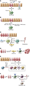
The recent clinical successes of immune checkpoint blockade and chimeric antigen receptor T cell therapies represent a turning point in cancer immunotherapy. These successes also underscore the importance of understanding basic tumor immunology for successful clinical translation in treating patients with cancer. The Reviews in this Review Series focus on current developments in cancer immunotherapy, highlight recent advances in our understanding of basic aspects of tumor immunology, and suggest how these insights can lead to the development of new immunotherapeutic strategies.
Authors
Yiping Yang
×Abstract
Cancer immunoediting explains the dual role by which the immune system can both suppress and/or promote tumor growth. Although cancer immunoediting was first demonstrated using mouse models of cancer, strong evidence that it occurs in human cancers is now accumulating. In particular, the importance of CD8+ T cells in cancer immunoediting has been shown, and more broadly in those tumors with an adaptive immune resistance phenotype. This Review describes the characteristics of the adaptive immune resistance tumor microenvironment and discusses data obtained in mouse and human settings. The role of other immune cells and factors influencing the effector function of tumor-specific CD8+ T cells is covered. We also discuss the temporal occurrence of cancer immunoediting in metastases and whether it differs from immunoediting in the primary tumor of origin.
Authors
Michele W.L. Teng, Jerome Galon, Wolf-Herman Fridman, Mark J. Smyth
×Abstract
Cancer development and its response to therapy are strongly influenced by innate and adaptive immunity, which either promote or attenuate tumorigenesis and can have opposing effects on therapeutic outcome. Chronic inflammation promotes tumor development, progression, and metastatic dissemination, as well as treatment resistance. However, cancer development and malignant progression are also associated with accumulation of genetic alterations and loss of normal regulatory processes, which cause expression of tumor-specific antigens and tumor-associated antigens (TAAs) that can activate antitumor immune responses. Although signals that trigger acute inflammatory reactions often stimulate dendritic cell maturation and antigen presentation, chronic inflammation can be immunosuppressive. This antagonism between inflammation and immunity also affects the outcome of cancer treatment and needs to be considered when designing new therapeutic approaches.
Authors
Shabnam Shalapour, Michael Karin
×Abstract
Our understanding of the role of myeloid-derived suppressor cells (MDSCs) in cancer is becoming increasingly complex. In addition to their eponymous role in suppressing immune responses, they directly support tumor growth, differentiation, and metastasis in a number of ways that are only now beginning to be appreciated. It is because of this increasingly complex role that these cells may become an important factor in the treatment of human cancer. In this Review, we discuss the most pertinent and controversial issues of MDSC biology and their role in promoting cancer progression and highlight how these cells may be used in the clinic, both as prognostic factors and as therapeutic targets.
Authors
Douglas Marvel, Dmitry I. Gabrilovich
×Abstract
The generation of an inflammatory environment is favorable and often decisive for the growth of both primary tumors and metastases. Tumor cells either express membrane molecules or release tumor-derived soluble factors able to alter myelopoiesis. Tumor-reprogrammed myeloid cells not only create a tolerogenic environment by blocking T cell functions and proliferation, but also directly drive tumor growth by promoting cancer stemness, angiogenesis, stroma deposition, epithelial-to-mesenchymal transition, and metastasis formation. In this Review, we discuss the interplay between immunosuppressive and protumoral myeloid cells and detail their immune-regulatory mechanisms, the molecular pathways involved in their differentiation, as well as their potential role as prognostic and diagnostic biomarkers and prospective targets for innovative approaches to treat tumor-bearing hosts.
Authors
Stefano Ugel, Francesco De Sanctis, Susanna Mandruzzato, Vincenzo Bronte
×Abstract
The relationship between cancer and the immune system is complex and provides unique therapeutic opportunities. Cytotoxic T lymphocyte antigen-4 (CTLA-4) is a regulatory molecule that suppresses T cell effector function following initial activation by costimulatory signals. Fully human monoclonal antibodies targeting CTLA-4 have been shown to increase T cell function and antitumor responses in patients with advanced metastatic melanoma. Responses observed with such immune checkpoint therapy can follow a different pattern from that seen with cytotoxic chemotherapy or targeted therapy and may continue after therapy is discontinued. In addition, the toxicities that are associated with anti–CTLA-4 therapy may differ from those of conventional therapies and consist of inflammatory events in parts of the body that do not contain cancerous cells. Early recognition of these inflammatory events and intervention is important, and the identification of predictive biomarkers continues to be an unfulfilled need in the field of immunotherapy. Combinatorial approaches with targeted therapies, radiation therapy, chemotherapy, or other immune checkpoint agonists/antagonists have the potential to increase the efficacy of CTLA-4 blockade.
Authors
Elizabeth Buchbinder, F. Stephen Hodi
×Abstract
Major progress has been made toward our understanding of the programmed death-1/programmed death ligand-1 (PD-1/PD-L1) pathway (referred to as the PD pathway). mAbs are already being used to block the PD pathway to treat human cancers (anti-PD therapy), especially advanced solid tumors. This therapy is based on principles that were discovered through basic research more than a decade ago, but the great potential of this pathway to treat a broad spectrum of advanced human cancers is just now becoming apparent. In this Review, we will briefly review the history and development of anti-PD therapy, from the original benchwork to the most up-to-date clinical results. We will then focus the discussion on three basic principles that define this unique therapeutic approach and highlight how anti-PD therapy is distinct from other immunotherapeutic approaches, namely tumor site immune modulation, targeting tumor-induced immune defects, and repairing ongoing (rather than generating de novo) tumor immunity. We believe that these fundamental principles set the standard for future immunotherapies and will guide our efforts to develop more efficacious and less toxic immune therapeutics to treat human cancers.
Authors
Lieping Chen, Xue Han
×Abstract
Twenty-five years after its inception, the genetic engineering of T cells is now a therapeutic modality pursued at an increasing number of medical centers. This immunotherapeutic strategy is predicated on gene transfer technology to instruct T lymphocytes to recognize and reject tumor cells. Chimeric antigen receptors (CARs) are synthetic receptors that mediate antigen recognition, T cell activation, and — in the case of second-generation CARs — costimulation to augment T cell functionality and persistence. We demonstrated over a decade ago that human T cells engineered with a CD19-specific CAR eradicated B cell malignancies in mice. Several phase I clinical trials eventually yielded dramatic results in patients with leukemia or lymphoma, especially acute lymphoblastic leukemia (ALL). This review recounts the milestones of CD19 CAR therapy and summarizes lessons learned from the CD19 paradigm.
Authors
Michel Sadelain
×Abstract
The clinical benefit of therapeutic cancer vaccines has been established. Whereas regression of lesions was shown for premalignant lesions caused by HPV, clinical benefit in cancer patients was mostly noted as prolonged survival. Suboptimal vaccine design and an immunosuppressive cancer microenvironment are the root causes of the lack of cancer eradication. Effective cancer vaccines deliver concentrated antigen to both HLA class I and II molecules of DCs, promoting both CD4 and CD8 T cell responses. Optimal vaccine platforms include DNA and RNA vaccines and synthetic long peptides. Antigens of choice include mutant sequences, selected cancer testis antigens, and viral antigens. Drugs or physical treatments can mitigate the immunosuppressive cancer microenvironment and include chemotherapeutics, radiation, indoleamine 2,3-dioxygenase (IDO) inhibitors, inhibitors of T cell checkpoints, agonists of selected TNF receptor family members, and inhibitors of undesirable cytokines. The specificity of therapeutic vaccination combined with such immunomodulation offers an attractive avenue for the development of future cancer therapies.
Authors
Cornelis J.M. Melief, Thorbald van Hall, Ramon Arens, Ferry Ossendorp, Sjoerd H. van der Burg
×Abstract
It is now well established that the immune system can recognize developing cancers and that therapeutic manipulation of immunity can induce tumor regression. The capacity to manifest remarkably durable responses in some patients has been ascribed in part to T cells that can (a) kill tumor cells directly, (b) orchestrate diverse antitumor immune responses, (c) manifest long-lasting memory, and (d) display remarkable specificity for tumor-derived proteins. This specificity stems from fundamental differences between cancer cells and their normal counterparts in that the former develop protein-altering mutations and undergo epigenetic and genetic alterations, resulting in aberrant protein expression. These events can result in formation of tumor antigens. The identification of mutated and aberrantly expressed self-tumor antigens has historically been time consuming and laborious. While mutant antigens are usually expressed in a tumor-specific manner, aberrantly expressed antigens are often shared between cancers and, therefore, in the past, have been the major focus of therapeutic cancer vaccines. However, advances in next-generation sequencing and epitope prediction now permit the rapid identification of mutant tumor neoantigens. This review focuses on a discussion of mutant tumor neoantigens and their use in personalizing cancer immunotherapies.
Authors
Matthew M. Gubin, Maxim N. Artyomov, Elaine R. Mardis, Robert D. Schreiber


-
Commentaries
×
Abstract
Iron-deficient individuals experience a loss of appetite that can be restored with iron supplementation. It has been proposed that iron influences the satiety hormone leptin; however, a direct link between iron and leptin has remained elusive. In this issue of the
JCI , Gao and colleagues demonstrate an inverse relationship between adipocyte iron and leptin that is mediated by iron-dependent activation of cAMP-responsive element binding protein (CREB), the transcription factor that represses leptin transcription. Together, the results of this study provide a mechanistic connection between dietary iron and the appetite-regulating hormone leptin.Authors
Nancy C. Andrews
×Abstract
Recent gene therapy progress has raised the possibility that vision loss caused by inherited retinal degeneration can be slowed or prevented. Unfortunately, patients are not usually diagnosed until enough degeneration has occurred that the deterioration in vision is noticeable. Therefore, effective gene therapy must halt degeneration to stabilize and preserve any remaining vision. Gene therapy methods currently in human clinical trials rely on subretinal or intravitreal injections of adeno-associated virus to deliver the therapeutic gene. To date, long-term results in patients treated with subretinal injections for Leber congenital amaurosis have been mixed. Proposed limitations include variability in the gene delivery method and a possible point of no return, at which treatment would be ineffective. In this issue of the
JCI , Koch et al. describe a well-controlled and precise mouse model for testing the ability of gene therapy to halt the progress of degeneration. Instead of viral-mediated therapeutic gene delivery, the authors induced expression of an integrated transgene at specific times during the course of photoreceptor degeneration. InPde6b -deficient retina, this strategy halted degeneration, even when more than 70% of photoreceptors had already degenerated. The results of this study demonstrate that retinal degeneration can be stopped, even at late stages of disease.Authors
James B. Hurley, Jennifer R. Chao
×Abstract
B cell precursor acute lymphoblastic leukemia (BCP ALL) is the most common malignancy in children. While treatments have improved remarkably over the past four decades, resistant disease and late effects that result from cytotoxic chemotherapy remain serious problems for individuals with BCP ALL. Improved genetic tools have led to the discovery of numerous somatic mutations associated with BCP ALL, leading to a framework for the genetic classification of BCP ALL. In this issue of the
JCI , Duque-Afonso et al. develop an accurate in vivo model for BCP ALL that recapitulates the key features of human disease, including acquired mutations in genes encoding PAX5 and components of the JAK/STAT pathway. The authors further show, as proof of principle, that this model can be used to evaluate the efficacy of drugs designed to target specific acquired mutations in patients with BCP ALL.Authors
Terry J. Fry, Peter D. Aplan
×Abstract
HIV-1 infection usually leads to systemic chronic inflammation that is associated with gut microbial translocation. The recently defined group 3 innate lymphoid cells (ILC3s) are critical for maintenance of intestinal barrier function; however, it is not clear whether and how HIV-1 infection influences the function of these cells. In this issue of the
JCI , Zhang and colleagues present compelling evidence that the survival and function of ILC3s are dramatically impaired by HIV-1 infection. The authors provide evidence that HIV-1 infection induces persistent activation of plasmacytoid dendritic cells (pDCs) and production of type I IFNs, which together increase expression of death receptor CD95 on ILC3s and thereby promote subsequent ILC3 apoptosis. Together, these results identify a mechanism that explains the impaired intestinal barrier function that results from chronic HIV-1 infection and shed light on the role of pDCs in HIV-1 immunopathogenesis and therapy.Authors
Xiaohuan Guo, Yang-Xin Fu


-
Research Articles
×
Abstract
The thiazide-sensitive NaCl cotransporter (NCC) is important for renal salt handling and blood-pressure homeostasis. The canonical NCC-activating pathway consists of With-No-Lysine (WNK) kinases and their downstream effector kinases SPAK and OSR1, which phosphorylate NCC directly. The upstream mechanisms that connect physiological stimuli to this system remain obscure. Here, we have shown that aldosterone activates SPAK/OSR1 via WNK1. We identified 2 alternatively spliced exons embedded within a proline-rich region of WNK1 that contain PY motifs, which bind the E3 ubiquitin ligase NEDD4-2. PY motif–containing WNK1 isoforms were expressed in human kidney, and these isoforms were efficiently degraded by the ubiquitin proteasome system, an effect reversed by the aldosterone-induced kinase SGK1. In gene-edited cells, WNK1 deficiency negated regulatory effects of NEDD4-2 and SGK1 on NCC, suggesting that WNK1 mediates aldosterone-dependent activity of the WNK/SPAK/OSR1 pathway. Aldosterone infusion increased proline-rich WNK1 isoform abundance in WT mice but did not alter WNK1 abundance in hypertensive
Nedd4-2 KO mice, which exhibit high baseline WNK1 and SPAK/OSR1 activity toward NCC. Conversely, hypotensiveSgk1 KO mice exhibited low WNK1 expression and activity. Together, our findings indicate that the proline-rich exons are modular cassettes that convert WNK1 into a NEDD4-2 substrate, thereby linking aldosterone and other NEDD4-2–suppressing antinatriuretic hormones to NCC phosphorylation status.Authors
Ankita Roy, Lama Al-Qusairi, Bridget F. Donnelly, Caroline Ronzaud, Allison L. Marciszyn, Fan Gong, Y.P. Christy Chang, Michael B. Butterworth, Núria M. Pastor-Soler, Kenneth R. Hallows, Olivier Staub, Arohan R. Subramanya
×Abstract
Wilson’s disease (WD) is an autosomal recessive disorder that results in accumulation of copper in the liver as a consequence of mutations in the gene encoding the copper-transporting P-type ATPase (ATP7B). WD is a chronic liver disorder, and individuals with the disease present with a variety of complications, including steatosis, cholestasis, cirrhosis, and liver failure. Similar to patients with WD,
Atp7b–/– mice have markedly elevated levels of hepatic copper and liver pathology. Previous studies have demonstrated that replacement of zinc in the DNA-binding domain of the estrogen receptor (ER) with copper disrupts specific binding to DNA response elements. Here, we found decreased binding of the nuclear receptors FXR, RXR, HNF4α, and LRH-1 to promoter response elements and decreased mRNA expression of nuclear receptor target genes inAtp7b–/– mice, as well as in adult and pediatric WD patients. Excessive hepatic copper has been described in progressive familial cholestasis (PFIC), and we found that similar to individuals with WD, patients with PFIC2 or PFIC3 who have clinically elevated hepatic copper levels exhibit impaired nuclear receptor activity. Together, these data demonstrate that copper-mediated nuclear receptor dysfunction disrupts liver function in WD and potentially in other disorders associated with increased hepatic copper levels.Authors
Clavia Ruth Wooton-Kee, Ajay K. Jain, Martin Wagner, Michael A. Grusak, Milton J. Finegold, Svetlana Lutsenko, David D. Moore
×Abstract
Mitochondrial homeostasis is critical for tissue health, and mitochondrial dysfunction contributes to numerous diseases, including heart failure. Here, we have shown that the transcription factor Kruppel-like factor 4 (KLF4) governs mitochondrial biogenesis, metabolic function, dynamics, and autophagic clearance. Adult mice with cardiac-specific
Klf4 deficiency developed cardiac dysfunction with aging or in response to pressure overload that was characterized by reduced myocardial ATP levels, elevated ROS, and marked alterations in mitochondrial shape, size, ultrastructure, and alignment. Evaluation of mitochondria isolated from KLF4-deficient hearts revealed a reduced respiration rate that is likely due to defects in electron transport chain complex I. Further, cardiac-specific, embryonicKlf4 deletion resulted in postnatal premature mortality, impaired mitochondrial biogenesis, and altered mitochondrial maturation. We determined that KLF4 binds to, cooperates with, and is requisite for optimal function of the estrogen-related receptor/PPARγ coactivator 1 (ERR/PGC-1) transcriptional regulatory module on metabolic and mitochondrial targets. Finally, we found that KLF4 regulates autophagy flux through transcriptional regulation of a broad array of autophagy genes in cardiomyocytes. Collectively, these findings identify KLF4 as a nodal transcriptional regulator of mitochondrial homeostasis.Authors
Xudong Liao, Rongli Zhang, Yuan Lu, Domenick A. Prosdocimo, Panjamaporn Sangwung, Lilei Zhang, Guangjin Zhou, Puneet Anand, Ling Lai, Teresa C. Leone, Hisashi Fujioka, Fang Ye, Mariana G. Rosca, Charles L. Hoppel, P. Christian Schulze, E. Dale Abel, Jonathan S. Stamler, Daniel P. Kelly, Mukesh K. Jain
×Abstract
Due to their ability to rapidly proliferate and produce effector cytokines, memory CD8+ T cells increase protection following reexposure to a pathogen. However, low inflammatory immunizations do not provide memory CD8+ T cells with a proliferation advantage over naive CD8+ T cells, suggesting that cell-extrinsic factors enhance memory CD8+ T cell proliferation in vivo. Herein, we demonstrate that inflammatory signals are critical for the rapid proliferation of memory CD8+ T cells following infection. Using murine models of viral infection and antigen exposure, we found that type I IFN–driven expression of IL-15 in response to viral infection prepares memory CD8+ T cells for rapid division independently of antigen reexposure by transiently inducing cell-cycle progression via a pathway dependent on mTOR complex-1 (mTORC1). Moreover, exposure to IL-15 allowed more rapid division of memory CD8+ T cells following antigen encounter and enhanced their protective capacity against viral infection. Together, these data reveal that inflammatory IL-15 promotes optimal responses by memory CD8+ T cells.
Authors
Martin J. Richer, Lecia L. Pewe, Lisa S. Hancox, Stacey M. Hartwig, Steven M. Varga, John T. Harty
×Abstract
Venous malformations (VMs) are composed of ectatic veins with scarce smooth muscle cell coverage. Activating mutations in the endothelial cell tyrosine kinase receptor TIE2 are a common cause of these lesions. VMs cause deformity, pain, and local intravascular coagulopathy, and they expand with time. Targeted pharmacological therapies are not available for this condition. Here, we generated a model of VMs by injecting HUVECs expressing the most frequent VM-causing TIE2 mutation, TIE2-L914F, into immune-deficient mice. TIE2-L914F–expressing HUVECs formed VMs with ectatic blood-filled channels that enlarged over time. We tested both rapamycin and a TIE2 tyrosine kinase inhibitor (TIE2-TKI) for their effects on murine VM expansion and for their ability to inhibit mutant TIE2 signaling. Rapamycin prevented VM growth, while TIE2-TKI had no effect. In cultured TIE2-L914F–expressing HUVECs, rapamycin effectively reduced mutant TIE2-induced AKT signaling and, though TIE2-TKI did target the WT receptor, it only weakly suppressed mutant-induced AKT signaling. In a prospective clinical pilot study, we analyzed the effects of rapamycin in 6 patients with difficult–to-treat venous anomalies. Rapamycin reduced pain, bleeding, lesion size, functional and esthetic impairment, and intravascular coagulopathy. This study provides a VM model that allows evaluation of potential therapeutic strategies and demonstrates that rapamycin provides clinical improvement in patients with venous malformation.
Authors
Elisa Boscolo, Nisha Limaye, Lan Huang, Kyu-Tae Kang, Julie Soblet, Melanie Uebelhoer, Antonella Mendola, Marjut Natynki, Emmanuel Seront, Sophie Dupont, Jennifer Hammer, Catherine Legrand, Carlo Brugnara, Lauri Eklund, Miikka Vikkula, Joyce Bischoff, Laurence M. Boon
×Abstract
The regulatory microRNA miR-150 is involved in the development of hemopathies and is downregulated in T-lymphomas, such as anaplastic large-cell lymphoma (ALCL) tumors. ALCL is defined by the presence or absence of translocations that activate the anaplastic lymphoma kinase (ALK), with nucleophosmin-ALK (NPM-ALK) fusions being the most common. Here, we compared samples of primary NPM-ALK(+) and NPM-ALK(–) ALCL to investigate the role of miR-150 downstream of NPM-ALK. Methylation of the
MIR150 gene was substantially elevated in NPM-ALK(+) biopsies and correlated with reduced miR-150 expression. In NPM-ALK(+) cell lines, DNA hypermethylation–mediated miR-150 repression required ALK-dependent pathways, as ALK inhibition restored miR-150 expression. Moreover, epigenetic silencing of miR-150 was due to the activation of STAT3, a major downstream substrate of NPM-ALK, in cooperation with DNA methyltransferase 1 (DNMT1). Accordingly, miR-150 repression was turned off following treatment with the DNMT inhibitor, decitabine. In murine NPM-ALK(+) xenograft models, miR-150 upregulation induced antineoplastic activity. Treatment of crizotinib-resistant NPM-ALK(+) KARPAS-299-CR06 cells with decitabine or ectopic miR-150 expression reduced viability and growth. Altogether, our results suggest that hypomethylating drugs, alone or in combination with other agents, may benefit ALK(+) patients harboring tumors resistant to crizotinib and other anti-ALK tyrosine kinase inhibitors (TKIs). Moreover, these results support further work on miR-150 in these and other ALK(+) malignancies.Authors
Coralie Hoareau-Aveilla, Thibaud Valentin, Camille Daugrois, Cathy Quelen, Géraldine Mitou, Samuel Quentin, Jinsong Jia, Salvatore Spicuglia, Pierre Ferrier, Monica Ceccon, Sylvie Giuriato, Carlo Gambacorti-Passerini, Pierre Brousset, Laurence Lamant, Fabienne Meggetto
×Abstract
Diarrhea is one of the troublesome complications of diabetes, and the underlying causes of this problem are complex. Here, we investigated whether altered electrolyte transport contributes to diabetic diarrhea. We found that the expression of Na+/H+ exchanger NHE3 and several scaffold proteins, including NHE3 regulatory factors (NHERFs), inositol trisphosphate (IP3) receptor-binding protein released with IP3 (IRBIT), and ezrin, was decreased in the intestinal brush border membrane (BBM) of mice with streptozotocin-induced diabetes. Treatment of diabetic mice with insulin restored intestinal NHE3 activity and fluid absorption. Molecular analysis revealed that NHE3, NHERF1, IRBIT, and ezrin form macrocomplexes, which are perturbed under diabetic conditions, and insulin administration reconstituted these macrocomplexes and restored NHE3 expression in the BBM. Silencing of NHERF1 or IRBIT prevented NHE3 trafficking to the BBM and insulin-dependent NHE3 activation. IRBIT facilitated the interaction of NHE3 with NHERF1 via protein kinase D2–dependent phosphorylation. Insulin stimulated ezrin phosphorylation, which enhanced the interaction of ezrin with NHERF1, IRBIT, and NHE3. Additionally, oral administration of lysophosphatidic acid (LPA) increased NHE3 activity and fluid absorption in diabetic mice via an insulin-independent pathway. Together, these findings indicate the importance of NHE3 in diabetic diarrhea and suggest LPA administration as a potential therapeutic strategy for management of diabetic diarrhea.
Authors
Peijian He, Luqing Zhao, Lixin Zhu, Edward J. Weinman, Roberto De Giorgio, Michael Koval, Shanthi Srinivasan, C. Chris Yun
×Abstract
Epigenetic regulators play critical roles in normal hematopoiesis, and the activity of these enzymes is frequently altered in hematopoietic cancers. The major type II protein arginine methyltransferase PRMT5 catalyzes the formation of symmetric dimethyl arginine and has been implicated in various cellular processes, including pluripotency and tumorigenesis. Here, we generated
Prmt5 conditional KO mice to evaluate the contribution of PRMT5 to adult hematopoiesis. Loss of PRMT5 triggered an initial but transient expansion of hematopoietic stem cells (HSCs); however,Prmt5 deletion resulted in a concurrent loss of hematopoietic progenitor cells (HPCs), leading to fatal BM aplasia. PRMT5-specific effects on hematopoiesis were cell intrinsic and depended on PRMT5 methyltransferase activity. We found that PRMT5-deficient hematopoietic stem and progenitor cells exhibited severely impaired cytokine signaling as well as upregulation of p53 and expression of its downstream targets. Together, our results demonstrate that PRMT5 plays distinct roles in the behavior of HSCs compared with HPCs and is essential for the maintenance of adult hematopoietic cells.Authors
Fan Liu, Guoyan Cheng, Pierre-Jacques Hamard, Sarah Greenblatt, Lan Wang, Na Man, Fabiana Perna, Haiming Xu, Madhavi Tadi, Luisa Luciani, Stephen D. Nimer
×Abstract
The identification of the molecular events responsible for strain emergence, enhanced virulence, and epidemicity has been a long-pursued goal in infectious diseases research. A recent analysis of 3,615 genomes of serotype M1 group A
Streptococcus strains (the so-called “flesh-eating” bacterium) identified a recombination event that coincides with the global M1 pandemic beginning in the early 1980s. Here, we have shown that the allelic variation that results from this recombination event, which replaces the chromosomal region encoding secreted NADase and streptolysin O, is the key driver of increased toxin production and enhanced infection severity of the M1 pandemic strains. Using isoallelic mutant strains, we found that 3 polymorphisms in this toxin gene region increase resistance to killing by human polymorphonuclear leukocytes, increase bacterial proliferation, and increase virulence in animal models of pharyngitis and necrotizing fasciitis. Genome sequencing of an additional 1,125 streptococcal strains and virulence studies revealed that a highly similar recombinational replacement event underlies an ongoing intercontinental epidemic of serotype M89 group AStreptococcus infections. By identifying the molecular changes that enhance upper respiratory tract fitness, increased resistance to innate immunity, and increased tissue destruction, we describe a mechanism that underpins epidemic streptococcal infections, which have affected many millions of people.Authors
Luchang Zhu, Randall J. Olsen, Waleed Nasser, Stephen B. Beres, Jaana Vuopio, Karl G. Kristinsson, Magnus Gottfredsson, Adeline R. Porter, Frank R. DeLeo, James M. Musser
×Abstract
Heterozygous mutations in the syntaxin-binding protein 1 (
STXBP1 ) gene, which encodes Munc18-1, a core component of the presynaptic membrane-fusion machinery, cause infantile early epileptic encephalopathy (Ohtahara syndrome), but it is unclear how a partial loss of Munc18-1 produces this severe clinical presentation. Here, we generated human ES cells designed to conditionally express heterozygous and homozygousSTXBP1 loss-of-function mutations and studied isogenic WT andSTXBP1 -mutant human neurons derived from these conditionally mutant ES cells. We demonstrated that heterozygousSTXBP1 mutations lower the levels of Munc18-1 protein and its binding partner, the t-SNARE-protein Syntaxin-1, by approximately 30% and decrease spontaneous and evoked neurotransmitter release by nearly 50%. Thus, our results confirm that using engineered human embryonic stem (ES) cells is a viable approach to studying disease-associated mutations in human neurons on a controlled genetic background, demonstrate that partialSTXBP1 loss of function robustly impairs neurotransmitter release in human neurons, and suggest that heterozygousSTXBP1 mutations cause early epileptic encephalopathy specifically through a presynaptic impairment.Authors
Christopher Patzke, Yan Han, Jason Covy, Fei Yi, Stephan Maxeiner, Marius Wernig, Thomas C. Südhof
×Abstract
Aging and increased amyloid burden are major risk factors for cognitive diseases such as Alzheimer’s disease (AD). Effective therapies for these diseases are lacking. Here, we evaluated mouse models of age-associated memory impairment and amyloid deposition to study transcriptome and cell type–specific epigenome plasticity in the brain and peripheral organs. We determined that aging and amyloid pathology are associated with inflammation and impaired synaptic function in the hippocampal CA1 region as the result of epigenetic-dependent alterations in gene expression. In both amyloid and aging models, inflammation was associated with increased gene expression linked to a subset of transcription factors, while plasticity gene deregulation was differentially mediated. Amyloid pathology impaired histone acetylation and decreased expression of plasticity genes, while aging altered H4K12 acetylation–linked differential splicing at the intron-exon junction in neurons, but not nonneuronal cells. Furthermore, oral administration of the clinically approved histone deacetylase inhibitor vorinostat not only restored spatial memory, but also exerted antiinflammatory action and reinstated epigenetic balance and transcriptional homeostasis at the level of gene expression and exon usage. This study provides a systems-level investigation of transcriptome plasticity in the hippocampal CA1 region in aging and AD models and suggests that histone deacetylase inhibitors should be further explored as a cost-effective therapeutic strategy against age-associated cognitive decline.
Authors
Eva Benito, Hendrik Urbanke, Binu Ramachandran, Jonas Barth, Rashi Halder, Ankit Awasthi, Gaurav Jain, Vincenzo Capece, Susanne Burkhardt, Magdalena Navarro-Sala, Sankari Nagarajan, Anna-Lena Schütz, Steven A. Johnsen, Stefan Bonn, Reinhardt Lührmann, Camin Dean, André Fischer
×Abstract
The genetic disorder Kabuki syndrome (KS) is characterized by developmental delay and congenital anomalies. Dominant mutations in the chromatin regulators lysine (K)–specific methyltransferase 2D (
KMT2D ) (also known asMLL2 ) and lysine (K)–specific demethylase 6A (KDM6A ) underlie the majority of cases. Although the functions of these chromatin-modifying proteins have been studied extensively, the physiological systems regulated by them are largely unknown. Using whole-exome sequencing, we identified a mutation inRAP1A that was converted to homozygosity as the result of uniparental isodisomy (UPD) in a patient with KS and a de novo, dominant mutation inRAP1B in a second individual with a KS-like phenotype. We elucidated a genetic and functional interaction between the respective KS-associated genes and their products in zebrafish models and patient cell lines. Specifically, we determined that dysfunction of known KS genes and the genes identified in this study results in aberrant MEK/ERK signaling as well as disruption of F-actin polymerization and cell intercalation. Moreover, these phenotypes could be rescued in zebrafish models by rebalancing MEK/ERK signaling via administration of small molecule inhibitors of MEK. Taken together, our studies suggest that the KS pathophysiology overlaps with the RASopathies and provide a potential direction for treatment design.Authors
Nina Bögershausen, I-Chun Tsai, Esther Pohl, Pelin Özlem Simsek Kiper, Filippo Beleggia, E. Ferda Percin, Katharina Keupp, Angela Matchan, Esther Milz, Yasemin Alanay, Hülya Kayserili, Yicheng Liu, Siddharth Banka, Andrea Kranz, Martin Zenker, Dagmar Wieczorek, Nursel Elcioglu, Paolo Prontera, Stanislas Lyonnet, Thomas Meitinger, A. Francis Stewart, Dian Donnai, Tim M. Strom, Koray Boduroglu, Gökhan Yigit, Yun Li, Nicholas Katsanis, Bernd Wollnik
×Abstract
Inherited thrombocytopenias are a group of disorders that are characterized by a low platelet count and are sometimes associated with excessive bleeding that ranges from mild to severe. We evaluated 36 unrelated patients and 17 family members displaying thrombocytopenia that were recruited to the UK Genotyping and Phenotyping of Platelets (GAPP) study. All patients had a history of excessive bleeding of unknown etiology. We performed platelet phenotyping and whole-exome sequencing (WES) on all patients and identified mutations in schlafen 14 (
SLFN14 ) in 12 patients from 3 unrelated families. Patients harboringSLFN14 mutations displayed an analogous phenotype that consisted of moderate thrombocytopenia, enlarged platelets, decreased ATP secretion, and a dominant inheritance pattern. Three heterozygous missense mutations were identified in affected family members and predicted to encode substitutions (K218E, K219N, and V220D) within an ATPase-AAA-4, GTP/ATP-binding region of SLFN14. Endogenous SLFN14 expression was reduced in platelets from all patients, and mutant SLFN14 expression was markedly decreased compared with that of WT SLFN14 when overexpressed in transfected cells. Electron microscopy revealed a reduced number of dense granules in affected patients platelets, correlating with a decreased ATP secretion observed in lumiaggregometry studies. These results identifySLFN14 mutations as cause for an inherited thrombocytopenia with excessive bleeding, outlining a fundamental role for SLFN14 in platelet formation and function.Authors
Sarah J. Fletcher, Ben Johnson, Gillian C. Lowe, Danai Bem, Sian Drake, Marie Lordkipanidzé, Isabel Sánchez Guiú, Ban Dawood, José Rivera, Michael A. Simpson, Martina E. Daly, Jayashree Motwani, Peter W. Collins, Steve P. Watson, Neil V. Morgan, on behalf of the UK Genotyping and Phenotyping of Platelets study group
×Mucosally transplanted mesenchymal stem cells stimulate intestinal healing by promoting angiogenesis
Abstract
Mesenchymal stem cell (MSC) therapy is an emerging field of regenerative medicine; however, it is often unclear how these cells mediate repair. Here, we investigated the use of MSCs in the treatment of intestinal disease and modeled abnormal repair by creating focal wounds in the colonic mucosa of prostaglandin-deficient mice. These wounds developed into ulcers that infiltrated the outer intestinal wall. We determined that penetrating ulcer formation in this model resulted from increased hypoxia and smooth muscle wall necrosis. Prostaglandin I2 (PGI2) stimulated VEGF-dependent angiogenesis to prevent penetrating ulcers. Treatment of mucosally injured WT mice with a VEGFR inhibitor resulted in the development of penetrating ulcers, further demonstrating that VEGF is critical for mucosal repair. We next used this model to address the role of transplanted colonic MSCs (cMSCs) in intestinal repair. Compared with intravenously injected cMSCs, mucosally injected cMSCs more effectively prevented the development of penetrating ulcers, as they were more efficiently recruited to colonic wounds. Importantly, mucosally injected cMSCs stimulated angiogenesis in a VEGF-dependent manner. Together, our results reveal that penetrating ulcer formation results from a reduction of local angiogenesis and targeted injection of MSCs can optimize transplantation therapy. Moreover, local MSC injection has potential for treating diseases with features of abnormal angiogenesis and repair.
Authors
Nicholas A. Manieri, Madison R. Mack, Molly D. Himmelrich, Daniel L. Worthley, Elaine M. Hanson, Lars Eckmann, Timothy C. Wang, Thaddeus S. Stappenbeck
×Abstract
BACKGROUND. Several lines of evidence suggest that male embryos may have greater vulnerability than female embryos to disordered inflammation; therefore, antiinflammatory drugs, such as low-dose aspirin (LDA), may alter the sex ratio. Here, we assessed the effect of LDA on male live birth and male offspring, incorporating pregnancy losses (n = 56) via genetic assessment, as part of a parallel-design, block-randomized, placebo-controlled trial of preconception LDA.METHODS. Participants (615 treated with LDA, 613 treated with placebo) ranged in age from 18 to 40 years of age, with 1 to 2 prior pregnancy losses. We estimated the intention-to-treat (ITT) risk ratio (RR) and 95% CI and assessed interaction with baseline high-sensitivity C-reactive protein (hsCRP) serum concentration — a marker of systemic inflammation.RESULTS. Among the 1,078 women who completed follow-up (535 treated with LDA, 543 treated with placebo), the male live birth ITT RR equaled 1.31 (95% CI: 1.07–1.59). With increasing tertile of hsCRP, the proportion of males at birth decreased in the placebo group, and the effect of LDA on male live birth increased (first tertile: 48% male in LDA vs. 52% in placebo, ITT RR = 0.97, 95% CI: 0.70–1.35; second tertile: 57% male in LDA vs. 43% in placebo, ITT RR = 1.36, 95% CI: 0.98–1.90; third tertile: 53% male in LDA vs. 35% in placebo, ITT RR = 1.70, 95% CI: 1.13–2.57;P interaction = 0.03). Analysis of pregnancy with male offspring yielded similar results.CONCLUSION. Initiation of LDA prior to conception restored numbers of male live births and pregnancy with male offspring among women with 1 to 2 prior pregnancy losses. Moreover, our data suggest that LDA modulates inflammation that would otherwise reduce the conception or survival of male embryos.TRIAL REGISTRATION. ClinicalTrials.gov NCT00467363.FUNDING. Intramural Research Program of the Eunice Kennedy Shriver National Institute of Child Health and Human Development, National Institutes of Health.Authors
Rose G. Radin, Sunni L. Mumford, Robert M. Silver, Laurie L. Lesher, Noya Galai, David Faraggi, Jean Wactawski-Wende, Janet M. Townsend, Anne M. Lynch, Hyagriv N. Simhan, Lindsey A. Sjaarda, Neil J. Perkins, Shvetha M. Zarek, Karen C. Schliep, Enrique F. Schisterman
×Abstract
Regulatory T cells (Tregs) have been shown to enhance immune reconstitution and prevent graft-versus-host disease (GVHD) after hematopoietic stem cell transplantation; however, it is unclear how Tregs mediate these effects. Here, we developed a model to examine the mechanism of Treg-dependent regulation of immune reconstitution. Lymphopenic mice were selectively reconstituted with Tregs prior to transfer of conventional CD4+ T cells. Full Treg reconstitution prevented the rapid oligoclonal proliferation that gives rise to pathogenic CD4 effector T cells, while preserving the slow homeostatic form of lymphopenia-induced peripheral expansion that repopulates a diverse peripheral T cell pool. Treg-mediated CTLA-4–dependent downregulation of CD80/CD86 on DCs was critical for inhibition of rapid proliferation and was a function of the Treg/DC ratio achieved by reconstitution. In an allogeneic BM transplant model, selective Treg reconstitution before T cell transfer also normalized DC costimulation and provided complete protection against GVHD. In contrast, cotransfer of Tregs was not protective. Our results indicate that achieving optimal recovery from lymphopenia should aim to improve early Treg reconstitution in order to increase the relative number of Tregs to DCs and thereby inhibit spontaneous oligoclonal T cell proliferation.
Authors
Holly A. Bolton, Erhua Zhu, Alexandra M. Terry, Thomas V. Guy, Woon-Puay Koh, Sioh-Yang Tan, Carl A. Power, Patrick Bertolino, Katharina Lahl, Tim Sparwasser, Elena Shklovskaya, Barbara Fazekas de St. Groth
×Abstract
Induced pluripotent stem cell–derived (iPS-derived) neural precursor cells may represent the ideal autologous cell source for cell-based therapy to promote remyelination and neuroprotection in myelin diseases. So far, the therapeutic potential of reprogrammed cells has been evaluated in neonatal demyelinating models. However, the repair efficacy and safety of these cells has not been well addressed in the demyelinated adult CNS, which has decreased cell plasticity and scarring. Moreover, it is not clear if these induced pluripotent–derived cells have the same reparative capacity as physiologically committed CNS-derived precursors. Here, we performed a side-by-side comparison of CNS-derived and skin-derived neural precursors in culture and following engraftment in murine models of adult spinal cord demyelination. Grafted induced neural precursors exhibited a high capacity for survival, safe integration, migration, and timely differentiation into mature bona fide oligodendrocytes. Moreover, grafted skin–derived neural precursors generated compact myelin around host axons and restored nodes of Ranvier and conduction velocity as efficiently as CNS-derived precursors while outcompeting endogenous cells. Together, these results provide important insights into the biology of reprogrammed cells in adult demyelinating conditions and support use of these cells for regenerative biomedicine of myelin diseases that affect the adult CNS.
Authors
Sabah Mozafari, Cecilia Laterza, Delphine Roussel, Corinne Bachelin, Antoine Marteyn, Cyrille Deboux, Gianvito Martino, Anne Baron-Van Evercooren
×Abstract
Juvenile ciliopathy syndromes that are associated with renal cysts and premature renal failure are commonly the result of mutations in the gene encoding centrosomal protein CEP290. In addition to centrosomes and the transition zone at the base of the primary cilium, CEP290 also localizes to the nucleus; however, the nuclear function of CEP290 is unknown. Here, we demonstrate that reduction of cellular CEP290 in primary human and mouse kidney cells as well as in zebrafish embryos leads to enhanced DNA damage signaling and accumulation of DNA breaks ex vivo and in vivo. Compared with those from WT mice, primary kidney cells from
Cep290 -deficient mice exhibited supernumerary centrioles, decreased replication fork velocity, fork asymmetry, and increased levels of cyclin-dependent kinases (CDKs). Treatment ofCep290 -deficient cells with CDK inhibitors rescued DNA damage and centriole number. Moreover, the loss of primary cilia that results from CEP290 dysfunction was rescued in 3D cell culture spheroids of primary murine kidney cells after exposure to CDK inhibitors. Together, our results provide a link between CEP290 and DNA replication stress and suggest CDK inhibition as a potential treatment strategy for a wide range of ciliopathy syndromes.Authors
Gisela G. Slaats, Joshua C. Saldivar, Julien Bacal, Michelle K. Zeman, Andrew C. Kile, Ann Marie Hynes, Shalabh Srivastava, Jekaterina Nazmutdinova, Krista den Ouden, Miriam S. Zagers, Veronica Foletto, Marianne C. Verhaar, Colin Miles, John A. Sayer, Karlene A. Cimprich, Rachel H. Giles
×Abstract
Acute lymphoblastic leukemia (ALL) is the most common childhood cancer; however, its genetic diversity limits investigation into the molecular pathogenesis of disease and development of therapeutic strategies. Here, we engineered mice that conditionally express the
E2A-PBX1 fusion oncogene, which results from chromosomal translocation t(1;19) and is present in 5% to 7% of pediatric ALL cases. The incidence of leukemia in these mice varied from 5% to 50%, dependent on the Cre-driving promoter (Cd19 ,Mb1 , orMx1 ) used to induceE2A-PBX1 expression. Two distinct but highly similar subtypes of B cell precursor ALLs that differed by their pre–B cell receptor (pre-BCR) status were induced and displayed maturation arrest at the pro-B/large pre–B II stages of differentiation, similar to human E2A-PBX1 ALL. Somatic activation ofE2A-PBX1 in B cell progenitors enhanced self-renewal and led to acquisition of multiple secondary genomic aberrations, including prominent spontaneous loss ofPax5 . In preleukemic mice, conditionalPax5 deletion cooperated withE2A-PBX1 to expand progenitor B cell subpopulations, increasing penetrance and shortening leukemia latency. Recurrent secondary activating mutations were detected in key signaling pathways, most notably JAK/STAT, that leukemia cells require for proliferation. These data support conditional E2A-PBX1 mice as a model of human ALL and suggest targeting pre-BCR signaling and JAK kinases as potential therapeutic strategies.Authors
Jesús Duque-Afonso, Jue Feng, Florian Scherer, Chiou-Hong Lin, Stephen H.K. Wong, Zhong Wang, Masayuki Iwasaki, Michael L. Cleary
×Abstract
Dietary iron supplementation is associated with increased appetite. Here, we investigated the effect of iron on the hormone leptin, which regulates food intake and energy homeostasis. Serum ferritin was negatively associated with serum leptin in a cohort of patients with metabolic syndrome. Moreover, the same inverse correlation was observed in mice fed a high-iron diet. Adipocyte-specific loss of the iron exporter ferroportin resulted in iron loading and decreased leptin, while decreased levels of hepcidin in a murine hereditary hemochromatosis (HH) model increased adipocyte ferroportin expression, decreased adipocyte iron, and increased leptin. Treatment of 3T3-L1 adipocytes with iron decreased leptin mRNA in a dose-dependent manner. We found that iron negatively regulates leptin transcription via cAMP-responsive element binding protein activation (CREB activation) and identified 2 potential CREB-binding sites in the mouse leptin promoter region. Mutation of both sites completely blocked the effect of iron on promoter activity. ChIP analysis revealed that binding of phosphorylated CREB is enriched at these two sites in iron-treated 3T3-L1 adipocytes compared with untreated cells. Consistent with the changes in leptin, dietary iron content was also directly related to food intake, independently of weight. These findings indicate that levels of dietary iron play an important role in regulation of appetite and metabolism through CREB-dependent modulation of leptin expression.
Authors
Yan Gao, Zhonggang Li, J. Scott Gabrielsen, Judith A. Simcox, Soh-hyun Lee, Deborah Jones, Bob Cooksey, Gregory Stoddard, William T. Cefalu, Donald A. McClain
×Abstract
Group 3 innate lymphoid cells (ILC3s) have demonstrated roles in promoting antibacterial immunity, maintaining epithelial barrier function, and supporting tissue repair. ILC3 alterations are associated with chronic inflammation and inflammatory disease; however, the characteristics and relevant regulatory mechanisms of this cell population in HIV-1 infection are poorly understood due in part to a lack of a robust model. Here, we determined that functional human ILC3s develop in lymphoid organs of humanized mice and that persistent HIV-1 infection in this model depletes ILC3s, as observed in chronic HIV-1–infected patients. In HIV-1–infected mice, effective antiretroviral therapy reversed the loss of ILC3s. HIV-1–dependent reduction of ILC3s required plasmacytoid dendritic cells (pDCs), IFN-I, and the CD95/FasL pathway, as targeted depletion or blockade of these prevented HIV-1–induced ILC3 depletion in vivo and in vitro, respectively. Finally, we determined that HIV-1 infection induces CD95 expression on ILC3s via a pDC- and IFN-I–dependent mechanism that sensitizes ILC3s to undergo CD95/FasL-mediated apoptosis. We conclude that chronic HIV-1 infection depletes ILC3s through pDC activation, induction of IFN-I, and CD95-mediated apoptosis.
Authors
Zheng Zhang, Liang Cheng, Juanjuan Zhao, Guangming Li, Liguo Zhang, Weiwei Chen, Weiming Nie, Natalia J. Reszka-Blanco, Fu-Sheng Wang, Lishan Su
×Abstract
Hereditary retinal degenerative diseases, such as retinitis pigmentosa (RP), are characterized by the progressive loss of rod photoreceptors followed by loss of cones. While retinal gene therapy clinical trials demonstrated temporary improvement in visual function, this approach has yet to achieve sustained functional and anatomical rescue after disease onset in patients. The lack of sustained benefit could be due to insufficient transduction efficiency of viral vectors (“too little”) and/or because the disease is too advanced (“too late”) at the time therapy is initiated. Here, we tested the latter hypothesis and developed a mouse RP model that permits restoration of the mutant gene in all diseased photoreceptor cells, thereby ensuring sufficient transduction efficiency. We then treated mice at early, mid, or late disease stages. At all 3 time points, degeneration was halted and function was rescued for at least 1 year. Not only do our results demonstrate that gene therapy effectively preserves function after the onset of degeneration, our study also demonstrates that there is a broad therapeutic time window. Moreover, these results suggest that RP patients are treatable, despite most being diagnosed after substantial photoreceptor loss, and that gene therapy research must focus on improving transduction efficiency to maximize clinical impact.
Authors
Susanne F. Koch, Yi-Ting Tsai, Jimmy K. Duong, Wen-Hsuan Wu, Chun-Wei Hsu, Wei-Pu Wu, Luis Bonet-Ponce, Chyuan-Sheng Lin, Stephen H. Tsang
×Abstract
BACKGROUND. The disrupted in schizophrenia 1 (DISC1 ) gene locus was originally identified in a Scottish pedigree with a high incidence of psychiatric disorders that is associated with a balanced t(1;11)(q42.1;q14.3) chromosomal translocation. Here, we investigated whether members of this family carrying the t(1;11)(q42.1;q14.3) translocation have a common brain-related phenotype and whether this phenotype is similar to that observed in schizophrenia (SCZ), using multivariate pattern recognition techniques.METHODS. We measured cortical thickness, cortical surface area, subcortical volumes, and regional cerebral blood flow (rCBF) in healthy controls (HC) (n = 24), patients diagnosed with SCZ (n = 24), patients diagnosed with bipolar disorder (BP) (n = 19), and members of the original Scottish family (n = 30) who were either carriers (T+) or noncarriers (T–) of theDISC1 translocation. Binary classification models were developed to assess the differences and similarities across groups.RESULTS. Based on cortical thickness, 72% of the T– group were assigned to the HC group, 83% of the T+ group were assigned to the SCZ group, and 45% of the BP group were classified as belonging to the SCZ group, suggesting high specificity of this measurement in predicting brain-related phenotypes. Shared brain-related phenotypes between SCZ and T+ individuals were found for cortical thickness only. Finally, a classification accuracy of 73% was achieved when directly comparing the pattern of cortical thickness of T+ and T– individuals.CONCLUSION. Together, the results of this study suggest that theDISC1 translocation may increase the risk of psychiatric disorders in this pedigree by affecting neurostructural phenotypes such as cortical thickness.FUNDING. This work was supported by the National Health Service Research Scotland, the Scottish Translational Medicine Research Collaboration, the Innovative Medicines Initiative (IMI), the Engineering and Physical Sciences Research Council (EPSRC), The Wellcome Trust, the National Institute of Health Research (NIHR), and Pfizer.Authors
Orla M. Doyle, Catherine Bois, Pippa Thomson, Liana Romaniuk, Brandon Whitcher, Steven C.R. Williams, Federico E. Turkheimer, Hreinn Stefansson, Andrew M. McIntosh, Mitul A. Mehta, Stephen M. Lawrie


Copyright © 2025 American Society for Clinical Investigation
ISSN: 0021-9738 (print), 1558-8238 (online)