
Intermediate filament–associated diseases
Series edited by Bishr Omary
Intermediate filaments (IFs) are one of the three major fibrillar cytoplasmic elements that make up the cytoskeleton. Cytoskeletal IFs in distinct cell types are formed from different members of a large family of proteins, the IF protein family, which also includes proteins that are present in the nucleus, where they are the main component of the nucleoskeleton. As discussed in this Review series, roles have been revealed for IFs in more than 80 human tissue-specific diseases.
Articles in series

Abstract
Intermediate filaments (IFs) are encoded by the largest gene family among the three major cytoskeletal protein groups. Unique IF compliments are expressed in selective cell types, and this expression is reflected in their involvement, upon mutation, as a cause of or predisposition to more than 80 human tissue-specific diseases. This Review Series covers diseases and functional and structural aspects pertaining to IFs and highlights the molecular and functional consequences of IF-associated diseases (IF-pathies). Exciting challenges and opportunities face the IF field, including developing both a better understanding of the pathogenesis of IF-pathies and targeted therapeutic approaches.
Authors
M. Bishr Omary

Abstract
It took more than 100 years before it was established that the proteins that form intermediate filaments (IFs) comprise a unified protein family, the members of which are ubiquitous in virtually all differentiated cells and present both in the cytoplasm and in the nucleus. However, during the past 2 decades, knowledge regarding the functions of these structures has been expanding rapidly. Many disease-related roles of IFs have been revealed. In some cases, the molecular mechanisms underlying these diseases reflect disturbances in the functions traditionally assigned to IFs, i.e., maintenance of structural and mechanical integrity of cells and tissues. However, many disease conditions seem to link to the nonmechanical functions of IFs, many of which have been defined only in the past few years.
Authors
John E. Eriksson, Thomas Dechat, Boris Grin, Brian Helfand, Melissa Mendez, Hanna-Mari Pallari, Robert D. Goldman

Abstract
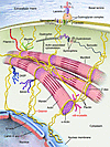
Intermediate filaments (IFs) are major constituents of the cytoskeleton and nuclear boundary in animal cells. They are of prime importance for the functional organization of structural elements. Depending on the cell type, morphologically similar but biochemically distinct proteins form highly viscoelastic filament networks with multiple nanomechanical functions. Besides their primary role in cell plasticity and their established function as cellular stress absorbers, recently discovered gene defects have elucidated that structural alterations of IFs can affect their involvement both in signaling and in controlling gene regulatory networks. Here, we highlight the basic structural and functional properties of IFs and derive a concept of how mutations may affect cellular architecture and thereby tissue construction and physiology.
Authors
Harald Herrmann, Sergei V. Strelkov, Peter Burkhard, Ueli Aebi

Abstract
Epidermolysis bullosa (EB) simplex is a rare genetic condition typified by superficial bullous lesions that result from frictional trauma to the skin. Most cases are due to dominantly acting mutations in either keratin 14 (K14) or K5, the type I and II intermediate filament (IF) proteins tasked with forming a pancytoplasmic network of 10-nm filaments in basal keratinocytes of the epidermis and in other stratified epithelia. Defects in K5/K14 filament network architecture cause basal keratinocytes to become fragile and account for their trauma-induced rupture. Here we review how laboratory investigations centered on keratin biology have deepened our understanding of the etiology and pathophysiology of EB simplex and revealed novel avenues for its therapy.
Authors
Pierre A. Coulombe, Michelle L. Kerns, Elaine Fuchs

Abstract
Simple epithelial keratins (SEKs) are found primarily in single-layered simple epithelia and include keratin 7 (K7), K8, K18–K20, and K23. Genetically engineered mice that lack SEKs or overexpress mutant SEKs have helped illuminate several keratin functions and served as important disease models. Insight into the contribution of SEKs to human disease has indicated that K8 and K18 are the major constituents of Mallory-Denk bodies, hepatic inclusions associated with several liver diseases, and are essential for inclusion formation. Furthermore, mutations in the genes encoding K8, K18, and K19 predispose individuals to a variety of liver diseases. Hence, as we discuss here, the SEK cytoskeleton is involved in the orchestration of several important cellular functions and contributes to the pathogenesis of human liver disease.
Authors
M. Bishr Omary, Nam-On Ku, Pavel Strnad, Shinichiro Hanada
Abstract
Muscle fiber deterioration resulting in progressive skeletal muscle weakness, heart failure, and respiratory distress occurs in more than 20 inherited myopathies. As discussed in this Review, one of the newly identified myopathies is desminopathy, a disease caused by dysfunctional mutations in desmin, a type III intermediate filament protein, or αB-crystallin, a chaperone for desmin. The range of clinical manifestations in patients with desminopathy is wide and may overlap with those observed in individuals with other myopathies. Awareness of this disease needs to be heightened, diagnostic criteria reliably outlined, and molecular testing readily available; this would ensure prevention of sudden death from cardiac arrhythmias and other complications.
Authors
Lev G. Goldfarb, Marinos C. Dalakas

Abstract
Intermediate filaments (IFs) are abundant structures found in most eukaryotic cells, including those in the nervous system. In the CNS, the primary components of neuronal IFs are α-internexin and the neurofilament triplet proteins. In the peripheral nervous system, a fifth neuronal IF protein known as peripherin is also present. IFs in astrocytes are primarily composed of glial fibrillary acidic protein (GFAP), although vimentin is also expressed in immature astrocytes and some mature astrocytes. In this Review, we focus on the IFs of glial cells (primarily GFAP) and neurons as well as their relationship to different neurodegenerative diseases.
Authors
Ronald K.H. Liem, Albee Messing

Abstract
The main function of the nuclear lamina, an intermediate filament meshwork lying primarily beneath the inner nuclear membrane, is to provide structural scaffolding for the cell nucleus. However, the lamina also serves other functions, such as having a role in chromatin organization, connecting the nucleus to the cytoplasm, gene transcription, and mitosis. In somatic cells, the main protein constituents of the nuclear lamina are lamins A, C, B1, and B2. Interest in the nuclear lamins increased dramatically in recent years with the realization that mutations in LMNA, the gene encoding lamins A and C, cause a panoply of human diseases (“laminopathies”), including muscular dystrophy, cardiomyopathy, partial lipodystrophy, and progeroid syndromes. Here, we review the laminopathies and the long strange trip from basic cell biology to therapeutic approaches for these diseases.
Authors
Howard J. Worman, Loren G. Fong, Antoine Muchir, Stephen G. Young

Abstract
Intermediate filaments (IFs) are a key component of the cytoskeleton in virtually all vertebrate cells, including those of the lens of the eye. IFs help integrate individual cells into their respective tissues. This Review focuses on the lens-specific IF proteins beaded filament structural proteins 1 and 2 (BFSP1 and BFSP2) and their role in lens physiology and disease. Evidence generated in studies in both mice and humans suggests a critical role for these proteins and their filamentous polymers in establishing the optical properties of the eye lens and in maintaining its transparency. For instance, mutations in both BFSP1 and BFSP2 cause cataract in humans. We also explore the potential role of BFSP1 and BFSP2 in aging processes in the lens.
Authors
Shuhua Song, Andrew Landsbury, Ralf Dahm, Yizhi Liu, Qingjiong Zhang, Roy A. Quinlan


Copyright © 2025 American Society for Clinical Investigation
ISSN: 0021-9738 (print), 1558-8238 (online)