Issue published February 1, 2011 Previous issue | Next issue

- Volume 121, Issue 2
Go to section:
On the cover: Making the beat go on and on

-
Personal perspective
×
Abstract
Epithelial-mesenchymal transition (EMT) has become widely accepted as a mechanism by which injured renal tubular cells transform into mesenchymal cells that contribute to the development of fibrosis in chronic renal failure. However, an increasing number of studies raise doubts about the existence of this process in vivo. Herein, we review and summarize both sides of this debate, but it is our view that unequivocal evidence supporting EMT as an in vivo process in kidney fibrosis is lacking.
Authors
Wilhelm Kriz, Brigitte Kaissling, Michel Le Hir


-
Review Series
×
Abstract
Huntington disease (HD) is a dominantly inherited neurodegenerative disorder that results from expansion of the polyglutamine repeat in the huntingtin (HTT) gene. There are currently no effective treatments for this devastating disease. Given its monogenic nature, disease modification therapies for HD should be theoretically feasible. Currently, pharmacological therapies aimed at disease modification by altering levels of HTT protein are in late-stage preclinical development. Here, we review current efforts to develop new treatments for HD based on our current understanding of HTT function and the main pathological mechanisms. We emphasize the need to enhance translational efforts and highlight the importance of aligning the clinical and basic research communities to validate existing hypotheses in clinical studies. Human and animal therapeutic trials are presented with an emphasis on cellular and molecular mechanisms relevant to disease progression.
Authors
Ignacio Munoz-Sanjuan, Gillian P. Bates
×Abstract
Recent studies have focused on understanding the neural mechanisms underlying the emergence of clinical signs and symptoms in early stage Huntington disease (HD). Although cell-based assays have focused on cell autonomous effects of mutant huntingtin, animal HD models have revealed alterations in the function of neuronal networks, particularly those linking the cerebral cortex and striatum. These findings are complemented by metabolic imaging studies of disease progression in premanifest subjects. Quantifying metabolic progression at the systems level may identify network biomarkers to aid in the objective assessment of new disease-modifying therapies and identify new regions that merit mechanistic study in HD models.
Authors
David Eidelberg, D. James Surmeier
×Abstract
Huntington disease (HD) is an autosomal dominant neurodegenerative disease with complete penetrance. Although the understanding of the cellular mechanisms that drive neurodegeneration in HD and account for the characteristic pattern of neuronal vulnerability is incomplete, defects in energy metabolism, particularly mitochondrial function, represent a common thread in studies of HD pathogenesis in humans and animal models. Here we review the clinical, biochemical, and molecular evidence of an energy deficit in HD and discuss the mechanisms underlying mitochondrial and related alterations.
Authors
Fanny Mochel, Ronald G. Haller
×Abstract
Huntington disease is an autosomal dominant neurodegenerative disorder caused by a toxic expansion in the CAG repeat region of the huntingtin gene. Oligonucleotide approaches based on RNAi and antisense oligonucleotides provide promising new therapeutic strategies for direct intervention through reduced production of the causative mutant protein. Allele-specific and simultaneous mutant and wild-type allele–lowering strategies are being pursued with local delivery to the brain, each with relative merits. Delivery remains a key challenge for translational success, especially with chronic therapy. The potential of disease-modifying oligonucleotide approaches for Huntington disease will be revealed as they progress into clinical trials.
Authors
Dinah W.Y. Sah, Neil Aronin


-
Commentaries
×
Abstract
Aberrant Notch1 signaling is implicated in several types of cancer. Therefore, Notch signaling pathways are important anticancer targets. Pan–Notch receptor inhibition is associated with numerous complications; thus, selective Notch receptor inhibition has been pursued. Studies have shown minimal side effects with short-term blockade of either Notch1 or its ligand Delta-like 4, but long-term side effects were not investigated. In this issue of the JCI, Liu et al. use mouse models to demonstrate the consequence of long-term Notch1 inhibition. They present evidence that chronic Notch1 inhibition leads to vascular tumors in the liver and decreased survival, which suggests that Notch1 therapies should be reevaluated.
Authors
Sandra W. Ryeom
×Abstract
Proteins that end with a CAAX sequence are targeted to cellular membranes by a series of posttranslational modifications that include prenylation, proteolysis, and carboxyl methylation. Two prenyltransferases modify CAAX proteins: farnesyltransferase and geranylgeranyltransferase type I (GGTase-I). Rho family GTPases that control the actin cytoskeleton and are therefore critical to inflammatory cell function are substrates for GGTase-I. In this issue of the JCI, Khan et al. examined mice in which GGTase-I was conditionally deleted in macrophages. Rather than obtunded cells, the authors found activated Rho proteins in fully functional macrophages that hypersecreted inflammatory cytokines and induced an erosive, inflammatory arthritis. This surprising result calls into question the role of protein geranylgeranylation in inflammatory cell signaling.
Authors
Mark R. Philips
×Abstract
In the normal heart, an insulating barrier separates the atria and ventricles. The only way in which electrical impulses can cross this barrier is via the atrioventricular (AV) node, which delays impulse conduction to ensure the forward flow of the blood. However, in some individuals, additional muscular bundles (accessory pathways) allow rapid conduction of electrical impulses from the atria to the ventricles, resulting in premature ventricular excitation and contraction. In this issue of the JCI, two independent research groups demonstrate that erroneous development of the embryonic AV canal, which performs a similar function to that of the adult AV node, is a novel mechanism by which accessory pathways can form.
Authors
Hiroshi Akazawa, Issei Komuro
×Abstract
The tumor stroma is critical in cancer progression; understanding its formation is therefore important biologically and therapeutically. In this issue of the JCI, Elkabets et al. report on the generation of data in mice that lead them to propose that certain tumors can stimulate the growth of a second otherwise quiescent or indolent tumor in the same animal by stimulating stromal formation. Granulin-expressing Sca+Kit– hematopoietic progenitor cells in the bone marrow of the tumor host were required to mediate this effect. These data shed new light on the importance of the bone marrow in tumor growth and the role of granulin in carcinogenesis.
Authors
Andrew Bateman
×Abstract
Liver X receptors (LXRs) are nuclear receptors involved in cholesterol homeostasis. Notably, they are also expressed by T cells and are involved in regulating T cell proliferation and differentiation. In this issue of the JCI, Cui et al. have elucidated the molecular mechanism underlying the effects of LXR activation on a subset of T cells known as Th17 cells in mice and humans. Specifically, they showed that LXR-induced Srebp-1 inhibits Il17 transcription by binding to the Il17 promoter through interaction with the aryl hydrocarbon receptor (Ahr), a transcription factor known to enhance Th17 cell responses.
Authors
Jennifer J. Heller, Ju Qiu, Liang Zhou
×Abstract
von Willebrand factor (vWF) has a key role in initiating platelet aggregation, and thereby thrombus formation, that is dependent on its ability to form appropriately sized multimers. Ultralarge multimers promote the formation of the microvascular thrombi that are hallmarks of the life-threatening condition thrombotic thrombocytopenic purpura (TTP). In this issue of the JCI, Chen et al. show that the drug N-acetylcysteine (NAC) can decrease the size of vWF multimers in vitro and in vivo, resolving thrombi in mice. These data suggest that NAC could potentially be used to treat thrombotic conditions such as TTP.
Authors
Michael C. Berndt, Robert K. Andrews


-
Letters
×
Abstract
Authors
Thomas Thum, Nelson Chau, Balkrishen Bhat, Shashi Kumar Gupta, Peter S. Linsley, Johann Bauersachs, Stefan Engelhardt
×Abstract
Authors
David M. Patrick, Sakari Kauppinen, Eva van Rooij, Eric N. Olson
×Abstract
Authors
Vadim Y. Taraban, John R. Ferdinand, Aymen Al-Shamkhani
×Abstract
Authors
Taylor H. Schreiber, Samia Q. Khan, Eckhard R. Podack


-
Research Articles
×
Notch signaling regulates murine atrioventricular conduction and the formation of accessory pathways
Abstract
Ventricular preexcitation, which characterizes Wolff-Parkinson-White syndrome, is caused by the presence of accessory pathways that can rapidly conduct electrical impulses from atria to ventricles, without the intrinsic delay characteristic of the atrioventricular (AV) node. Preexcitation is associated with an increased risk of tachyarrhythmia, palpitations, syncope, and sudden death. Although the pathology and electrophysiology of preexcitation syndromes are well characterized, the developmental mechanisms are poorly understood, and few animal models that faithfully recapitulate the human disorder have been described. Here we show that activation of Notch signaling in the developing myocardium of mice can produce fully penetrant accessory pathways and ventricular preexcitation. Conversely, inhibition of Notch signaling in the developing myocardium resulted in a hypoplastic AV node, with specific loss of slow-conducting cells expressing connexin-30.2 (Cx30.2) and a resulting loss of physiologic AV conduction delay. Taken together, our results suggest that Notch regulates the functional maturation of AV canal embryonic myocardium during the development of the specialized conduction system. Our results also show that ventricular preexcitation can arise from inappropriate patterning of the AV canal–derived myocardium.
Authors
Stacey Rentschler, Brett S. Harris, Laura Kuznekoff, Rajan Jain, Lauren Manderfield, Min Min Lu, Gregory E. Morley, Vickas V. Patel, Jonathan A. Epstein
×Abstract
Ventricular preexcitation, a feature of Wolff-Parkinson-White syndrome, is caused by accessory myocardial pathways that bypass the annulus fibrosus. This condition increases the risk of atrioventricular tachycardia and, in the presence of atrial fibrillation, sudden death. The developmental mechanisms underlying accessory pathway formation are poorly understood but are thought to primarily involve malformation of the annulus fibrosus. Before birth, slowly conducting atrioventricular myocardium causes a functional atrioventricular activation delay in the absence of the annulus fibrosus. This myocardium remains present after birth, suggesting that the disturbed development of the atrioventricular canal myocardium may mediate the formation of rapidly conducting accessory pathways. Here we show that myocardium-specific inactivation of T-box 2 (Tbx2), a transcription factor essential for atrioventricular canal patterning, leads to the formation of fast-conducting accessory pathways, malformation of the annulus fibrosus, and ventricular preexcitation in mice. The accessory pathways ectopically express proteins required for fast conduction (connexin-40 [Cx40], Cx43, and sodium channel, voltage-gated, type V, α [Scn5a]). Additional inactivation of Cx30.2, a subunit for gap junctions with low conductance expressed in the atrioventricular canal and unaffected by the loss of Tbx2, did not affect the functionality of the accessory pathways. Our results suggest that malformation of the annulus fibrosus and preexcitation arise from the disturbed development of the atrioventricular myocardium.
Authors
Wim T.J. Aanhaanen, Bastiaan J.D. Boukens, Aleksander Sizarov, Vincent Wakker, Corrie de Gier-de Vries, Antoni C. van Ginneken, Antoon F.M. Moorman, Ruben Coronel, Vincent M. Christoffels
×Abstract
Reliable diagnostic strategies for individuals with cancer demand practical methods for highly sensitive and specific detection of tumor cells. Amplification of genomic regions that include putative oncogenes is common in tumor cells of various types. Genomic array platforms offer the opportunity to identify and precisely map amplified genomic regions (ampGRs). The stable existence of these tumor cell–specific genomic aberrations during and after therapy, in theory, make ampGRs optimal targets for cancer diagnostics. In this study, we mapped ampGRs around the proto-oncogene MYCN of human neuroblastomas using a high-resolution tiling array (HR-TA). Based on the HR-TA data, we were able to precisely describe the telomeric and centromeric borders of the ampGRs and deduce virtual fusion sites of the joined ampGRs (amplicon fusion sites [AFSs]). These AFSs served as blueprints for the subsequent design of AFS bridging PCR assays (AFS-PCRs). Strikingly, these assays were absolutely tumor cell specific and capable of detecting 1 tumor cell in 1 × 10⁶ to 8 × 10⁶ control cells. We successfully proved the in vivo practicability of AFS-PCR by detecting and quantifying the specific AFS DNA of human MYCN-amplified neuroblastomas in the patients’ corresponding peripheral blood and bone marrow samples. Thus, we believe AFS-PCR could become a powerful and nevertheless feasible personalized diagnostic tool applicable to a large number of cancer patients, including children with MYCN-amplified neuroblastomas.
Authors
Axel Weber, Sylvia Taube, Sven Starke, Eckhard Bergmann, Nina Merete Christiansen, Holger Christiansen
×Abstract
Worldwide rates of systemic fungal infections, including three of the major pathogens responsible for such infections in North America (Coccidioides posadasii, Histoplasma capsulatum, and Blastomyces dermatitidis), have soared recently, spurring interest in developing vaccines. The development of Th1 cells is believed to be crucial for protective immunity against pathogenic fungi, whereas the role of Th17 cells is vigorously debated. In models of primary fungal infection, some studies have shown that Th17 cells mediate resistance, while others have shown that they promote disease pathology. Here, we have shown that Th1 immunity is dispensable and that fungus-specific Th17 cells are sufficient for vaccine-induced protection against lethal pulmonary infection with B. dermatitidis in mice. Further, vaccine-induced Th17 cells were necessary and sufficient to protect against the three major systemic mycoses in North America. Mechanistically, Th17 cells engendered protection by recruiting and activating neutrophils and macrophages to the alveolar space, while the induction of Th17 cells and acquisition of vaccine immunity unexpectedly required the adapter molecule Myd88 but not the fungal pathogen recognition receptor Dectin-1. These data suggest that human vaccines against systemic fungal infections should be designed to induce Th17 cells if they are to be effective.
Authors
Marcel Wüthrich, Benjamin Gern, Chiung Yu Hung, Karen Ersland, Nicole Rocco, John Pick-Jacobs, Kevin Galles, Hanna Filutowicz, Thomas Warner, Michael Evans, Garry Cole, Bruce Klein
×Abstract
E-series resolvins are antiinflammatory and pro-resolving lipid mediators derived from the ω-3 polyunsaturated fatty acid eicosapentaenoic acid (EPA) that actively clear inflammation to promote tissue homeostasis. Aspirin, in addition to exerting antithrombotic actions, also triggers the biosynthesis of these specialized pro-resolving mediators. Here, we used metabolomic profiling to investigate the biosynthesis of E-series resolvins with specific chiral chemistry in serum from human subjects and present evidence for new 18S series resolvins. Aspirin increased endogenous formation of 18S-hydroxyeicosapentaenoate (18S-HEPE) compared with 18R-HEPE, a known resolvin precursor. Human recombinant 5-lipoxygenase used both enantiomers as substrates, and recombinant LTA4 hydrolase (LTA4H) converted chiral 5S(6)-epoxide–containing intermediates to resolvin E1 and 18S-resolvin E1 (RvE1 and 18S-RvE1, respectively). 18S-RvE1 bound to the leukocyte GPCRs ChemR23 and BLT1 with increased affinity and potency compared with the R-epimer, but was more rapidly inactivated than RvE1 by dehydrogenase. Like RvE1, 18S-RvE1 enhanced macrophage phagocytosis of zymosan, E. coli, and apoptotic neutrophils and reduced both neutrophil infiltration and proinflammatory cytokines in murine peritonitis. These results demonstrate two parallel stereospecific pathways in the biosynthesis of E-series resolvins, 18R- and 18S-, which are antiinflammatory, pro-resolving, and non-phlogistic and may contribute to the beneficial actions of aspirin and ω-3 polyunsaturated fatty acids.
Authors
Sungwhan F. Oh, Padmini S. Pillai, Antonio Recchiuti, Rong Yang, Charles N. Serhan
×Abstract
Transplantation of allogeneic stem cells into the early gestational fetus, a treatment termed in utero hematopoietic cell transplantation (IUHCTx), could potentially overcome the limitations of bone marrow transplants, including graft rejection and the chronic immunosuppression required to prevent rejection. However, clinical use of IUHCTx has been hampered by poor engraftment, possibly due to a host immune response against the graft. Since the fetal immune system is relatively immature, we hypothesized that maternal cells trafficking into the fetus may pose the true barrier to effective IUHCTx. Here, we have demonstrated that there is macrochimerism of maternal leukocytes in the blood of unmanipulated mouse fetuses, with substantial increases in T cell trafficking after IUHCTx. To determine the contribution of these maternal lymphocytes to rejection after IUHCTx, we bred T and/or B cell–deficient mothers to wild-type fathers and performed allogeneic IUHCTx into the immunocompetent fetuses. There was a marked improvement in engraftment if the mother lacked T cells but not B cells, indicating that maternal T cells are the main barrier to engraftment. Furthermore, when the graft was matched to the mother, there was no difference in engraftment between syngeneic and allogeneic fetal recipients. Our study suggests that the clinical success of IUHCTx may be improved by transplanting cells matched to the mother.
Authors
Amar Nijagal, Marta Wegorzewska, Erin Jarvis, Tom Le, Qizhi Tang, Tippi C. MacKenzie
×Abstract
Thrombotic thrombocytopenic purpura (TTP) is a life-threatening disease characterized by systemic microvascular thrombosis caused by adhesion of platelets to ultra-large vWF (ULVWF) multimers. These multimers accumulate because of a deficiency of the processing enzyme ADAMTS13. vWF protein forms long multimers from homodimers that first form through C-terminal disulfide bonds and then join through their N termini by further disulfide bonding. N-acetylcysteine (NAC) is an FDA-approved drug that has long been used to treat chronic obstructive lung disease and acetaminophen toxicity and is known to function in the former disorder by reducing mucin multimers. Here, we examined whether NAC could reduce vWF multimers, which polymerize in a manner similar to mucins. In vitro, NAC reduced soluble plasma-type vWF multimers in a concentration-dependent manner and rapidly degraded ULVWF multimer strings extruded from activated ECs. The effect was preceded by reduction of the intrachain disulfide bond encompassing the platelet-binding A1 domain. NAC also inhibited vWF-dependent platelet aggregation and collagen binding. Injection of NAC into ADAMTS13-deficient mice led to the rapid resolution of thrombi produced by ionophore treatment of the mesenteric venules and reduced plasma vWF multimers. These results suggest that NAC may be a rapid and effective treatment for patients with TTP.
Authors
Junmei Chen, Adili Reheman, Francisca C. Gushiken, Leticia Nolasco, Xiaoyun Fu, Joel L. Moake, Heyu Ni, José A. López
×Genetic rescue of nonclassical ERα signaling normalizes energy balance in obese Erα-null mutant mice
Abstract
In addition to its role in reproduction, estradiol-17β is critical to the regulation of energy balance and body weight. Estrogen receptor α–null (Erα–/–) mutant mice develop an obese state characterized by decreased energy expenditure, decreased locomotion, increased adiposity, altered glucose homeostasis, and hyperleptinemia. Such features are reminiscent of the propensity of postmenopausal women to develop obesity and type 2 diabetes. The mechanisms by which ERα signaling maintains normal energy balance, however, have remained unclear. Here we used knockin mice that express mutant ERα that can only signal through the noncanonical pathway to assess the role of nonclassical ERα signaling in energy homeostasis. In these mice, we found that nonclassical ERα signaling restored metabolic parameters dysregulated in Erα–/– mutant mice to normal or near-normal values. The rescue of body weight and metabolic function by nonclassical ERα signaling was mediated by normalization of energy expenditure, including voluntary locomotor activity. These findings indicate that nonclassical ERα signaling mediates major effects of estradiol-17β on energy balance, raising the possibility that selective ERα agonists may be developed to reduce the risks of obesity and metabolic disturbances in postmenopausal women.
Authors
Cheryl J. Park, Zhen Zhao, Christine Glidewell-Kenney, Milos Lazic, Pierre Chambon, Andrée Krust, Jeffrey Weiss, Deborah J. Clegg, Andrea Dunaif, J. Larry Jameson, Jon E. Levine
×Abstract
Androgenetic alopecia (AGA), also known as common baldness, is characterized by a marked decrease in hair follicle size, which could be related to the loss of hair follicle stem or progenitor cells. To test this hypothesis, we analyzed bald and non-bald scalp from AGA individuals for the presence of hair follicle stem and progenitor cells. Cells expressing cytokeratin15 (KRT15), CD200, CD34, and integrin, α6 (ITGA6) were quantitated via flow cytometry. High levels of KRT15 expression correlated with stem cell properties of small cell size and quiescence. These KRT15hi stem cells were maintained in bald scalp samples. However, CD200hiITGA6hi and CD34hi cell populations — which both possessed a progenitor phenotype, in that they localized closely to the stem cell–rich bulge area but were larger and more proliferative than the KRT15hi stem cells — were markedly diminished. In functional assays, analogous CD200hiItga6hi cells from murine hair follicles were multipotent and generated new hair follicles in skin reconstitution assays. These findings support the notion that a defect in conversion of hair follicle stem cells to progenitor cells plays a role in the pathogenesis of AGA.
Authors
Luis A. Garza, Chao-Chun Yang, Tailun Zhao, Hanz B. Blatt, Michelle Lee, Helen He, David C. Stanton, Lee Carrasco, Jeffrey H. Spiegel, John W. Tobias, George Cotsarelis
×Abstract
To be of therapeutic use, autologous stem cells derived from patients with inherited genetic disorders require genetic modification via gene repair or insertion. Here, we present proof of principle that, for diseases associated with dominant alleles (gain-of-function or haploinsufficient loss-of-function), disease allele–free ES cells can be derived from afflicted individuals without genome manipulation. This approach capitalizes on the derivation of uniparental cells, such as parthenogenetic (PG) ES cell lines from disease allele–free gametes. Diploid mammalian uniparental embryos with only maternally (oocyte-) or paternally (sperm-)derived genomes fail early in development due to the nonequivalence of parental genomes caused by genomic imprinting. However, these uniparental embryos develop to the blastocyst stage, allowing the derivation of ES cell lines. Using a mouse model for dominant beta-thalassemia, we developed disease allele–free PG ES cell lines from the oocytes of affected animals. Phenotype correction was obtained in donor-genotype recipients after transplantation of in vitro hematopoietic ES cell derivatives. This genetic correction strategy without gene targeting is potentially applicable to any dominant disease. It could also be the sole approach for larger or more complex mutations that cannot be corrected by homologous recombination.
Authors
Sigrid Eckardt, N. Adrian Leu, Ashley Yanchik, Seigo Hatada, Michael Kyba, K. John McLaughlin
×Abstract
RHO family proteins are important for the function of inflammatory cells. They are modified with a 20-carbon geranylgeranyl lipid in a process catalyzed by protein geranylgeranyltransferase type I (GGTase-I). Geranylgeranylation is viewed as essential for the membrane targeting and activity of RHO proteins. Consequently, inhibiting GGTase-I to interfere with RHO protein activity has been proposed as a strategy to treat inflammatory disorders. However, here we show that mice lacking GGTase-I in macrophages develop severe joint inflammation resembling erosive rheumatoid arthritis. The disease was initiated by the GGTase-I–deficient macrophages and was transplantable and reversible in bone marrow transplantation experiments. The cells accumulated high levels of active GTP-bound RAC1, CDC42, and RHOA, and RAC1 remained associated with the plasma membrane. Moreover, GGTase-I deficiency activated p38 and NF-κB and increased the production of proinflammatory cytokines. The results challenge the view that geranylgeranylation is essential for the activity and localization of RHO family proteins and suggest that reduced geranylgeranylation in macrophages can initiate erosive arthritis.
Authors
Omar M. Khan, Mohamed X. Ibrahim, Ing-Marie Jonsson, Christin Karlsson, Meng Liu, Anna-Karin M. Sjogren, Frida J. Olofsson, Mikael Brisslert, Sofia Andersson, Claes Ohlsson, Lillemor Mattsson Hultén, Maria Bokarewa, Martin O. Bergo
×Abstract
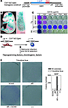
Repair of cartilage injury with hyaline cartilage continues to be a challenging clinical problem. Because of the limited number of chondrocytes in vivo, coupled with in vitro de-differentiation of chondrocytes into fibrochondrocytes, which secrete type I collagen and have an altered matrix architecture and mechanical function, there is a need for a novel cell source that produces hyaline cartilage. The generation of induced pluripotent stem (iPS) cells has provided a tool for reprogramming dermal fibroblasts to an undifferentiated state by ectopic expression of reprogramming factors. Here, we show that retroviral expression of two reprogramming factors (c-Myc and Klf4) and one chondrogenic factor (SOX9) induces polygonal chondrogenic cells directly from adult dermal fibroblast cultures. Induced cells expressed marker genes for chondrocytes but not fibroblasts, i.e., the promoters of type I collagen genes were extensively methylated. Although some induced cell lines formed tumors when subcutaneously injected into nude mice, other induced cell lines generated stable homogenous hyaline cartilage–like tissue. Further, the doxycycline-inducible induction system demonstrated that induced cells are able to respond to chondrogenic medium by expressing endogenous Sox9 and maintain chondrogenic potential after substantial reduction of transgene expression. Thus, this approach could lead to the preparation of hyaline cartilage directly from skin, without generating iPS cells.
Authors
Kunihiko Hiramatsu, Satoru Sasagawa, Hidetatsu Outani, Kanako Nakagawa, Hideki Yoshikawa, Noriyuki Tsumaki
×Abstract
Th17 cells are a subset of CD4+ T cells with an important role in clearing certain bacterial and fungal pathogens. However, they have also been implicated in autoimmune diseases such as multiple sclerosis. Exposure of naive CD4+ T cells to IL-6 and TGF-β leads to Th17 cell differentiation through a process in which many proteins have been implicated. We report here that ectopic expression of liver X receptor (LXR) inhibits Th17 polarization of mouse CD4+ T cells, while LXR deficiency promotes Th17 differentiation in vitro. LXR activation in mice ameliorated disease in the experimental autoimmune encephalomyelitis (EAE) model of multiple sclerosis, whereas LXR deficiency exacerbated disease. Further analysis revealed that Srebp-1, which is encoded by an LXR target gene, mediated the suppression of Th17 differentiation by binding to the E-box element on the Il17 promoter, physically interacting with aryl hydrocarbon receptor (Ahr) and inhibiting Ahr-controlled Il17 transcription. The putative active site (PAS) domain of Ahr and the N-terminal acidic region of Srebp-1 were essential for this interaction. Additional analyses suggested that similar LXR-dependent mechanisms were operational during human Th17 differentiation in vitro. This study reports what we believe to be a novel signaling pathway underlying LXR-mediated regulation of Th17 cell differentiation and autoimmunity.
Authors
Guoliang Cui, Xia Qin, Lili Wu, Yuebo Zhang, Xiaoyan Sheng, Qiwen Yu, Hongguang Sheng, Beili Xi, Jingwu Z. Zhang, Ying Qin Zang
×Abstract
Activation of NF-κB and 5-lipoxygenase–mediated (5-LO–mediated) biosynthesis of the lipid mediator leukotriene B4 (LTB4) are pivotal components of host defense and inflammatory responses. However, the role of LTB4 in mediating innate immune responses elicited by specific TLR ligands and cytokines is unknown. Here we have shown that responses dependent on MyD88 (an adaptor protein that mediates signaling through all of the known TLRs, except TLR3, as well as IL-1β and IL-18) are reduced in mice lacking either 5-LO or the LTB4 receptor BTL1, and that macrophages from these mice are impaired in MyD88-dependent activation of NF-κB. This macrophage defect was associated with lower basal and inducible expression of MyD88 and reflected impaired activation of STAT1 and overexpression of the STAT1 inhibitor SOCS1. Expression of MyD88 and responsiveness to the TLR4 ligand LPS were decreased by Stat1 siRNA silencing in WT macrophages and restored by Socs1 siRNA in 5-LO–deficient macrophages. These results uncover a pivotal role in macrophages for the GPCR BLT1 in regulating activation of NF-κB through Stat1-dependent expression of MyD88.
Authors
Carlos H. Serezani, Casey Lewis, Sonia Jancar, Marc Peters-Golden
×Abstract
Type 1 or invariant NKT (iNKT) cell agonists, epitomized by α-galactosylceramide, protect against cancer largely by IFN-γ–dependent mechanisms. Here we describe what we believe to be a novel IFN-γ–independent mechanism induced by β-mannosylceramide, which also defines a potentially new class of iNKT cell agonist, with an unusual β-linked sugar. Like α-galactosylceramide, β-mannosylceramide directly activates iNKT cells from both mice and humans. In contrast to α-galactosylceramide, protection by β-mannosylceramide was completely dependent on NOS and TNF-α, neither of which was required to achieve protection with α-galactosylceramide. Moreover, at doses too low for either alone to protect, β-mannosylceramide synergized with α-galactosylceramide to protect mice against tumors. These results suggest that treatment with β-mannosylceramide provides a distinct mechanism of tumor protection that may allow efficacy where other agonists have failed. Furthermore, the ability of β-mannosylceramide to synergize with α-galactosylceramide suggests treatment with this class of iNKT agonist may provide protection against tumors in humans.
Authors
Jessica J. O’Konek, Petr Illarionov, Deborah Stewart Khursigara, Elena Ambrosino, Liat Izhak, Bernard F. Castillo II, Ravinder Raju, Maryam Khalili, Hee-Yong Kim, Amy R. Howell, Gurdyal S. Besra, Steven A. Porcelli, Jay A. Berzofsky, Masaki Terabe
×Abstract
Inherited immunodeficiency disorders can be caused by mutations in any one of a large number of genes involved in the function of immune cells. Here, we describe two families with an autosomal recessive inherited immunodeficiency disorder characterized by increased susceptibility to infection and autoimmunity. Genetic linkage studies mapped the disorder to chromosomal region 14q11.2, and a homozygous guanine-to-adenine substitution was identified at the last base of exon 3 immediately following the translational termination codon in the TCRα subunit constant gene (TRAC). RT-PCR analysis in the two affected individuals revealed impaired splicing of the mRNA, as exon 3 was lost from the TRAC transcript. The mutant TCRα chain protein was predicted to lack part of the connecting peptide domain and all of the transmembrane and cytoplasmic domains, which have a critical role in the regulation of the assembly and/or intracellular transport of TCR complexes. We found that T cells from affected individuals were profoundly impaired for surface expression of the TCRαβ complex. We believe this to be the first report of a disease-causing human TRAC mutation. Although the absence of TCRαβ+ T cells in the affected individuals was associated with immune dysregulation and autoimmunity, they had a surprising level of protection against infection.
Authors
Neil V. Morgan, Sarah Goddard, Tony S. Cardno, David McDonald, Fatimah Rahman, Dawn Barge, Andrew Ciupek, Anna Straatman-Iwanowska, Shanaz Pasha, Mary Guckian, Graham Anderson, Aarnoud Huissoon, Andrew Cant, Warren P. Tate, Sophie Hambleton, Eamonn R. Maher
×Abstract
Tissue homeostasis and remodeling are processes that involve high turnover of biological macromolecules. Many of the waste molecules that are by-products or degradation intermediates of biological macromolecule turnover enter the circulation and are subsequently cleared by liver sinusoidal endothelial cells (LSEC). Besides the mannose receptor, stabilin-1 and stabilin-2 are the major scavenger receptors expressed by LSEC. To more clearly elucidate the functions of stabilin-1 and -2, we have generated mice lacking stabilin-1, stabilin-2, or both stabilin-1 and -2 (Stab1–/–Stab2–/– mice). Mice lacking either stabilin-1 or stabilin-2 were phenotypically normal; however, Stab1–/–Stab2–/– mice exhibited premature mortality and developed severe glomerular fibrosis, while the liver showed only mild perisinusoidal fibrosis without dysfunction. Upon kidney transplantation into WT mice, progression of glomerular fibrosis was halted, indicating the presence of profibrotic factors in the circulation of Stab1–/–Stab2–/– mice. While plasma levels of known profibrotic cytokines were unaltered, clearance of the TGF-β family member growth differentiation factor 15 (GDF-15) was markedly impaired in Stab1–/–Stab2–/– mice but not in either Stab1–/– or Stab2–/– mice, indicating that it is a common ligand of both stabilin-1 and stabilin-2. These data lead us to conclude that stabilin-1 and -2 together guarantee proper hepatic clearance of potentially noxious agents in the blood and maintain tissue homeostasis not only in the liver but also distant organs.
Authors
Kai Schledzewski, Cyrill Géraud, Bernd Arnold, Shijun Wang, Hermann-Josef Gröne, Tibor Kempf, Kai C. Wollert, Beate K. Straub, Peter Schirmacher, Alexandra Demory, Hiltrud Schönhaber, Alexei Gratchev, Lisa Dietz, Hermann-Josef Thierse, Julia Kzhyshkowska, Sergij Goerdt
×Abstract
Post-mortem analyses of brains from patients with Parkinson disease who received fetal mesencephalic transplants show that α-synuclein–containing (α-syn–containing) Lewy bodies gradually appear in grafted neurons. Here, we explored whether intercellular transfer of α-syn from host to graft, followed by seeding of α-syn aggregation in recipient neurons, can contribute to this phenomenon. We assessed α-syn cell-to-cell transfer using microscopy, flow cytometry, and high-content screening in several coculture model systems. Coculturing cells engineered to express either GFP– or DsRed-tagged α-syn resulted in a gradual increase in double-labeled cells. Importantly, α-syn–GFP derived from 1 neuroblastoma cell line localized to red fluorescent aggregates in other cells expressing DsRed–α-syn, suggesting a seeding effect of transmitted α-syn. Extracellular α-syn was taken up by cells through endocytosis and interacted with intracellular α-syn. Next, following intracortical injection of recombinant α-syn in rats, we found neuronal uptake was attenuated by coinjection of an endocytosis inhibitor. Finally, we demonstrated in vivo transfer of α-syn between host cells and grafted dopaminergic neurons in mice overexpressing human α-syn. In summary, intercellularly transferred α-syn interacts with cytoplasmic α-syn and can propagate α-syn pathology. These results suggest that α-syn propagation is a key element in the progression of Parkinson disease pathology.
Authors
Christian Hansen, Elodie Angot, Ann-Louise Bergström, Jennifer A. Steiner, Laura Pieri, Gesine Paul, Tiago F. Outeiro, Ronald Melki, Pekka Kallunki, Karina Fog, Jia-Yi Li, Patrik Brundin
×Abstract
Amyotrophic lateral sclerosis (ALS) and frontotemporal lobar degeneration (FTLD) are characterized by cytoplasmic protein aggregates in the brain and spinal cord that include TAR-DNA binding protein 43 (TDP-43). TDP-43 is normally localized in the nucleus with roles in the regulation of gene expression, and pathological cytoplasmic aggregates are associated with depletion of nuclear protein. Here, we generated transgenic mice expressing human TDP-43 with a defective nuclear localization signal in the forebrain (hTDP-43-ΔNLS), and compared them with mice expressing WT hTDP-43 (hTDP-43-WT) to determine the effects of mislocalized cytoplasmic TDP-43 on neuronal viability. Expression of either hTDP-43-ΔNLS or hTDP-43-WT led to neuron loss in selectively vulnerable forebrain regions, corticospinal tract degeneration, and motor spasticity recapitulating key aspects of FTLD and primary lateral sclerosis. Only rare cytoplasmic phosphorylated and ubiquitinated TDP-43 inclusions were seen in hTDP-43-ΔNLS mice, suggesting that cytoplasmic inclusions were not required to induce neuronal death. Instead, neurodegeneration in hTDP-43 and hTDP-43-ΔNLS–expressing neurons was accompanied by a dramatic downregulation of the endogenous mouse TDP-43. Moreover, mice expressing hTDP-43-ΔNLS exhibited profound changes in gene expression in cortical neurons. Our data suggest that perturbation of endogenous nuclear TDP-43 results in loss of normal TDP-43 function(s) and gene regulatory pathways, culminating in degeneration of selectively vulnerable affected neurons.
Authors
Lionel M. Igaz, Linda K. Kwong, Edward B. Lee, Alice Chen-Plotkin, Eric Swanson, Travis Unger, Joe Malunda, Yan Xu, Matthew J. Winton, John Q. Trojanowski, Virginia M.-Y. Lee
×Abstract
Both mucosal and systemic immune responses are required for preventing or containing HIV transmission and chronic infection. However, currently described vaccination approaches are largely ineffective in inducing both mucosal and systemic responses. In this study, we found that the ubiquitin-editing enzyme A20 — an inducible feedback inhibitor of the TNFR, RIG-I, and TLR signaling pathways that broadly controls the maturation, cytokine production, and immunostimulatory potency of DCs — restricted systemically immunized DCs to induce both robust mucosal and systemic HIV-specific cellular and humoral responses. Mechanistic studies revealed that A20 regulated DC production of retinoic acid and proinflammatory cytokines, inhibiting the expression of gut-homing receptors on T and B cells. Furthermore, A20-silenced, hyperactivated DCs exhibited an enhanced homing capacity to draining and gut-associated lymphoid tissues (GALTs) after systemic administration. Thus, this study provides insights into the role of A20 in innate immunity. This work may allow the development of an efficient HIV vaccination strategy that is capable of inducing both robust systemic and mucosal anti-HIV cellular and humoral responses.
Authors
Bangxing Hong, Xiao-Tong Song, Lisa Rollins, Lindsey Berry, Xue F. Huang, Si-Yi Chen
×Abstract
Kaposi sarcoma–associated herpesvirus (KSHV; also known as HHV8) is the causative agent of two B cell tumors, multicentric Castleman disease (MCD) and primary effusion lymphoma (PEL). However, little is known about the nature of the specific B cell subtype(s) most susceptible to infection. Identifying these cells would provide direct insight into KSHV transmission and virus-induced transformation. To identify this subset and to determine whether infection alters its cellular phenotype, we exposed human tonsillar cells to KSHV and characterized infected cells using high-throughput multispectral imaging flow cytometry (MIFC). Stable expression of the virally encoded latency-associated nuclear antigen (LANA), a marker of latent KSHV infection, was observed predominantly in cells expressing the l light chain of the B cell receptor. These LANA+ B cells proliferated and exhibited similarities to the cells characteristic of MCD (IgMl-expressing plasmablasts), including blasting morphology with elevated expression of Ki67, variable expression of CD27, and high levels of IgM and IL-6 receptor. Furthermore, the proportion of infected cells showing a blasting phenotype increased upon addition of exogenous IL-6. Our data lead us to propose that oral transmission of KSHV involves the latent infection of a subset of tonsillar IgMl-expressing B cells, which then proliferate as they acquire the plasmablast phenotype characteristic of MCD.
Authors
Lynn M. Hassman, Thomas J. Ellison, Dean H. Kedes
×Abstract
Collagen V, broadly expressed as α1(V)2α2(V) heterotrimers that regulate collagen fibril geometry and strength, also occurs in some tissues, such as white adipose tissue (WAT), pancreatic islets, and skeletal muscle, as the poorly characterized α1(V) α2(V) α3(V) heterotrimer. Here, we investigate the role of α3(V) collagen chains by generating mice with a null allele of the α3(V) gene Col5a3 (Col5a3–/– mice). Female Col5a3–/– mice had reduced dermal fat and were resistant to high-fat diet–induced weight gain. Male and female mutant mice were glucose intolerant, insulin-resistant, and hyperglycemic, and these metabolic defects worsened with age. Col5a3–/– mice demonstrated decreased numbers of pancreatic islets, which were more susceptible to streptozotocin-induced apoptosis, and islets isolated from mutant mice displayed blunted glucose-stimulated insulin secretion. Moreover, Col5a3–/– WAT and skeletal muscle were defective in glucose uptake and mobilization of intracellular GLUT4 glucose transporter to the plasma membrane in response to insulin. Our results underscore the emerging view of the importance of ECM to the microenvironments that inform proper development/functioning of specialized cells, such as adipocytes, β cells, and skeletal muscle.
Authors
Guorui Huang, Gaoxiang Ge, Dingyan Wang, Bagavathi Gopalakrishnan, Delana H. Butz, Ricki J. Colman, Andras Nagy, Daniel S. Greenspan
×Abstract
Systemic instigation is a process by which endocrine signals sent from certain tumors (instigators) stimulate BM cells (BMCs), which are mobilized into the circulation and subsequently foster the growth of otherwise indolent carcinoma cells (responders) residing at distant anatomical sites. The identity of the BMCs and their specific contribution or contributions to responder tumor growth have been elusive. Here, we have demonstrated that Sca1+cKit– hematopoietic BMCs of mouse hosts bearing instigating tumors promote the growth of responding tumors that form with a myofibroblast-rich, desmoplastic stroma. Such stroma is almost always observed in malignant human adenocarcinomas and is an indicator of poor prognosis. We then identified granulin (GRN) as the most upregulated gene in instigating Sca1+cKit– BMCs relative to counterpart control cells. The GRN+ BMCs that were recruited to the responding tumors induced resident tissue fibroblasts to express genes that promoted malignant tumor progression; indeed, treatment with recombinant GRN alone was sufficient to promote desmoplastic responding tumor growth. Further, analysis of tumor tissues from a cohort of breast cancer patients revealed that high GRN expression correlated with the most aggressive triple-negative, basal-like tumor subtype and reduced patient survival. Our data suggest that GRN and the unique hematopoietic BMCs that produce it might serve as novel therapeutic targets.
Authors
Moshe Elkabets, Ann M. Gifford, Christina Scheel, Bjorn Nilsson, Ferenc Reinhardt, Mark-Anthony Bray, Anne E. Carpenter, Karin Jirström, Kristina Magnusson, Benjamin L. Ebert, Fredrik Pontén, Robert A. Weinberg, Sandra S. McAllister
×Abstract
The role of the Notch signaling pathway in tumor development is complex, with Notch1 functioning either as an oncogene or as a tumor suppressor in a context-dependent manner. To further define the role of Notch1 in tumor development, we systematically surveyed for tumor suppressor activity of Notch1 in vivo. We combined the previously described Notch1 intramembrane proteolysis–Cre (Nip1::Cre) allele with a floxed Notch1 allele to create a mouse model for sporadic, low-frequency loss of Notch1 heterozygosity. Through this approach, we determined the cell types most affected by Notch1 loss. We report that the loss of Notch1 caused widespread vascular tumors and organism lethality secondary to massive hemorrhage. These findings reflected a cell-autonomous role for Notch1 in suppressing neoplasia in the vascular system and provide a model by which to explore the mechanism of neoplastic transformation of endothelial cells. Importantly, these results raise concerns regarding the safety of chronic application of drugs targeting the Notch pathway, specifically those targeting Notch1, because of mechanism-based toxicity in the endothelium. Our strategy also can be broadly applied to induce sporadic in vivo loss of heterozygosity of any conditional alleles in progenitors that experience Notch1 activation.
Authors
Zhenyi Liu, Ahu Turkoz, Erin N. Jackson, Joseph C. Corbo, John A. Engelbach, Joel R. Garbow, David R. Piwnica-Worms, Raphael Kopan
×Abstract
The p53 tumor suppressor, a central mediator of chemosensitivity in normal cells, is functionally inactivated in many human cancers. Therefore, a central challenge in human cancer therapy is the identification of pathways that control tumor cell survival and chemosensitivity in the absence of functional p53. The p53-related transcription factors p63 and p73 exhibit distinct functions — p73 mediates chemosensitivity while p63 promotes proliferation and cell survival — and are both overexpressed in squamous cell carcinomas (SCCs). However, how p63 and p73 interact functionally and govern the balance between prosurvival and proapoptotic programs in SCC remains elusive. Here, we identify a microRNA-dependent mechanism of p63/p73 crosstalk that regulates p53-independent survival of both human and murine SCC. We first discovered that a subset of p63-regulated microRNAs target p73 for inhibition. One of these, miR-193a-5p, expression of which was repressed by p63, was activated by proapoptotic p73 isoforms in both normal cells and tumor cells in vivo. Chemotherapy caused p63/p73-dependent induction of this microRNA, thereby limiting chemosensitivity due to microRNA-mediated feedback inhibition of p73. Importantly, inhibiting miR-193a interrupted this feedback and thereby suppressed tumor cell viability and induced dramatic chemosensitivity both in vitro and in vivo. Thus, we have identified a direct, microRNA-dependent regulatory circuit mediating inducible chemoresistance, whose inhibition may provide a new therapeutic opportunity in p53-deficient tumors.
Authors
Benjamin Ory, Matthew R. Ramsey, Catherine Wilson, Douangsone D. Vadysirisack, Nicole Forster, James W. Rocco, S. Michael Rothenberg, Leif W. Ellisen


Copyright © 2025 American Society for Clinical Investigation
ISSN: 0021-9738 (print), 1558-8238 (online)