Issue published June 15, 2003 Previous issue | Next issue
- Volume 111, Issue 12
Go to section:
-
Science in Medicine
×
Abstract
Authors
Daniel J. Rader, Jonathan Cohen, Helen H. Hobbs


-
Commentaries
×
Abstract
Apoptosis signal–regulating kinase 1 (ASK1) is an upstream activator of JNK and p38 MAPK signaling cascades. Evidence now shows that the ASK1-interacting protein, AIP1, plays an important role in TNF-α–induced ASK1 activation by facilitating dissociation from its inhibitor.
Authors
M. Eugenia Guicciardi, Gregory J. Gores
×Abstract
Microarray analysis of the expression profiles of lung tissue in two murine models of asthma revealed high levels of arginase I and arginase II activity, in association with IL-4 and IL-13 overexpression, suggesting that arginine pathways are critical in the pathogenesis of asthma.
Authors
Donata Vercelli
×Abstract
Diabetic retinopathy, the most frequent complication of diabetes and leading cause of vision loss, involves vascular and neural damage in the retina. Insulin and IGF-1 signaling are now shown to contribute to retinal neovascularization, in part, by modulating the expression of various vascular mediators.
Authors
Sarah K. Bronson, Chad E.N. Reiter, Thomas W. Gardner
×Abstract
Little is known about the effect of an individual’s immune history on his or her response to an allogeneic tissue transplant. An important study now reveals that individuals harboring virally-induced memory T cells that are cross reactive with donor alloantigen are resistant to conventional strategies designed to induce transplant tolerance.
Authors
David H. Sachs


-
Research Articles
×
Abstract
Infection of neonatal mice with some reovirus strains produces a disease similar to infantile biliary atresia, but previous attempts to correlate reovirus infection with this disease have yielded conflicting results. We used isogenic reovirus strains T3SA– and T3SA+, which differ solely in the capacity to bind sialic acid as a coreceptor, to define the role of sialic acid in reovirus encephalitis and biliary tract infection in mice. Growth in the intestine was equivalent for both strains following peroral inoculation. However, T3SA+ spread more rapidly from the intestine to distant sites and replicated to higher titers in spleen, liver, and brain. Strikingly, mice infected with T3SA+ but not T3SA– developed steatorrhea and bilirubinemia. Liver tissue from mice infected with T3SA+ demonstrated intense inflammation focused at intrahepatic bile ducts, pathology analogous to that found in biliary atresia in humans, and high levels of T3SA+ antigen in bile duct epithelial cells. T3SA+ bound 100-fold more efficiently than T3SA– to human cholangiocarcinoma cells. These observations suggest that the carbohydrate-binding specificity of a virus can dramatically alter disease in the host and highlight the need for epidemiologic studies focusing on infection by sialic acid–binding reovirus strains as a possible contributor to the pathogenesis of neonatal biliary atresia.
Authors
Erik S. Barton, Bryan E. Youree, Daniel H. Ebert, J. Craig Forrest, Jodi L. Connolly, Tibor Valyi-Nagy, Kay Washington, J. Denise Wetzel, Terence S. Dermody
×Abstract
Both insulin and IGF-1 have been implicated in control of retinal endothelial cell growth, neovascularization, and diabetic retinopathy. To precisely define the role of insulin and IGF-1 signaling in endothelium in these processes, we have used the oxygen-induced retinopathy model to study mice with a vascular endothelial cell–specific knockout of the insulin receptor (VENIRKO) or IGF-1 receptor (VENIFARKO). Following relative hypoxia, VENIRKO mice show a 57% decrease in retinal neovascularization as compared with controls. This is associated with a blunted rise in VEGF, eNOS, and endothelin-1. By contrast, VENIFARKO mice show only a 34% reduction in neovascularization and a very modest reduction in mediator generation. These data indicate that both insulin and IGF-1 signaling in endothelium play a role in retinal neovascularization through the expression of vascular mediators, with the effect of insulin being most important in this process.
Authors
Tatsuya Kondo, David Vicent, Kiyoshi Suzuma, Masashi Yanagisawa, George L. King, Martin Holzenberger, C. Ronald Kahn
×Abstract
Vascular endothelial growth factor (VEGF) is an angiogenic protein with therapeutic potential in ischemic disorders, including stroke. VEGF confers neuroprotection and promotes neurogenesis and cerebral angiogenesis, but the manner in which these effects may interact in the ischemic brain is poorly understood. We produced focal cerebral ischemia by middle cerebral artery occlusion for 90 minutes in the adult rat brain and measured infarct size, neurological function, BrdU labeling of neuroproliferative zones, and vWF-immunoreactive vascular profiles, without and with intracerebroventricular administration of VEGF on days 1–3 of reperfusion. VEGF reduced infarct size, improved neurological performance, enhanced the delayed survival of newborn neurons in the dentate gyrus and subventricular zone, and stimulated angiogenesis in the striatal ischemic penumbra, but not the dentate gyrus. We conclude that in the ischemic brain VEGF exerts an acute neuroprotective effect, as well as longer latency effects on survival of new neurons and on angiogenesis, and that these effects appear to operate independently. VEGF may, therefore, improve histological and functional outcome from stroke through multiple mechanisms.
Authors
Yunjuan Sun, Kunlin Jin, Lin Xie, Jocelyn Childs, Xiao Ou Mao, Anna Logvinova, David A. Greenberg
×Abstract
Previously we showed that neuropeptide Y (NPY), a sympathetic vasoconstrictor neurotransmitter, stimulates endothelial cell migration, proliferation, and differentiation in vitro. Here, we report on NPY’s actions, receptors, and mediators in ischemic angiogenesis. In rats, hindlimb ischemia stimulates sympathetic NPY release (attenuated by lumbar sympathectomy) and upregulates NPY-Y2 (Y2) receptor and a peptidase forming Y2/Y5-selective agonist. Exogenous NPY at physiological concentrations also induces Y5 receptor, stimulates neovascularization, and restores ischemic muscle blood flow and performance. NPY-mediated ischemic angiogenesis is not prevented by a selective Y1 receptor antagonist but is reduced in Y2–/– mice. Nonischemic muscle vascularity is also lower in Y2–/– mice, whereas it is increased in NPY-overexpressing rats compared with their WT controls. Ex vivo, NPY-induced aortic sprouting is markedly reduced in Y2–/– aortas and spontaneous sprouting is severely impaired in NPY–/– mice. NPY-mediated aortic sprouting, but not cell migration/proliferation, is blocked by an antifetal liver kinase 1 antibody and abolished in mice null for eNOS. Thus, NPY mediates neurogenic ischemic angiogenesis at physiological concentrations by activating Y2/Y5 receptors and eNOS, in part due to release of VEGF. NPY’s effectiveness in revascularization and restoring function of ischemic tissue suggests its therapeutic potential in ischemic conditions.
Authors
Edward W. Lee, Mieczyslaw Michalkiewicz, Joanna Kitlinska, Ivana Kalezic, Hanna Switalska, Peter Yoo, Amarin Sangkharat, Hong Ji, Lijun Li, Teresa Michalkiewicz, Milos Ljubisavljevic, Hakan Johansson, Derrick S. Grant, Zofia Zukowska
×Abstract
Asthma is on the rise despite intense, ongoing research underscoring the need for new scientific inquiry. In an effort to provide unbiased insight into disease pathogenesis, we took an approach involving expression profiling of lung tissue from mice with experimental asthma. Employing asthma models induced by different allergens and protocols, we identified 6.5% of the tested genome whose expression was altered in an asthmatic lung. Notably, two phenotypically similar models of experimental asthma were shown to have distinct transcript profiles. Genes related to metabolism of basic amino acids, specifically the cationic amino acid transporter 2, arginase I, and arginase II, were particularly prominent among the asthma signature genes. In situ hybridization demonstrated marked staining of arginase I, predominantly in submucosal inflammatory lesions. Arginase activity was increased in allergen-challenged lungs, as demonstrated by increased enzyme activity, and increased levels of putrescine, a downstream product. Lung arginase activity and mRNA expression were strongly induced by IL-4 and IL-13, and were differentially dependent on signal transducer and activator of transcription 6. Analysis of patients with asthma supported the importance of this pathway in human disease. Based on the ability of arginase to regulate generation of NO, polyamines, and collagen, these results provide a basis for pharmacologically targeting arginine metabolism in allergic disorders.
Authors
Nives Zimmermann, Nina E. King, Johanne Laporte, Ming Yang, Anil Mishra, Sam M. Pope, Emily E. Muntel, David P. Witte, Anthony A. Pegg, Paul S. Foster, Qutayba Hamid, Marc E. Rothenberg
×Abstract
In a strategy to specifically target complement inhibitors to sites of complement activation and disease, recombinant fusion proteins consisting of a complement inhibitor linked to a C3 binding region of complement receptor (CR) 2 were prepared and characterized. Natural ligands for CR2 are C3 breakdown products deposited at sites of complement activation. Fusion proteins were prepared consisting of a human CR2 fragment linked to either the N terminus or C terminus of soluble forms of the membrane complement inhibitors decay accelerating factor (DAF) or CD59. The targeted complement inhibitors bound to C3-opsonized cells, and all were significantly more effective (up to 20-fold) than corresponding untargeted inhibitors at protecting target cells from complement. CR2 fusion proteins also inhibited CR3-dependent adhesion of U937 cells to C3 opsonized erythrocytes, indicating a second potential anti-inflammatory mechanism of CR2 fusion proteins, since CR3 is involved in endothelial adhesion and diapedesis of leukocytes at inflammatory sites. Finally, the in vivo validity of the targeting strategy was confirmed by the demonstration that CR2-DAF, but not soluble DAF, targets to the kidney in mouse models of lupus nephritis that are associated with renal complement deposition.
Authors
Hongbin Song, Chun He, Christian Knaak, Joel M. Guthridge, V. Michael Holers, Stephen Tomlinson
×Abstract
Many strategies have been proposed to induce tolerance to transplanted tissue in rodents; however, few if any have shown equal efficacy when tested in nonhuman primate transplant models. We hypothesized that a critical distinction between specific pathogen-free mice and nonhuman primates or human patients is their acquired immune history. Here, we show that a heterologous immune response — specifically, virally induced alloreactive memory — is a potent barrier to tolerance induction. A critical threshold of memory T cells is needed to promote rejection, and CD8+ “central” memory T cells are primarily responsible. Finally, treatment with deoxyspergualin, an inhibitor of NF-κB translocation, together with costimulation blockade, synergistically impairs memory T cell activation and promotes antigen-specific tolerance of memory. These data offer a potential explanation for the difficulty encountered when inducing tolerance in nonhuman primates and human patients and provide insight into the signaling pathways essential for memory T cell activation and function.
Authors
Andrew B. Adams, Matthew A. Williams, Thomas R. Jones, Nozomu Shirasugi, Megan M. Durham, Susan M. Kaech, E. John Wherry, Thandi Onami, J. Gibson Lanier, Kenneth E. Kokko, Thomas C. Pearson, Rafi Ahmed, Christian P. Larsen
×Abstract
Graves disease, a common organ-specific autoimmune disease affecting humans, differs from all other autoimmune diseases in being associated with target organ hyperfunction rather than organ damage. Clinical thyrotoxicosis is directly caused by autoantibodies that activate the thyrotropin receptor (TSHR). The etiology of Graves disease is multifactorial, with nongenetic factors playing an important role. Of the latter, there is the intriguing possibility that the molecular structure of the target antigen contributes to the development of thyroid-stimulatory autoantibodies (TSAb’s). Among the glycoprotein hormone receptors, only the TSHR undergoes intramolecular cleavage into disulfide-linked subunits with consequent shedding of some of the extracellular, autoantibody-binding A subunits. Functional autoantibodies do not arise to the noncleaving glycoprotein hormone receptors. Recently, TSAb’s were found to preferentially recognize shed, rather than attached, A subunits. Here we use a new adenovirus-mediated animal model of Graves disease to show that goiter and hyperthyroidism occur to a much greater extent when the adenovirus expresses the free A subunit as opposed to a genetically modified TSHR that cleaves minimally into subunits. These data show that shed A subunits induce or amplify the immune response leading to hyperthyroidism and provide new insight into the etiology of Graves disease.
Authors
Chun-Rong Chen, Pavel Pichurin, Yuji Nagayama, Francesco Latrofa, Basil Rapoport, Sandra M. McLachlan
×Abstract
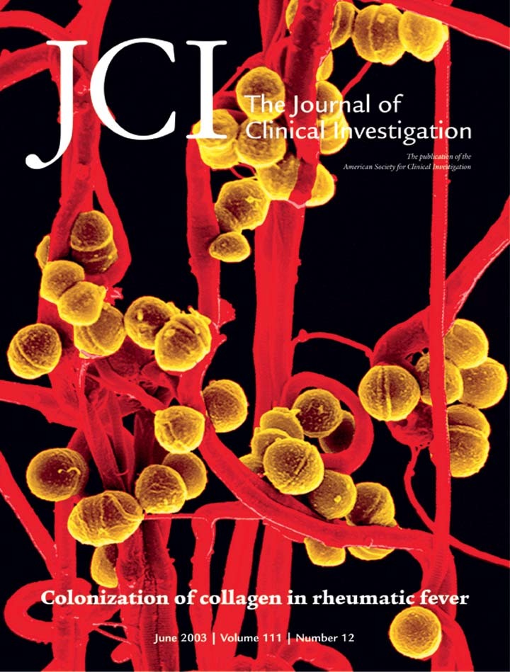
Acute rheumatic fever is a serious autoimmune sequel of Streptococcus pyogenes infection. This study shows that serotype M3 and M18 S. pyogenes isolated during outbreaks of rheumatic fever have the unique capability to bind and aggregate human basement membrane collagen type IV. M3 protein is identified as collagen-binding factor of M3 streptococci, whereas M18 isolates bind collagen through a hyaluronic acid capsule, revealing a novel function for M3 protein and capsule. Following in vivo mouse passage, conversion of a nonencapsulated and collagen-binding negative M1 S. pyogenes into an encapsulated, collagen-binding strain further supports the crucial role of capsule in mediating collagen binding. Collagen binding represents a novel colonization mechanism, as it is demonstrated that S. pyogenes bind to collagen matrix in vitro and in vivo. Moreover, immunization of mice with purified recombinant M3 protein led to the generation of anti–collagen type IV antibodies. Finally, sera from acute rheumatic fever patients had significantly increased titers of anti–collagen type IV antibodies as compared with healthy controls. These findings may suggest a link between the potential of rheumatogenic S. pyogenes isolates to bind collagen, and the presence of collagen-reactive autoantibodies in the serum of rheumatic fever patients, which may form a basis for post-streptococcal rheumatic disease. These anti-collagen antibodies may form a basis for poststreptococcal rheumatic disease.
Authors
Katrin Dinkla, Manfred Rohde, Wouter T.M. Jansen, Edward L. Kaplan, Gursharan S. Chhatwal, Susanne R. Talay
×Abstract
Mitochondrial neurogastrointestinal encephalomyopathy (MNGIE) is an autosomal recessive disorder caused by loss-of-function mutations in the gene encoding thymidine phosphorylase (TP). This deficiency of TP leads to increased circulating levels of thymidine (deoxythymidine, dThd) and deoxyuridine (dUrd) and has been associated with multiple deletions and depletion of mitochondrial DNA (mtDNA). Here we describe 36 point mutations in mtDNA of tissues and cultured cells from MNGIE patients. Thirty-one mtDNA point mutations (86%) were T-to-C transitions, and of these, 25 were preceded by 5′-AA sequences. In addition, we identified a single base-pair mtDNA deletion and a TT-to-AA mutation. Next-nucleotide effects and dislocation mutagenesis may contribute to the formation of these mutations. These results provide the first demonstration that alterations of nucleoside metabolism can induce multiple sequence-specific point mutations in humans. We hypothesize that, in patients with TP deficiency, increased levels of dThd and dUrd cause mitochondrial nucleotide pool imbalances, which, in turn, lead to mtDNA abnormalities including site-specific point mutations.
Authors
Yutaka Nishigaki, Ramon Martí, William C. Copeland, Michio Hirano
×Abstract
IL-10 is a pleiotropic cytokine that inhibits several immune parameters, including Th1 cell–mediated immune responses, antigen presentation, and antigen-specific T cell proliferation. Recent data implicate IL-10 as a mediator of suppression of cell-mediated immunity induced by exposure to UVB radiation (280–320 nm). To investigate the effects of IL-10 on the cutaneous immune system, we engineered transgenic mice that overexpress viral IL-10 (vIL-10) in the epidermis. vIL-10 transgenic mice demonstrated a reduced number of I-A+ epidermal and dermal cells and fewer I-A+ hapten-bearing cells in regional lymph nodes after hapten painting of the skin. Reduced CD80 and CD86 expression by I-A+ epidermal cells was also observed. vIL-10 transgenic mice demonstrated a smaller delayed-type hypersensitivity response to allogeneic cells upon challenge but had normal contact hypersensitivity to an epicutaneously applied hapten. Fresh epidermal cells from vIL-10 transgenic mice showed a decreased ability to stimulate allogeneic T cell proliferation, as did splenocytes. Additionally, chronic exposure of mice to UVB radiation led to the development of fewer skin tumors in vIL-10 mice than in WT controls, and vIL-10 transgenic mice had increased splenic NK cell activity against YAC-1targets. These findings support the concept that IL-10 is an important regulator of cutaneous immune function.
Authors
Wanhong Ding, Stefan Beissert, Liang Deng, Edward Miranda, Christopher Cassetty, Kristina Seiffert, Kristina L. Campton, Zhengmin Yan, George F. Murphy, Jeffrey A. Bluestone, Richard D. Granstein
×Abstract
TNF-α activates ASK1 in part by dissociating 14-3-3 from apoptosis signal–regulating kinase 1 (ASK1). In the present study, we identified a novel Ras GTPase-activating protein (Ras-GAP) as an ASK1-interacting protein (AIP1). AIP1 binds to the C-terminal domain of ASK1 via a lysine-rich cluster within the N-terminal C2 domain. AIP1 exists in a closed form through an intramolecular interaction between the N-terminus and the C-terminus, and TNF-α induces unfolding of AIP1 leading to association of AIP1 with ASK1. Thus, the N-terminus of AIP1 containing the C2 and GAP domains constitutively binds to ASK1 and facilitates the release of 14-3-3 from ASK1. In contrast to 14-3-3, AIP1 binds preferentially to dephosphorylated ASK1. Recruited AIP1 enhances ASK1-induced JNK activation, and the ASK1 binding and the GAP activity of AIP1 are critical for AIP1-enhanced ASK1 activation. Furthermore, TNF-induced ASK1/JNK activation is significantly blunted in cells where AIP1 is knocked down by RNA interference. These data suggest that AIP1 mediates TNF-α–induced ASK1 activation by facilitating dissociation of inhibitor 14-3-3 from ASK1, a novel mechanism by which TNF-α activates ASK1.
Authors
Rong Zhang, Xiangrong He, Weimin Liu, Meng Lu, Jer-Tsong Hsieh, Wang Min


Copyright © 2025 American Society for Clinical Investigation
ISSN: 0021-9738 (print), 1558-8238 (online)