
Oncology
Abstract
Hepatocellular carcinoma (HCC) is heterogeneous, and hepatocyte plasticity is linked to poorer patient outcomes. A subset of human HCC harboring Tuberous Sclerosis Complex 1 (TSC1) mutations exhibits more aggressive behavior. TFEB is a master regulator of lysosomal biogenesis and cell fate. We analyzed human normal and HCC tissue arrays for TFEB and CK19 expression, as well as bulk and single-cell RNA-seq datasets from mouse and human HCC, to define TFEB-associated transcriptional programs. We performed biochemical, histological, metabolomic, and transcriptomic analyses in liver-specific Tsc1 knockout (L-Tsc1 KO) and L-Tsc1,Tfeb double KO (DKO) mice. Loss of hepatic Tsc1 led to increased phosphorylation of S6 and 4EBP1, with paradoxical increases in TFEB nuclear translocation and activation. L-Tsc1 KO mice showed increased hepatocyte plasticity, decreased HFN4α, increased YAP1 activation, and spontaneous HCC with increased SOX9 and CK19-positive biliary epithelial cell (BEC)-like cells at 8-12 months. Deletion of Tfeb dampened hepatic metabolic reprogramming and hepatocyte fate changes and inhibited tumor progression in L-Tsc1 KO mice. Increased TFEB activity was associated with increased YAP and SOX9 gene expression and high-grade malignant HCC in humans. These findings indicate that loss of hepatic TSC1 leads to non-canonical TFEB activation, promoting hepatocyte plasticity and tumor heterogeneity associated with high-grade malignancy in both mouse and human HCC.
Authors
Chen Zhang, Xiaojuan Chao, Sha Neisha Williams, Xiaoli Wei, Anthony DiGirolamo, Alisha Bajracharya, Lichun Ma, Ming Huang, Nicholas Dunn, Wanqing Liu, Kaito Ueda, Masayuki Sugimoto, Andrea Ballabio, Hong-Min Ni, Wen-Xing Ding
Abstract
Authors
Yashika Parashar, Zsofia Sztupinszki, Aurel György Prósz, Xiaolu Wang, Pratyusha Bala, Shweta Kiran Cavale, Chinedu Ukaegbu, Sapna Syngal, Asaf Maoz, Leah H. Biller, Ramona Lim, Matthew B. Yurgelun, Zoltan Szallasi, Nilay S. Sethi
Abstract
Background: Despite therapeutic advances in the early-stage triple negative breast cancer (TNBC) setting, residual disease (RD) following neoadjuvant therapy remains a key predictor of a worse prognosis and is a major obstacle to improving patient outcomes. Methods: To better characterize RD and identify survival associated features, we performed comprehensive transcriptomic profiling of 340 pre-treatment stage II/III TNBCs and 70 matched post-treatment RD samples from the randomized CALGB 40603 (Alliance) Phase 2 clinical trial. Preclinical treatment strategies mimicking RD patients were explored using Antibody Drug Conjugate (ADC) treatment in patient derived xenograft (PDX) mouse models. Results: Our study shows that prognostic genomic features measured prior to treatment may differ from prognostic features measured after treatment from RD specimens. Specifically, we identified that patients with a genomic PAM50 subtype of Basal-like in RD specimens have a poor survival outcome and their matching pre-treatment tumors are characterized by elevated chromosomal amplifications of oncogenic drivers (i.e., MYC, CDK6, and CCND1) as well as significantly reduced B- and T-cell expression features. Paired analyses of Basal-like RD and matched pre-treatment tumors reveal further lymphocyte depletion in the RD, along with lower expression of MHC class I and interferon signaling, indicating an immune-cold RD microenvironment. Treatment of a Basal-like and conventional chemotherapy-resistant PDX model, resembling Basal-like RD, with sacituzumab govitecan or trastuzumab deruxtecan produced a marked antitumor response. Conclusion: RD biology differs from pre-treatment tumors, with Basal-like subtype RD following neoadjuvant chemotherapy being immune cold and associated with poor survival. Pre-clinical modeling suggests that this high-risk group may benefit from adjuvant ADC therapy. Trial registration: ClinicalTrials.gov NCT00861705 Funding: National Cancer Institute (NCI) U10CA180821 (Alliance for Clinical Trials in Oncology) NCI U24CA176171 (Alliance for Clinical Trials in Oncology) NCI UG1CA233373 (Alliance for Clinical Trials in Oncology) NCI Breast SPORE program P50-CA058223 (CMP) Susan G. Komen SAC-160074 (CMP, PDR) Breast Cancer Research Foundation BCRF-23-127 (CMP) NCI R01-CA229409 (CMP) UNC LCCC Triple Negative Breast Cancer Center (CMP)
Authors
Patrick D. Rädler, Brooke M. Felsheim, Aranzazu Fernandez-Martinez, Adam D. Pfefferle, Michele C. Hayward, Baljit Singh, William Sikov, Lisa A. Carey, Charles M. Perou
Abstract
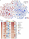
Fibroblast growth factor receptor 3 (FGFR3) is one of the most frequently altered genes in bladder cancer, primarily through activating mutations that drive oncogenesis and are enriched in luminal tumors. However, the underlying gene regulatory network (GRN) remains poorly characterized. Here, we constructed an FGFR3-mutated GRN using a bottom-up bioinformatics approach, integrating transcriptomic data from bladder cancer cell lines, FGFR3-mutated tumors, and FGFR3 perturbation experiments in human and mouse models. Using publicly available CRISPR/Cas9 screening data, we identified transcription factors from this GRN that regulate the viability of FGFR3-mutated cells, with a focus on p63 (TP63). We showed that FGFR3 activation upregulates p63 in patient-derived xenografts and cell lines, while single-cell RNA sequencing revealed heterogeneous p63 activation associated with basal differentiation. Functional studies, including TP63 knockdown in FGFR3-dependent in vitro and in vivo models and RNA-seq along with p63 ChIP-seq, demonstrated that p63 directly promotes cell proliferation and migration and uncovered a positive feedback loop between FGFR3 and p63. Together, these findings support p63 as a protumorigenic regulator in FGFR3-mutated tumors despite their luminal differentiation and provide a detailed FGFR3-driven GRN, offering insights into FGFR3-induced oncogenic dependency and potential strategies to circumvent resistance to FGFR inhibitors.
Authors
Aura Moreno-Vega, Macarena Zambrano, Lilia Estrada-Virrueta, Xiangyu Meng, Julia Puig, Helene Neyret-Kahn, Mingjun Shi, Florent Dufour, Guerric Gilbert, Ke Li, Clarice Groeneveld, Jacqueline Fontugne, Mercedes Pérez-Escavy, Wajdi Dhifli, Clément Hua, Luc Cabel, Clémentine Krucker, Laura Tanguy, Sia Viborg Lindskrog, Claire Beraud, Yanina V. Langle, Tao Ye, Fariza Tahi, Irwin Davidson, Jesus M. Paramio, Lars Dyrskjøt, Yves Allory, Philippe Lluel, Ana Maria Eiján, Mohamed Elati, François Radvanyi, Catalina Lodillinsky, Isabelle Bernard-Pierrot

Abstract
TRAIL is a TNF family ligand that trimerizes TRAIL-R1 (DR4) or TRAIL-R2 (DR5) to induce apoptosis, necroptosis, and/or NF-κB activation in receptor-bearing cells. We previously identified TRAILshort as a splice variant of TRAIL that lacks cysteine 230, cannot trimerize, and acts as a dominant-negative ligand that blocks TRAIL-mediated apoptosis. TRAILshort is expressed on cell surfaces and within extracellular vesicles, enabling it to confer TRAIL resistance to both producing and bystander cells. In this study, we showed that elevated TRAILshort levels were associated with chronic viral infections, cancer, and autoimmune diseases, suggesting a link to impaired immune regulation. Using unbiased phosphoproteomics and mechanistic studies, we demonstrated that TRAILshort binding to DR5 recruited and activated the phosphatase Src homology region 2 domain–containing phosphatase 1 (SHP-1), leading to zeta-chain-associated protein kinase 70 (ZAP-70) dephosphorylation, disruption of ZAP-70–CD3ζ interactions, and impaired T cell receptor signaling, thereby reducing T cell activation, proliferation, and cytokine production in response to antigen or CD3/CD28 ligation. Genetic or pharmacologic SHP-1 inhibition reverses these effects. In humanized mouse models, TRAILshort promoted the persistence of transformed mouse embryonic fibroblasts (MEFs) and L428 and antagonized CD19-directed CAR T cell activity, revealing TRAILshort as an immunomodulator of T cell function with therapeutic implications, including blocking TRAILshort to restore T cell immunity or delivering TRAILshort to enforce tolerance.
Authors
Shahrzad Jalali, Sekar Natesampillai, Zilin Nie, Ying Zhang, Ismail Can, Aswath P. Chandrasekar, Brianna M. Hameister, Cristina Correia, Tuantuan V. Zhao, Dong-Gi Mun, Enrique Garcia-Rivera, Robert Matson, Ashton Krogman, Mark A. Maynes, Robin Batchelor, Dileep D. Monie, Hu Li, Atta Behfar, Saad S. Kenderian, Akhilesh Pandey, Stephen M. Ansell, Timucin Taner, Cornelia Weyand, Daniel D. Billadeau, Andrew D. Badley

Abstract
Authors
Zhichuan Zhu, Yusha Liu, Yu Deng, Zhijun Li, Albert S. Baldwin, Pengda Liu
Abstract
Resistance to CDK4/6 inhibitors (CDK4/6i) combined with endocrine therapy presents a major barrier to improving outcomes in ER+ breast cancer. We identified FADD phosphorylation at Ser194 (phospho-FADD) as a mediator of CDK4/6i resistance in the models examined. Phospho-FADD acted as a pseudosubstrate inhibitor of the APC/C-Cdh1 complex, promoting G1/S transition and bypassing the canonical CDK4/6-Rb-E2F pathway. This CDK4/6-independent pathway was associated with PI3K hyperactivation. Clinical relevance of this bypass pathway was supported by increased phospho-FADD and pAKT in 73% of paired patient biopsies at post-treatment recurrence, while the remaining cases exhibited high baseline phospho-FADD and pAKT with intrinsic non-response. Inhibition of FADD phosphorylation with the CK1α degrader DEG-77, or PI3K inhibition, restored CDK4/6i sensitivity in resistant cells. In CDK4/6i-refractory xenografts, CK1α degradation combined with CDK4/6i resulted in profound tumor regressions and increased progression-free survival compared with either single agent, including complete tumor regressions in 92% of tumors. These findings support CK1α-mediated FADD phosphorylation as a targetable resistance mechanism in a subset of CDK4/6i-resistant ER+/HER2– breast cancer.
Authors
Sahezeel Awadia, Elizabeth K. Ziemke, Nicole M. Curnutt, Emily Kirk, Julianne Thomas, Anna Zimmerman, Maya J. Mileski, Sundaresh Ram, Reine Abou Zeidane, Craig Galban, Christina M. Woo, Corey W. Speers, Judith Leopold, Alnawaz Rehemtulla
Abstract
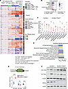
Human carcinomas often gain aggressive characteristics and escape cell-type specific treatment regimens through cryptic shifts in lineage states. However, the underlying mechanisms that govern lineage plasticity in carcinomas are undefined. Here in this study, we found that PAX5, a neural/lymphatic transcription factor, contributed to neuroendocrine (NE) lineage transition. PAX5 was highly expressed in aggressive human NE carcinoma cells and tissues but not in non-NE cancer cells and tissues. Deletion of Pax5 in Rb1fl/fl;Trp53fl/fl mice caused a reduction of tumor vessels, loss of NE morphologic features and decreased expression of ASCL1, NCAM, and SYP, whereas ectopic expression of PAX5 in CC10-rtTA;TetO-hEGFRex19del/T790M mice adenocarcinomas and in LNCAP prostate cancer xenografts induces an angiogenic microenvironment and NE morphology. Importantly, antiangiogenic drugs reduced NE features of Rb1fl/fl;Trp53fl/fl tumors and blocked PAX5-induced NE transformation. These studies demonstrate an essential role of angiogenic microenvironment in transition/maintenance of NE lineage, suggesting that targeting PAX5 and its downstream signaling may modulate lineage transitions responsible for treatment failure in both SCNCs and adenocarcinomas.
Authors
Ailing Wu, Yujie Hao, Xuemiao Yan, Junrong Liu, Lin Wang, Yan Jin, Wenxu Liu, Xiyue Chen, Yuan Jiang, Luc Girard, Zhiqun Shang, Jun Yan, Zhenfa Zhang, Wenchen Gong, Yuanjie Niu, Benjamin J. Drapkin, John D. Minna, Lance S. Terada, Zhenyi Ma, Zhe Liu
Abstract
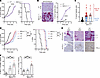
Antimetabolites, chemotherapy targeting nucleotide biosynthesis, are among the oldest and most widely used cancer treatments, yet resistance remains a daunting barrier, especially in the fight against B cell lymphomas. However, the underlying mechanisms of this resistance have long remained elusive. Using an innovative, integrated omics approach, we unexpectedly identified that the accumulation of dipeptides and upregulation of the dipeptide transporter SLC15A3 underlie resistance to nucleotide deficiency in a Myc-driven large B cell lymphoma mouse model. A similar mechanism occurred after long treatment of human B cell lymphoma cells with the chemotherapeutic purine synthesis inhibitor 6-mercaptopurine (6MP). Mechanistically, we demonstrated that dipeptides containing essential amino acids activated the growth and survival mTOR complex 1 (mTORC1) signaling pathway. Notably, SLC15A3 specifically interacted with mTOR on the lysosome, boosting mTORC1 activity selectively in resistant lymphoma cells but not in parental cancer cells. Silencing SLC15A3 diminished mTORC1 activity and restored resistant lymphoma sensitivity to 6MP. Strikingly, resistant lymphomas, but not primary tumors, exhibited heightened sensitivity to the clinical mTOR inhibitor, rapamycin, in culture and in vivo. We extended these findings in human lymphoma biopsies, which revealed increased SLC15A3 expression following antimetabolite therapy. Together, our study uncovered a metabolic adaptation that fuels cancer resistance to nucleotide deficiency and positions the mTORC1 inhibitor, rapamycin, as a potential therapeutic strategy for transforming the management of chemotherapy-resistant lymphomas.
Authors
Haojun Yang, Vincenzo Andrea Zingaro, Kevin Boardman, Ashish Noronha, Ekin Guney, Lingru Xue, Saishma Hoigebazar, Isabelle Liu, Sohit Miglani, Siyu Chen, Hieu Vu, Kwun Wah Wen, Hao G. Nguyen, Hani Goodarzi, Ralph J. DeBerardinis, Davide Ruggero
Abstract
Resistance to antiangiogenics is a major challenge in cancer therapy. These agents can either normalize or exacerbate tumor vascular abnormality and hypoxia. The mechanisms of resistance remain unclear in the latter setting. By integrating data from mouse models and clinical trials, we showed that hypoxia-inducing anti-VEGF therapy upregulated programmed cell death ligand 1 (PD-L1), yet failed to sensitize tumors to PD-L1 blockade. Mechanistically, early hypoxic stress triggered epithelial osteopontin (SPP1) production, which recruited monocytes and skewed macrophages toward M2 states, suppressing T cell cytotoxicity. Pharmacological SPP1 depletion impeded the development of hypoxia, reduced M2 infiltration, restored T cell activity, and enabled synergy between antiangiogenics and anti–PD-L1. Genetic dissection — tumor-epithelial Spp1-KO grafts and bone marrow chimeras generated by lethal irradiation and reconstitution with Spp1–/– or WT hematopoietic donors — showed that myeloid SPP1 contributed only marginally compared with epithelial SPP1. These findings identified SPP1 as a central mediator of resistance to hypoxia-inducing antiangiogenics, contributed to a comprehensive model of antiangiogenic resistance, and supported SPP1-targeted strategies to personalize immunotherapy and antiangiogenic therapy according to tumor hypoxia.
Authors
Jose Luis Ruiz-Sepulveda, Maria J. Bueno, Silvana Mouron, Veronica Jimenez-Renard, Manuel Muñoz, Manuel Moradiellos, Leonardo D. Garma, Luis García-Jimeno, Adam W. Watson, Ghassan Mouneimne, Solip Park, Rebeca Jimeno, Miguel Quintela-Fandino


Copyright © 2026 American Society for Clinical Investigation
ISSN: 0021-9738 (print), 1558-8238 (online)