Go to JCI Insight
  • About
  • Editors
  • Consulting Editors
  • For authors
  • Alerts
  • Advertising/recruitment
  • Subscribe
  • Contact
  • Current Issue
  • Past Issues
  • By specialty
    • COVID-19
    • Cardiology
    • Gastroenterology
    • Immunology
    • Metabolism
    • Nephrology
    • Neuroscience
    • Oncology
    • Pulmonology
    • Vascular biology
    • All ...
  • Videos
    • Conversations with Giants in Medicine
    • Author's Takes
  • Reviews
    • View all reviews ...
    • 100th Anniversary of Insulin's Discovery (Jan 2021)
    • Hypoxia-inducible factors in disease pathophysiology and therapeutics (Oct 2020)
    • Latency in Infectious Disease (Jul 2020)
    • Immunotherapy in Hematological Cancers (Apr 2020)
    • Big Data's Future in Medicine (Feb 2020)
    • Mechanisms Underlying the Metabolic Syndrome (Oct 2019)
    • Reparative Immunology (Jul 2019)
    • View all review series ...
  • Viewpoint
  • Collections
    • Recently published
    • In-Press Preview
    • Commentaries
    • Concise Communication
    • Editorials
    • Viewpoint
    • Top read articles
  • Clinical Medicine
  • JCI This Month
    • Current issue
    • Past issues

  • Current issue
  • Past issues
  • Specialties
  • Reviews
  • Review series
  • Conversations with Giants in Medicine
  • Author's Takes
  • Recently published
  • In-Press Preview
  • Commentaries
  • Concise Communication
  • Editorials
  • Viewpoint
  • Top read articles
  • About
  • Editors
  • Consulting Editors
  • For authors
  • Alerts
  • Advertising/recruitment
  • Subscribe
  • Contact
Long–echo time MR spectroscopy for skeletal muscle acetylcarnitine detection
Lucas Lindeboom, … , Patrick Schrauwen, Vera B. Schrauwen-Hinderling
Published October 1, 2014
Citation Information: J Clin Invest. 2014;124(11):4915-4925. https://doi.org/10.1172/JCI74830.
View: Text | PDF
Technical Advance Metabolism

Long–echo time MR spectroscopy for skeletal muscle acetylcarnitine detection

  • Text
  • PDF
Abstract

Animal models suggest that acetylcarnitine production is essential for maintaining metabolic flexibility and insulin sensitivity. Because current methods to detect acetylcarnitine involve biopsy of the tissue of interest, noninvasive alternatives to measure acetylcarnitine concentrations could facilitate our understanding of its physiological relevance in humans. Here, we investigated the use of long–echo time (TE) proton magnetic resonance spectroscopy (1H-MRS) to measure skeletal muscle acetylcarnitine concentrations on a clinical 3T scanner. We applied long-TE 1H-MRS to measure acetylcarnitine in endurance-trained athletes, lean and obese sedentary subjects, and type 2 diabetes mellitus (T2DM) patients to cover a wide spectrum in insulin sensitivity. A long-TE 1H-MRS protocol was implemented for successful detection of skeletal muscle acetylcarnitine in these individuals. There were pronounced differences in insulin sensitivity, as measured by hyperinsulinemic-euglycemic clamp, and skeletal muscle mitochondrial function, as measured by phosphorus-MRS (31P-MRS), across groups. Insulin sensitivity and mitochondrial function were highest in trained athletes and lowest in T2DM patients. Skeletal muscle acetylcarnitine concentration showed a reciprocal distribution, with mean acetylcarnitine concentration correlating with mean insulin sensitivity in each group. These results demonstrate that measuring acetylcarnitine concentrations with 1H-MRS is feasible on clinical MR scanners and support the hypothesis that T2DM patients are characterized by a decreased formation of acetylcarnitine, possibly underlying decreased insulin sensitivity.

Authors

Lucas Lindeboom, Christine I. Nabuurs, Joris Hoeks, Bram Brouwers, Esther Phielix, M. Eline Kooi, Matthijs K.C. Hesselink, Joachim E. Wildberger, Robert D. Stevens, Timothy Koves, Deborah M. Muoio, Patrick Schrauwen, Vera B. Schrauwen-Hinderling

×

Figure 8

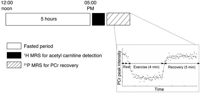
Outline of MR measurements performed.

Options: View larger image (or click on image) Download as PowerPoint
Outline of MR measurements performed.
Subjects were fasted for 5 hours b...
Subjects were fasted for 5 hours before measuring the acetylcarnitine concentration at rest, using our long-TE 1H-MRS protocol. After the acetylcarnitine measurement, an ergometer was placed in the scanner to perform exercise during 31P-MRS. The time course of the 31P-MRS measurement is given in more detail. Subjects performed exercise for 4 minutes in the scanner, after which the PCr peak recovery was monitored for 5 minutes.
Follow JCI:
Copyright © 2021 American Society for Clinical Investigation
ISSN: 0021-9738 (print), 1558-8238 (online)

Sign up for email alerts