
Advertisement
Commentary
Open Access |  10.1172/JCI210090
The alpha and the omega (6 lipids): discovering dietary drivers of heterotopic ossification
Peyton L. Carpen1 and Matthew B. Greenblatt1,2
1Department of Pathology and Laboratory Medicine, Weill Cornell Medical College, New York, New York, USA.
2Skeletal Health and Orthopedic Research Program, Hospital for Special Surgery, New York, New York, USA.
Address correspondence to: Matthew B. Greenblatt, Weill Cornell Medical College, LC 925, 1300 York Ave., New York, New York 10065, USA. Email: mag3003@med.cornell.edu.
Find articles by Carpen, P. in: PubMed | Google Scholar
1Department of Pathology and Laboratory Medicine, Weill Cornell Medical College, New York, New York, USA.
2Skeletal Health and Orthopedic Research Program, Hospital for Special Surgery, New York, New York, USA.
Address correspondence to: Matthew B. Greenblatt, Weill Cornell Medical College, LC 925, 1300 York Ave., New York, New York 10065, USA. Email: mag3003@med.cornell.edu.
Find articles by Greenblatt, M. in: PubMed | Google Scholar
Published September 1, 2026 - More info
J Clin Invest. 2026;136(17):e210090. https://doi.org/10.1172/JCI210090.
© 2026 Carpen et al. This work is licensed under the Creative Commons Attribution 4.0 International License. To view a copy of this license, visit http://creativecommons.org/licenses/by/4.0/.

Related article:
Abstract
Obesity is associated with impaired wound healing, but the mechanisms linking excess adiposity to aberrant tissue repair remain unresolved. Heterotopic ossification (HO) is a severe example of pathologic tissue repair in which mesenchymal progenitor cells (MPCs) undergo aberrant osteochondral differentiation within soft tissue, leading to joint contractures and pain. Here, we show that accumulation of dietary omega-6 (ω-6) lipids in the injury site is a key mechanism linking obesity to HO. Specifically, in mice fed a high-fat diet (HFD), injured tissues were enriched in linoleic and arachidonic acids, providing substrate for myeloid COX-2–dependent prostaglandin E2 (PGE2) production. PGE2 then drove a transcriptional program in MPCs that promoted osteochondral differentiation. An isocaloric, low linoleic acid HFD reduced HO despite comparable obesity, demonstrating that dietary lipid composition, rather than adiposity alone, drove pathological repair. Clinical data mirrored these findings, showing that obesity conferred increased HO risk, and COX-2 inhibition reduced HO exclusively in obese patients. Together, these findings identify injury site ω-6 lipid enrichment as the key signal linking the diet to MPC reprogramming, pointing to dietary lipid modulation as an actionable strategy to limit HO in obesity.
Authors
Stefanie L. Moye, Monisha Mittal, Tarun Srinivasan, Sneha Korlakunta, Chase A. Pagani, Ayelet Dar, Oromo Geshow, Dylan Feist, Lauren G. Zacharias, Zhao Li, Aaron W. James, Gerta Hoxhaj, Andrew M. Smith, Katherine A. Gallagher, Thomas P. Mathews, Robert J. Tower, Benjamin Levi
-
Dietary omega-6 lipids as drivers of HO
Extending these efforts, in this issue of the JCI, Moye et al. identified dietary omega-6 lipids as drivers of sporadic HO risk (9). Their work builds upon prior retrospective clinical studies finding a link between obesity and HO risk (10). The authors first substantiated this link between obesity and HO, mostly notably by directly establishing that high-fat diet–exposed mice display increased HO in a widely utilized model of burn- and tenotomy-associated HO that arises in tendon tissue. Moye et al. hypothesized that alterations in metabolism likely underlie the dietary effect on HO and performed metabolomics of the incipient HO site. Notably, they observed enrichment of omega-6 polyunsaturated fatty acids, including arachidonic acid and linoleic acid, a precursor for arachidonic acid, in the high-fat diet–exposed animals. This enrichment was coupled with local increases in a range of chemokines and cytokines, including IL-6 and TNF. These omega-6 fatty acids played a causal role in driving HO, as an isocaloric diet with a low omega-6/omega-3 fatty acid ratio had a protective effect in the murine HO model. Importantly, the use of an isocaloric control diet also clarified that this dietary effect on HO is driven directly by omega-6 fatty acids and not by general caloric surplus (Figure 1).
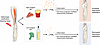 Figure 1
Dietary omega-6 fatty acids drive HO. Moye et al. (9) identified obesity as a risk factor for developing HO, a condition in which bone forms in soft tissue sporadically after limb injury. In a mouse model, depleting dietary intake of omega-6 fatty acids ameliorated HO by reducing local production of PGE2, indicating that dietary or pharmacological interventions could be developed for patients at risk of HO.
In obesity and in the setting of a high-fat and sugar-enriched Western diet, tissues accumulate omega-6 lipids, which can be converted to proinflammatory eicosanoids, including prostaglandins, thereby altering both immune and connective tissue cellular homeostasis. In particular, the omega-6 lipid arachidonic acid is converted to prostaglandins by the cyclooxygenases COX-1 and COX-2. Moye et al. found that the arachidonic acid enriched at the injury site was converted locally into prostanoids including PGE2 via COX-2–expressing myeloid cells. Consistent with past studies indicating that PGE2 has osteoanabolic activity, this PGE2 acted on local connective tissue progenitors to induce endochondral ossification (11).
In line with COX-2 being a mechanistic link between dietary omega-6 fatty acids and HO, treating the mouse HO model with the COX-2–specific inhibitor celecoxib attenuated HO development. While NSAIDs have been widely studied as a means to prevent HO formation (see ref. 12 for a meta-analysis of these studies), these findings refine the mechanistic context for their activity, placing NSAIDs downstream of a specific dietary pathway driving HO. Moreover, these findings suggest that NSAIDs will be most effective in obese patients at risk of HO, where these dietary omega-6 fatty acid pathways are most active, potentially explaining the observation that NSAIDs are only partially effective in preventing HO development. Moye et al. offer evidence to support this model. Analysis of an aggregated medical records database found that a prescription for celecoxib, a COX-2 inhibitor, significantly reduced HO incidence in obese patients who underwent total joint arthroplasty but did not significantly alter HO incidence in nonobese patients who underwent total joint arthroplasty.


Copyright © 2026 American Society for Clinical Investigation
ISSN: 0021-9738 (print), 1558-8238 (online)