
Advertisement
Review Series
Open Access |  10.1172/JCI207632
Expanding the therapeutic scope of PARP inhibitors in breast cancer: current indications and future directions
Charlotte S. Walmsley,1,2 Adela Rodriguez,1,2,3 Panagiotis A. Konstantinopoulos,1,2 Geoffrey I. Shapiro,1,2 Sara M. Tolaney,1,2 Judy E. Garber,1,2,3 and Filipa Lynce1,2
1Department of Medical Oncology, Dana-Farber Cancer Institute, Boston, Massachusetts, USA.
2Harvard Medical School, Boston, Massachusetts, USA.
3Division of Cancer Genetics and Prevention, Dana-Farber Cancer Institute, Boston, Massachusetts, USA.
Address correspondence to: Filipa Lynce, 450 Brookline Avenue, Boston, Massachusetts 02215, USA. Phone: 617.632.3000; Email: filipa_lynce@dfci.harvard.edu.
Find articles by Walmsley, C. in: PubMed | Google Scholar
1Department of Medical Oncology, Dana-Farber Cancer Institute, Boston, Massachusetts, USA.
2Harvard Medical School, Boston, Massachusetts, USA.
3Division of Cancer Genetics and Prevention, Dana-Farber Cancer Institute, Boston, Massachusetts, USA.
Address correspondence to: Filipa Lynce, 450 Brookline Avenue, Boston, Massachusetts 02215, USA. Phone: 617.632.3000; Email: filipa_lynce@dfci.harvard.edu.
Find articles by Rodriguez, A. in: PubMed | Google Scholar
1Department of Medical Oncology, Dana-Farber Cancer Institute, Boston, Massachusetts, USA.
2Harvard Medical School, Boston, Massachusetts, USA.
3Division of Cancer Genetics and Prevention, Dana-Farber Cancer Institute, Boston, Massachusetts, USA.
Address correspondence to: Filipa Lynce, 450 Brookline Avenue, Boston, Massachusetts 02215, USA. Phone: 617.632.3000; Email: filipa_lynce@dfci.harvard.edu.
Find articles by Konstantinopoulos, P. in: PubMed | Google Scholar
1Department of Medical Oncology, Dana-Farber Cancer Institute, Boston, Massachusetts, USA.
2Harvard Medical School, Boston, Massachusetts, USA.
3Division of Cancer Genetics and Prevention, Dana-Farber Cancer Institute, Boston, Massachusetts, USA.
Address correspondence to: Filipa Lynce, 450 Brookline Avenue, Boston, Massachusetts 02215, USA. Phone: 617.632.3000; Email: filipa_lynce@dfci.harvard.edu.
Find articles by
Shapiro, G.
in:
PubMed
|
Google Scholar
|

1Department of Medical Oncology, Dana-Farber Cancer Institute, Boston, Massachusetts, USA.
2Harvard Medical School, Boston, Massachusetts, USA.
3Division of Cancer Genetics and Prevention, Dana-Farber Cancer Institute, Boston, Massachusetts, USA.
Address correspondence to: Filipa Lynce, 450 Brookline Avenue, Boston, Massachusetts 02215, USA. Phone: 617.632.3000; Email: filipa_lynce@dfci.harvard.edu.
Find articles by Tolaney, S. in: PubMed | Google Scholar
1Department of Medical Oncology, Dana-Farber Cancer Institute, Boston, Massachusetts, USA.
2Harvard Medical School, Boston, Massachusetts, USA.
3Division of Cancer Genetics and Prevention, Dana-Farber Cancer Institute, Boston, Massachusetts, USA.
Address correspondence to: Filipa Lynce, 450 Brookline Avenue, Boston, Massachusetts 02215, USA. Phone: 617.632.3000; Email: filipa_lynce@dfci.harvard.edu.
Find articles by
Garber, J.
in:
PubMed
|
Google Scholar
|
1Department of Medical Oncology, Dana-Farber Cancer Institute, Boston, Massachusetts, USA.
2Harvard Medical School, Boston, Massachusetts, USA.
3Division of Cancer Genetics and Prevention, Dana-Farber Cancer Institute, Boston, Massachusetts, USA.
Address correspondence to: Filipa Lynce, 450 Brookline Avenue, Boston, Massachusetts 02215, USA. Phone: 617.632.3000; Email: filipa_lynce@dfci.harvard.edu.
Find articles by Lynce, F. in: PubMed | Google Scholar
Published October 1, 2026 - More info
J Clin Invest. 2026;136(19):e207632. https://doi.org/10.1172/JCI207632.
© 2026 Walmsley et al. This work is licensed under the Creative Commons Attribution 4.0 International License. To view a copy of this license, visit http://creativecommons.org/licenses/by/4.0/.

-
PARPi use in gBRCA1/2-mutated breast cancer
BRCA1 and BRCA2 have multiple critical roles in maintaining genomic integrity, including repairing double-strand DNA breaks through HRR, protecting replication forks, and preventing replication fork–associated gaps (Figure 1) (7–11). Germline mutations in BRCA1/2 result in hereditary cancer syndromes with elevated risks of breast cancer, ovarian cancer, and multiple other malignancies (12, 13). Defective DNA repair leads to genomic instability, cell cycle dysregulation, and unchecked proliferation. However, it also creates a therapeutic vulnerability that can be exploited by targeted anticancer therapies.
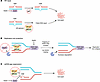 Figure 1
BRCA function in maintenance of genomic stability — lethality with PARP inhibition. Schematic illustrating the key functions of BRCA1/2 in maintaining genomic stability and the mechanisms underlying synthetic lethality with PARP inhibition. BRCA1/2 play critical roles in maintaining genomic integrity through three major mechanisms. (A) Homologous recombination–mediated (HR-mediated) repair of DNA double-strand breaks. PARP is recruited to sites of single-strand DNA breaks and facilitates their repair. In HR-deficient cells lacking functional BRCA1/2, double-strand break repair is compromised, resulting in genomic instability and ultimately cell death. (B) Protection of stalled replication forks from nucleolytic degradation. BRCA1 and BRCA2 also play essential roles in protecting stalled replication forks from degradation. During replication stress induced by PARPi treatment, PARP-DNA complexes become trapped on DNA, causing replication fork stalling. BRCA1 and BRCA2 facilitate loading of RAD51 onto stalled replication forks, preventing nucleases, such as MRE11, from degrading newly synthesized DNA. In the absence of BRCA1 or BRCA2, RAD51 loading fails to occur, leaving replication forks vulnerable to nucleolytic degradation and resulting in replication fork collapse and cytotoxicity. (C) Suppression of single-stranded DNA gap formation during DNA replication. Both PARP and BRCA1/2 suppress single-stranded DNA gap formation during DNA replication. PARP inhibition in BRCA1/2-mutant cells results in loss of both gap suppression mechanisms, leading to the accumulation of single-stranded DNA gaps and ultimately cell death. BER, base excision repair; ssDNA, single-stranded DNA; SSB, single-strand break.
Poly-ADP-ribosylation is a critical process for multiple DNA repair functions, including the recruitment and regulation of key base excision repair factors, such as DNA polymerase-β, XRCC1, and DNA ligase IIIα, as well as the promotion of chromatin relaxation (14–16). Poly-ADP-ribosylation is carried out by enzymes belonging to the ADP-ribosyltransferase superfamily, including PARP1 and PARP2. The primary targets of all currently approved PARPis, PARP1 and PARP2, are activated upon DNA damage to function as posttranslational modifiers within the BER pathway (17–19).
PARPis impair DNA damage repair pathways by targeting PARP enzymes, ultimately leading to cell death in HRR-deficient (HRD) tumors (20, 21). Multiple mechanisms have been proposed to underlie the synthetic lethality between PARP inhibition and BRCA1/2 deficiency, including inhibition of PARP-mediated BER (22), trapping of PARP-DNA complexes leading to replication fork collapse (23), and accumulation of single-strand DNA gaps at replication forks (24). Two PARPis, olaparib and talazoparib, are FDA approved for the treatment of breast cancer.
For early-stage disease, FDA approval of olaparib was based on results from the randomized phase III OlympiA trial, which evaluated 1 year of adjuvant olaparib versus placebo in patients with gBRCA1/2 mutations and high-risk human epidermal growth factor receptor 2–negative (HER2-negative) breast cancer. At 4 years of follow-up, olaparib significantly improved invasive disease-free survival (82.7% vs. 75.4%; HR 0.63), distant disease-free survival (86.5% vs. 79.1%; HR 0.57), and overall survival (OS) (89.8% vs. 86.4%; HR 0.68; P = 0.01), compared with placebo (25).
In the OlympiAD trial, olaparib significantly prolonged progression-free survival (PFS) compared with chemotherapy (7.0 vs. 4.2 months; HR 0.58; P < 0.001) in patients with metastatic breast cancer (MBC) harboring gBRCA1/2 mutations (26), without a corresponding OS benefit (27). However, a follow-up analysis suggested a greater OS benefit when using olaparib in the first-line setting compared with chemotherapy (22.6 vs. 14.7 months; HR 0.51; 95% CI: 0.29–0.90) (28). Similarly, in the EMBRACA trial, talazoparib demonstrated a 3-month PFS benefit compared with standard chemotherapy (8.6 vs. 5.6 months, HR 0.54, P < 0.001), with significant improvements in patient-reported quality of life and breast symptoms, and a numeric but not significant OS benefit (29). The pattern of PFS benefit without a corresponding OS benefit in the metastatic setting may be explained by underpowered OS analysis, subsequent postprogression therapies, and, particularly in EMBRACA, subsequent PARPi use in the chemotherapy arm (27, 29). Both agents were generally well tolerated, with adverse events rarely leading to treatment discontinuation. Olaparib was associated mostly with gastrointestinal symptoms, fatigue, and hematologic toxicities. Talazoparib was associated with a higher incidence of severe hematologic adverse events, likely due to higher PARP-trapping activity (27, 30).
-
PARPi use beyond gBRCA1/2-mutated breast cancer
HRD can arise through several mechanisms, including inherited germline variants, acquired somatic mutations, epigenetic alterations, and transcriptomic changes of HRR pathway genes (31). Efforts to better define and identify “BRCAness,” a state of HRD in tumors without gBRCA1/2 mutations, has emerged as a major area of investigation with important therapeutic implications (Figure 2) (32).
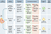 Figure 2
Approaches to HRD detection in breast cancer. A schematic overview of the genomic and functional strategies used to identify HRD in breast tumors. HRD can arise from a variety of mechanisms, including germline and somatic mutations in HR genes such as BRCA1/2 and PALB2, as well as transcriptomic and epigenetic changes. Methods of HRD detection can be broadly categorized as (i) cause-based biomarkers, such as germline or somatic mutation testing, which are supported by strong biological rationale and extensive clinical validation but identify only a subset of HRD tumors; (ii) consequence-based biomarkers that capture genomic scarring, LOH, large-scale transitions, and telomeric allelic imbalance, which offer strong clinical validation in ovarian cancer but are often expensive and may not reflect current HR status; and (iii) dynamic biomarkers that assess real-time functional HR status, such as RAD51 foci detection and transcriptome signatures, which are promising but lack clinical validation and assay standardization. LST, large-scale state transition; TAI, telomeric allelic imbalance. *Myriad Genetics MyChoice CDx is FDA approved as a companion diagnostic for selection of patients with HRD-positive, advanced ovarian cancer who may benefit from first-line maintenance treatment with a PARPi. Although genomic scar assays are widely used in breast cancer research, no HRD assay is currently FDA approved for patient selection in breast cancer.
sBRCA1/2 mutations occur spontaneously in approximately 3% of breast cancers (33–35). These somatic mutations closely resemble gBRCA1/2 mutations in their mutation type (including deletions, insertions, single nucleotide variants, and exon deletions), as well as degree of biallelic inactivation, genome-wide LOH, large chromosomal events, and telomeric imbalance (34–38). However, sBRCA1/2 mutations occur less frequently than gBRCA1/2 mutations and are generally diagnosed in older patients (35).
Growing evidence suggests efficacy of PARPis in patients with sBRCA1/2 mutations and other HRR deficiencies (Supplemental Table 1; supplemental material available online with this article; https://doi.org/10.1172/JCI207632DS1). The phase II Olaparib Expanded trial evaluated olaparib in MBC patients with sBRCA1/2 mutations or germline mutations in other HRR genes. For patients with sBRCA1/2 mutations, the study reported an objective response rate (ORR) of 50% with a median PFS (mPFS) of 6.3 months. Analysis of an expansion cohort confirmed sustained activity, with an ORR of 37% (11/30) and a mPFS of 5.5 months (39, 40). Similar findings were shown in a small cohort of patients with metastatic triple-negative breast cancer (TNBC) (41).
Additional prospective data are anticipated from an ongoing phase II trial evaluating talazoparib in MBC patients with sBRCA1/2 mutations (42). In line with new findings, the National Comprehensive Cancer Network Clinical Practice Guidelines for Breast Cancer were updated to include a statement on the potential efficacy of olaparib for sBRCA1/2 mutations in patients with MBC (43).
Mutations in other HRR genes. With the rapid expansion of germline and somatic tumor genetic testing, attention has shifted beyond BRCA1/2 to other HRR gene alterations as potential biomarkers of PARPi sensitivity. Among these is PALB2, which assists in the nuclear localization and stability of BRCA2, thus enabling its homologous recombination and checkpoint functions (44, 45). Inactivation of PALB2 confers HRD, leaving tumors vulnerable to PARP inhibition (46).
Consistent with this biological rationale, early clinical studies have shown meaningful activity of PARPis in patients with germline PALB2 (gPALB2) mutations, though available data remain limited. In the phase II Olaparib Expanded trial, 11 patients with gPALB2 mutations achieved a mPFS of 13.3 months and an ORR of 82%, with similarly favorable outcomes in the expansion cohort (PFS 9.6 months, ORR 75%) (39). Additional evidence comes from the Talazoparib Beyond BRCA trial, which evaluated talazoparib in patients with solid tumors and wild-type BRCA1/2 harboring mutations in other HRR genes. Among 6 patients with gPALB2 mutations, the mPFS was 6.9 months with an ORR of 50%, and all 5 patients with breast cancer achieved tumor shrinkage as best response (47).
In contrast, clinical activity of PARPis in tumors harboring mutations in other HRR genes appears less consistent (48). In the Olaparib Expanded Trial, no responses were observed in patients harboring CHEK2 (n = 10) or ATM (n = 11) mutations (40). A study from Hinic and colleagues showed that CHEK2-deficient tumors do not exhibit mutational signatures consistent with HRD, offering a mechanistic explanation for the absence of meaningful PARPi activity in this population (48). The lack of benefit observed in patients with ATM mutations may be explained by the variability of these mutations, particularly monoallelic or missense mutations, to confer ATM protein loss, as well as the compensatory pathways capable of maintaining homologous recombination (49, 50). However, preclinical evidence in other disease groups suggests ATM-deficient tumors may be more sensitive to PARPis in the appropriate circumstances, including when combined with ATR inhibition (51), or in the presence of an ASXL1 mutation (52), highlighting the complexity of translating HRR genotypes into clinically actionable biomarkers. Despite this, clinical studies have not demonstrated a clear benefit in breast cancer (53).
HRD genomic scar assays. Beyond somatic and germline mutations in HRR genes, multiple additional mechanisms can cause HRD. This complexity has driven efforts to create technologies that capture HRD across the genome aimed at predicting sensitivity to PARPis. Tumors with HRD depend on error-prone alternative pathways to resolve double-strand DNA breaks, resulting in characteristic genomic features, including large-scale chromosomal rearrangements detectable by genomic scar assays and distinct somatic mutational patterns identified through mutational signature profiling (54, 55).
Genomic scarring associated with HRD is measured through three primary mechanisms: LOH, TAI, and LSTs (56–58). Abnormalities in each of these categories can be quantified and integrated to generate a composite assessment of HRD. Multiple clinical assays have been developed to detect these genomic signatures of defective DNA repair, including the myChoice CDx test by Myriad Genetics and the FoundationFocus CDxBRCA LOH test (59, 60).
The myChoice HRD test includes BRCA1 and BRCA2 gene sequencing and quantification of LOH, TAI, and LST to create a Genomic Instability Score. This assay is FDA approved in ovarian cancer to guide the use of olaparib with bevacizumab in the first-line maintenance setting (61–66). In breast cancer, Telli and colleagues demonstrated that a combined HRD score — including LOH, TAI, and LST — significantly predicted pathologic complete response (pCR) to neoadjuvant platinum chemotherapy in patients with early-stage TNBC, even those without BRCA1/2 mutations (61). The FoundationFocus CDxBRCA LOH test, which integrates LOH scoring with HRR gene mutation analysis, was used in the ARIEL2 and ARIEL3 trials in patients with ovarian cancer to study the efficacy of the PARPi rucaparib (67, 68). In the ARIEL2 trial, patients with BRCA1/2 mutations or high LOH scores achieved a significantly longer PFS with rucaparib compared with those with low LOH scores. Thus far, neither assay has received FDA approval as a companion diagnostic to guide PARPi use in breast cancer, though this remains an area of strong clinical interest.
HRD mutational signatures. Somatic mutation analysis and HRD mutational signatures represent an alternative approach to quantifying HRD and identifying patients who may be sensitive to PARPis. HRDetect is a mutational signature–based test that combines six genomic signatures derived from high-depth whole-genome sequencing data to predict tumor BRCAness (69). Development of the HRDetect model consisted of initial training with gBRCA1/2-null samples used as positive controls, followed by validation with independent cohorts of breast, ovarian, and pancreatic tumor samples. In a cohort of 560 breast cancer tumors, the HRDetect test identified all 22 patients with gBRCA1/2 mutations, as well as 22 patients with sBRCA1/2 mutations and 47 tumors with functional BRCA1/2 deficiency without identifiable mutations. The phase II RIO trial evaluated rucaparib in 43 patients with untreated TNBC and demonstrated greater PARPi sensitivity in patients classified as HRD positive by HRDetect testing, as reflected by decreased circulating tumor DNA levels following treatment (70).
Additional clinical evidence supporting HRD-associated mutational signatures as predictive biomarkers of PARPi sensitivity comes from a retrospective cohort study by Batalini and colleagues. Using a clinico-genomic database linked to clinical outcomes, the investigators analyzed 177 patients with MBC treated with PARPis and found that patients with HRD-positive mutational signatures experienced significantly longer PFS than their HRD-negative counterparts (6.3 vs. 2.8 months; HR 0.62, 95% CI 0.42–0.92) (71). While these findings support its potential utility, the HRDetect algorithm has yet to be prospectively validated as a biomarker for predicting PARPi response, and its reliance on whole-genome sequencing may limit widespread implementation in routine clinical practice (72).
A major limitation of mutational signatures and genomic scar assays is their insensitivity to known mechanisms of PARPi resistance. For instance, when HRR proficiency is restored through secondary reversion mutations or DNA end resection rewiring, the genomic alterations accumulated during the period of HRD remain embedded in the tumor genome (73, 74). As a result, scar-based and mutational signature assays may continue to classify these tumors as HRD despite functional restoration of HRR proficiency (75). Functional biomarkers that directly assess real-time HRR competency, including transcriptome signatures and RAD51 foci formation assays (74), may help overcome this limitation by more accurately capturing dynamic changes in DNA repair capacity.
Transcriptome signature/gene expression profiling. By measuring functional HRR status, transcriptomic assays may better capture the dynamic nature of BRCAness, including adaptive changes associated with treatment resistance and tumor progression. Multiple signatures have been developed to quantify transcriptional HRD (76–78). In an early proof-of-concept study, Konstantinopoulos and colleagues used a microarray gene expression profile to characterize BRCA-like and non-BRCA-like tumors in 61 patients with epithelial ovarian cancer and demonstrated an association between BRCA-like expression and sensitivity to platinum chemotherapy and PARPis (79). Notably, 2 of 4 patients who underwent longitudinal sample collection exhibited alterations in their BRCAness profiles concomitant with the development of platinum resistance during treatment, underscoring the relevance of real-time evaluation of HRD status.
In breast cancer, Kang and colleagues developed a transcriptomic HRD signature that outperformed genomic HRD assays and BRCA1/2 mutation status in predicting sensitivity to multiple PARPis across cell lines and patient cohorts (78). These findings were further supported by results from an arm of I-SPY2, a phase II adaptive randomized clinical trial that used a gene expression–based diagnostic signature to identify tumors with a BRCA1ness phenotype. In the neoadjuvant setting, patients received either an oral PARPi combined with chemotherapy or standard chemotherapy alone. The study demonstrated a significant association between BRCA1ness and pCR, with the BRCA1ness signature predicting a higher likelihood of pCR in the veliparib–carboplatin arm (P = 0.03; OR, 3.2) but not in the carboplatin-alone arm (P = 0.45) (80). Although these studies demonstrate encouraging associations between transcriptomic HRD signatures and therapeutic response, additional validation in prospective clinical trials is necessary to establish their role in guiding PARPi therapy.
Protein expression. Protein levels of DNA damage response genes, particularly ATM, have been studied using immunohistochemistry (IHC) as a surrogate marker for HRD status (81). In metastatic gastric cancer, a clinical trial assessing combination therapy with a PARPi and paclitaxel demonstrated improved OS among patients whose tumors lacked ATM protein expression by IHC compared with those retaining ATM expression (82). However, follow-up analysis failed to reproduce this survival benefit (83). To date, similar IHC-based studies have not been systematically conducted in breast cancer populations, in part because IHC analysis has increasingly been replaced by other approaches for HRD characterization.
Functional assays: RAD51 foci detection. The RAD51 foci assay has emerged as the most clinically utilized and validated functional test for predicting PARPi response in breast cancer. RAD51 is a critical recombinase protein for HRR, forming RAD51 nucleoprotein filaments at sites of DNA double-strand breaks during the S or G2 phase of the cell cycle. The absence of RAD51 foci formation after DNA damage is indicative of deficient HRR (84).
Several testing strategies have been developed to assess RAD51 foci in breast cancer, including fresh tissue approaches, FFPE tissue–compatible tests, liquid biopsies, patient-derived xenograft–based (PDX-based) testing, and molecular profiling (85–89). Pellegrino and colleagues evaluated HRR genes, a genomic HRD test, and RAD51 foci detection across 103 breast cancer PDX models. RAD51 foci assessment was the most accurate predictor of PARPi response, achieving a predictive accuracy of 95% compared with 71% for genomic HRD analysis and 67% for HRR gene mutation analysis (89). Consistent findings were reported by Castroviejo-Bermejo and colleagues, in which RAD51 scores tracked more closely with PARPi response than genomic HRD scores in breast cancer PDX models (90).
Supporting the clinical relevance of RAD51, a post hoc biomarker analysis of the phase II GeparOLA trial in patients with early-stage HER2-negative breast cancer receiving neoadjuvant PARPi therapy combined with paclitaxel demonstrated that low RAD51 score was associated with higher rates of pCR to PARPi-based or platinum-based therapies (OR 12.03, 95% CI 2.60–55.73, P = 0.002) (91). Similarly, the phase II RADIOLA trial evaluating olaparib in patients with HER2-negative MBC showed a substantial improvement in ORR in RAD51-low compared with RAD51-high tumors (68.3% vs. 21.4%, P = 0.002) among patients harboring germline or somatic BRCA1/2, PALB2, or RAD51C/D mutations (92).
As a functional test that theoretically detects real-time HRR status, RAD51 foci detection is also an intriguing strategy for predicting PARPi resistance in patients who may have restored HRR functionality (93). A study by Cruz and colleagues evaluated PARPi response in 20 breast cancer PDX models harboring gBRCA1/2 mutations that had demonstrated variable response to PARPi therapy. High RAD51 foci correlated with poor PARPi response, regardless of the causal mechanism restoring HRR function (84). Taken together, these studies support RAD51 foci detection as a robust, functional biomarker capable of capturing real-time HRD status. However, several barriers have prevented clinical validation and implementation, including lack of standardized cutoffs, solid tumor tissue requirements, assay failure rates, and reliance on manual immunofluorescence scoring by trained pathologists (94–97).
Epigenetic assays: BRCA methylation. Epigenetic changes may also contribute to HRD tumors, including hypermethylation of BRCA1 and RAD51 promoter regions that lead to reduced gene expression. Hypermethylation within the BRCA1 promoter is estimated to occur in 9%–14% of breast cancers, whereas RAD51C promoter hypermethylation remains relatively uncommon (98–101). Notably, BRCA1 hypermethylation in breast and ovarian cancer is largely mutually exclusive with germline or somatic BRCA1/2 mutations, suggesting strong selective pressure to inactivate BRCA1 function through either genetic mutation or epigenetic silencing in these tumors (102, 103).
Clinical data supporting the use of BRCA1 methylation as a biomarker to guide PARPi therapy remain sparse. In a small study of patients with TNBC treated with PARPi-based therapy, those with BRCA1 promoter methylation or gBRCA1/2 mutations experienced a significantly improved 6-month PFS compared with patients with low BRCA1 methylation levels (P = 0.009). Notably, 1 patient with high BRCA1 promoter methylation (>80%) achieved a complete response (104). In contrast, the phase II COMETA-Breast trial, which enrolled 11 patients with advanced TNBC harboring confirmed sBRCA1/2 promoter methylation in the absence of gBRCA1/2 mutations, did not meet its predefined efficacy endpoints. Treatment with olaparib resulted in a low ORR of 9% and a mPFS of 2 months (105).
Article tools
- Download citation information
- Send a comment
- Terms of use
- Standard abbreviations
- Need help? Email the journal
Review Series
Emerging therapeutic strategies in breast cancer
-
Toward targeted therapeutics for lobular breast cancer
-
Antibody-drug conjugates in breast cancer: redefining targeted therapy
-
Expanding the therapeutic scope of PARP inhibitors in breast cancer: current indications and future directions
-
Breast cancer immunogenicity as a spectrum: evolving concepts and therapeutic implications
Metrics
Go to
- Top
- Abstract
- Introduction
- PARPi use in gBRCA1/2-mutated breast cancer
- PARPi use beyond gBRCA1/2-mutated breast cancer
- Mechanisms of resistance to PARP inhibition
- Novel PARPi combinations
- Conclusions and future directions
- Author contributions
- Conflict of interest
- Supplemental material
- Acknowledgments
- Footnotes
- References
- Version history


Copyright © 2026 American Society for Clinical Investigation
ISSN: 0021-9738 (print), 1558-8238 (online)